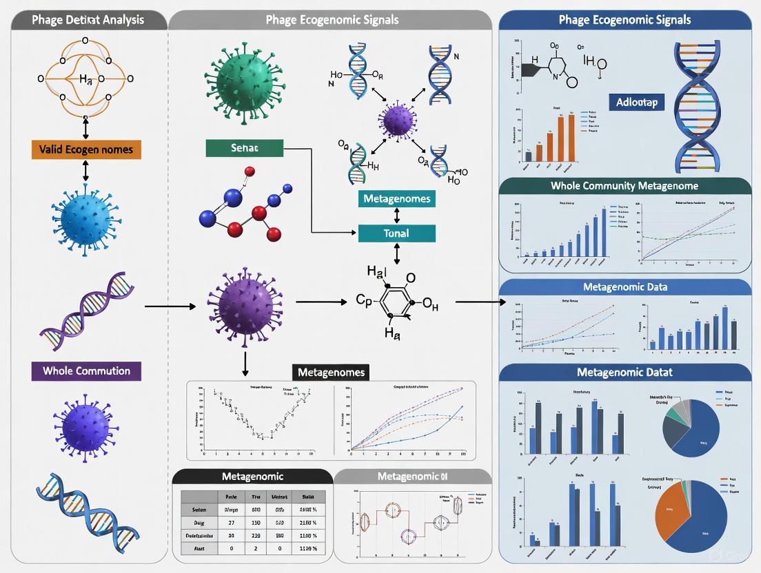
Validating Phage Ecogenomic Signals in Metagenomes: A Guide for Robust Detection and Analysis
The accurate identification and validation of bacteriophage sequences within whole-community metagenomes is a critical, yet challenging, step in understanding viral ecology, phage-host dynamics, and their implications for human health and...
Validating Phage Ecogenomic Signals in Metagenomes: A Guide for Robust Detection and Analysis
Abstract
The accurate identification and validation of bacteriophage sequences within whole-community metagenomes is a critical, yet challenging, step in understanding viral ecology, phage-host dynamics, and their implications for human health and biotechnology. This article provides a comprehensive framework for researchers and drug development professionals, addressing the entire workflow from foundational concepts to advanced validation. We explore the expanding diversity of phages, including jumbo phages, revealed by metagenomic surveys. The guide critically assesses current computational tools, from homology-based to machine learning approaches, and outlines best practices for benchmarking their performance. Furthermore, we detail strategies for in silico and experimental validation of phage signals, including host assignment and the use of viromes as validation standards. This synthesis aims to empower robust and reproducible phage ecogenomics, facilitating the translation of metagenomic signals into biological insights and therapeutic opportunities.
Unveiling the Viral Dark Matter: Foundations of Phage Diversity and Metagenomic Signals
Bacteriophages, or phages, are the most abundant and diverse biological entities on Earth. They play a pivotal role in shaping microbial communities through predation, horizontal gene transfer, and modulation of host metabolism. Recent advances in metagenomic sequencing have unveiled an unprecedented diversity of phage genomes, revealing expansive viral "dark matter" that had previously eluded characterization. Within this diversity, two groups stand out as particularly significant: jumbo phages with large genomic repertoires that blur the boundaries between viruses and cellular life, and crAss-like phages that dominate the human gut virome. Understanding the genomic landscape of these phages is critical for elucidating their ecological functions and potential applications in medicine and biotechnology. This review synthesizes current knowledge on phage genomic diversity, with a specific focus on validating phage ecogenomic signals within complex whole-community metagenomes—a fundamental challenge in viral ecology.
The Expanding Universe of Phage Genomic Diversity
Global Cataloguing Efforts and Quantitative Diversity
Large-scale metagenomic studies have dramatically expanded our catalog of phage genomes. The construction of unified, high-quality genome resources from diverse habitats has enabled systematic ecological and evolutionary insights previously hampered by fragmented data with significant habitat-specific biases [1]. One such effort analyzed 59,652,008 putative viral sequences from multiple environments to create a curated database of 741,692 phage genomes with ≥50% completeness (PGD50) [1]. This resource revealed that 28.96% (214,814) of these phage genomes clustered into 158,522 species-level viral clusters without any representation in existing databases, highlighting the substantial novelty being uncovered [1].
Table 1: Phage Diversity Across Different Habitats and Host Systems
| Habitat/Host System | Number of vOTUs/vMAGs | Notable Phage Groups | Key Genomic Features | Reference |
|---|---|---|---|---|
| Human Gut | 3738 complete genomes (451 genera) | "Flandersviridae", "Quimbyviridae", "Gratiaviridae" | Catalases, iron-sequestering enzymes, DGRs, isoprenoid pathway enzymes | [2] |
| Pig Gut | 12,896 high-confidence vOTUs | crAss-like phages (533 vOTUs) | Anti-CRISPR genes, CAZymes (lysozymes), alternative genetic codes | [3] [4] |
| Mouse Gut | 977 high-confidence vOTUs | Novel clades with high prevalence | Cas-harboring jumbophages | [3] |
| Cynomolgus Macaque Gut | 1,480 high-confidence vOTUs | crAss-like phages | 55.88% have connections to human microbiota | [3] |
| Honey Bee Gut (Individual Bees) | 1,069 vOTUs from 49 bees | Modular phage-bacteria interaction networks | High strain-level diversity correlated with bacterial hosts | [5] |
| Oral Cavity | 189,859 representative sequences | 3,416 huge phages (>200 kbp) | Anti-defense genes, AMGs, virulence factors | [6] |
| Human Breast Milk | 7 primary phage families | Herelleviridae, Myoviridae, Podoviridae | Vertical mother-to-infant transmission | [7] |
The honey bee gut microbiome has emerged as a powerful model system for studying phage-bacteria interactions due to its relative simplicity and well-characterized bacterial community. Research on 49 individual bees revealed 1,069 viral operational taxonomic units (vOTUs) with a highly modular phage-bacteria interaction network structure, where viral and bacterial diversity were strongly correlated, particularly at the strain level [5]. This correlation underscores the importance of strain-level resolution when studying phage-bacteria diversity patterns, as phage specificity often occurs at this taxonomic level rather than at the species level [5].
Jumbo Phages: Genomic Giants with Expanded Capabilities
Jumbo phages, typically defined by genomes exceeding 200 kbp, represent a fascinating frontier in phage genomics. These genomic giants encode expanded functional repertoires that may include metabolic genes, defense systems, and transcriptional machinery typically associated with cellular organisms. A comprehensive analysis of oral phages identified 3,416 "huge phages" with genome sizes >200 kbp, demonstrating their presence in diverse body sites [6].
Particularly noteworthy are cas-harboring jumbophages discovered in mammalian guts, which encode CRISPR-Cas systems potentially used in competition with other mobile genetic elements or host defenses [3]. These findings challenge traditional views of phages as simple genetic parasites and suggest more complex evolutionary relationships with their bacterial hosts.
Jumbo phages often manipulate host metabolism in sophisticated ways. Some "Flandersviridae" phages, for instance, encode enzymes of the isoprenoid pathway, a lipid biosynthesis pathway not previously known to be manipulated by phages [2]. Similarly, numerous phages across different families encode catalases and iron-sequestering enzymes that may enhance cellular tolerance to reactive oxygen species, potentially providing protection to their bacterial hosts under oxidative stress [2].
CrAss-like Phages: Ubiquitous Colonizers of the Mammalian Gut
Since its discovery in 2014, the crAss-like phage family has emerged as one of the most abundant and widespread viral groups in the human gut. Recent research has expanded our understanding of their diversity, host interactions, and distribution across mammalian species.
In pig guts, crAss-like phages are distributed across four well-known family-level clusters (Alpha, Beta, Zeta, and Delta) but are notably absent from Gamma and Epsilon clusters [4]. Genomic analysis of 533 pig crAss-like phage vOTUs revealed that 149 utilize alternative genetic codes, while approximately 64.73% of their genes lack functional annotations, highlighting significant gaps in understanding their functional potential [4].
These phages primarily infect bacteria in the Bacteroidetes phylum, particularly Prevotella, Parabacteroides, and UBA4372 [4]. Interestingly, interactions between crAss-like phages and Prevotella copri may influence fat deposition in pigs, suggesting potential applications in agricultural science [4]. Unlike the high prevalence observed in human populations, pig crAss-like vOTUs generally exhibit low prevalence across populations, indicating greater heterogeneity in their compositions [4].
Table 2: Comparative Genomic Features of crAss-like Phages Across Mammals
| Feature | Human Gut | Pig Gut | Cynomolgus Macaque Gut |
|---|---|---|---|
| Cluster Distribution | All known clusters | Alpha, Beta, Zeta, Delta (no Gamma, Epsilon) | Similar to human with animal-specific characteristics |
| Genome Size Range | ~70-100 kbp | >70 kbp | Similar to human |
| Host Range | Primarily Bacteroidetes | Prevotella, Parabacteroides, UBA4372 | Primarily Bacteroidetes |
| Prevalence | High, ubiquitous | Low prevalence, heterogeneous | 55.88% connected to human microbiota |
| Unique Features | Carrier state lifestyle | Alternative genetic codes, anti-CRISPR proteins, CAZymes | Animal-specific clusters |
Methodological Framework: Validating Ecogenomic Signals in Metagenomes
Experimental Workflows for Phage Genome Recovery
The accurate identification and characterization of phage genomes from metagenomic data requires sophisticated computational workflows that integrate multiple complementary approaches. The standard pipeline involves sequential steps of quality control, assembly, viral sequence identification, quality filtering, and host assignment [3] [8].
Figure 1: Workflow for phage genome recovery from metagenomic data. Critical steps include quality assessment tools like CheckV for estimating completeness and removing contaminating host sequences, followed by multiple viral identification tools to maximize recovery of diverse phage types.
The recovery of high-quality viral genomes requires stringent quality control measures. As demonstrated in studies of mammalian gut viromes, contigs are typically filtered to retain only those with ≥90% completeness as assessed by CheckV, while removing those with potential contamination or questionable quality warnings [3]. For species-level clustering, 95% average nucleotide identity (ANI) and 85% alignment fraction (AF) across the shorter sequence are widely adopted standards [3] [5].
The Marker-MAGu pipeline represents an innovative approach for simultaneous profiling of phage and bacterial communities from whole-community metagenomes [9]. This method identifies essential phage genes (involved in virion structure, genome packaging, and replication) and integrates them with bacterial marker genes from MetaPhlAn, enabling trans-kingdom taxonomic profiling from the same metagenomic dataset [9]. When applied to 12,262 longitudinal samples from 887 children, this approach demonstrated that phage communities change more quickly than bacterial communities, with most phages persisting for shorter durations [9].
The Scientist's Toolkit: Essential Research Reagents and Solutions
Table 3: Essential Computational Tools for Phage Metagenomics
| Tool Name | Function | Key Features | Applicability |
|---|---|---|---|
| VirSorter2 | Viral sequence identification | Modular outputs, detects diverse phage types | Metagenomic and single-genome data |
| DeepVirFinder | Viral sequence identification | k-mer based machine learning approach | Metagenomic data, novel phage detection |
| CheckV | Viral genome quality assessment | Estimates completeness, removes host contamination | Quality control for viral genomes |
| PhaMer | Viral sequence identification | Transformer model for metagenomic prediction | Handling fragmented metagenomic data |
| geNomad | Viral taxonomy & identification | Viral taxon markers for ICTV lineages | Taxonomic classification |
| BACPHLIP | Lifestyle prediction | Classifies virulent vs. temperate phages | Ecological inference |
| CRISPR spacer matching | Host prediction | Identifies protospacers matching bacterial CRISPR arrays | Host-phage interaction mapping |
| Marker-MAGu | Trans-kingdom profiling | Simultaneous detection of phages and bacteria | Whole-community metagenomic analysis |
Ecogenomic Signatures: Validating Phage-Habitat Associations
The concept of ecogenomic signatures refers to the habitat-specific genetic patterns that can distinguish microbial ecosystems. Research has demonstrated that individual phages can encode clear habitat-related signals diagnostic of underlying microbiomes [10]. For example, the gut-associated φB124-14 phage encodes an ecogenomic signature that can segregate metagenomes according to environmental origin and distinguish contaminated environmental metagenomes from uncontaminated datasets [10].
This approach was validated through comparative analysis of the relative representation of phage-encoded gene homologs in metagenomic datasets from different habitats. The φB124-14 phage showed significantly greater representation in human gut viromes compared to environmental datasets, while cyanophage SYN5 displayed the opposite pattern—greater representation in marine environments [10]. These distinct ecogenomic signatures persisted even when analyzing whole-community metagenomes, though the effects were less pronounced than in viral fraction metagenomes [10].
The power of ecogenomic signatures extends to clinical applications. In the TEDDY study, the addition of phage taxonomic profiles improved the ability to discriminate samples geographically over bacterial taxonomic profiles alone [9]. Furthermore, temporal dynamics of phage and bacterial communities differed during the second year of life for children later diagnosed with type 1 diabetes, suggesting that phage ecogenomic signatures may serve as early indicators of disease susceptibility [9].
The landscape of phage genomic diversity encompasses extraordinary variation, from the genomic giants represented by jumbo phages to the ubiquitous crAss-like phages that dominate mammalian guts. Methodological advances in metagenomic analysis have enabled the recovery of increasingly complete and accurate phage genomes, revealing novel taxa and unexpected genomic features. The validation of phage ecogenomic signatures in whole-community metagenomes represents a particularly promising frontier for both basic microbial ecology and applied biotechnology. As reference databases continue to expand and analytical methods improve, we anticipate that phage ecogenomic signatures will find increasing applications in source tracking, disease diagnostics, and therapeutic development. The integration of phage data with bacterial community profiles will provide a more complete understanding of microbiome dynamics and their impact on human and animal health.
The vast universe of bacteriophages (phages) represents one of the most significant frontiers in microbial ecology, yet it remains largely unexplored. Metagenomics has emerged as a powerful discovery engine, enabling researchers to probe this universe by identifying phage sequences within complex microbial communities without the need for cultivation [11]. A critical hypothesis driving this research is that individual phages encode discernible, habitat-associated ecogenomic signatures—genetic patterns diagnostic of their underlying microbial ecosystems [10]. For instance, the gut-associated phage ϕB124-14 encodes a specific suite of genes whose homologs are significantly enriched in human gut-derived metagenomes compared to those from other environments [10]. Validating these signals in whole community metagenomes is paramount, as it allows for the direct study of phage-host dynamics and integrated prophages, moving beyond the limitations of purified viromes [11]. This guide objectively compares the performance of modern bioinformatic tools designed to detect these phage sequences, providing a framework for researchers to validate ecogenomic signals and expand the known phage universe.
The development of numerous computational tools for phage identification has created a need for systematic benchmarking. Independent studies have evaluated these tools on standardized datasets to assess their precision, recall, F1 scores, and robustness to various challenges [12] [13]. The table below summarizes the key performance metrics of leading tools on a benchmark of artificial contigs derived from RefSeq genomes.
Table 1: Performance of Phage Identification Tools on RefSeq Artificial Contigs
| Tool | Primary Approach | Reported F1 Score | Reported Precision | Reported Recall |
|---|---|---|---|---|
| VIBRANT | Gene-based / Machine Learning | 0.93 | — | — |
| VirSorter2 | Gene-based / Machine Learning | 0.93 | — | — |
| Kraken2 | k-mer-based / Reference Database | 0.86 (on Mock Community) | 0.96 (on Mock Community) | — |
| DeepVirFinder | k-mer-based / Machine Learning | — | — | — |
| VirFinder | k-mer-based / Machine Learning | — | — | — |
| Seeker | Sequence Composition / Machine Learning | — | — | — |
| PPR-Meta | Sequence Composition / Machine Learning | (High FPs on shuffled sequences) | — | — |
| MetaPhinder | Homology / Reference Database | — | — | — |
| viralVerify | Gene-based / Machine Learning | — | — | — |
The performance of these tools can vary significantly based on the benchmark. For example, Kraken2 achieved a notably high F1 score of 0.86 on a mock community benchmark, largely due to its exceptional precision of 0.96 [12] [11]. In contrast, some tools, most notably PPR-Meta, have been shown to call a high number of false positives on randomly shuffled sequences, indicating a potential lack of specificity [12] [11].
Generally, a trade-off exists between different methodological approaches. Homology-based tools (e.g., VirSorter, VIBRANT, VirSorter2, viralVerify) typically demonstrate low false positive rates and robustness to eukaryotic contamination [13]. Conversely, tools relying on sequence composition (e.g., VirFinder, DeepVirFinder, Seeker) often show higher sensitivity, which allows them to detect phages with less representation in reference databases, but may be more susceptible to certain biases [13]. These differences lead to strikingly dissimilar outputs when applied to real metagenomes; in one evaluation of human gut data, nearly 80% of contigs flagged as phage were identified by only a single tool [13].
Experimental Protocols for Tool Benchmarking
To ensure fair and reproducible comparisons, benchmarking studies follow rigorous experimental protocols. The methodologies below outline the creation of key datasets used to evaluate tool performance.
RefSeq Artificial Contigs and Mock Community Benchmark
This protocol tests a tool's ability to correctly identify known phage sequences and reject non-viral sequences in a controlled setting [11].
True-Positive Set Creation:
- Source: All complete phage genomes deposited in RefSeq during a specific timeframe (e.g., Jan 2020 - Aug 2021) are downloaded.
- Quality Control: Genomes are dereplicated against older RefSeq data and the training sets of the tools being benchmarked to prevent overfitting.
- Fragmentation: Quality-controlled genomes are uniformly fragmented into contigs of sizes ranging from 1 kbp to 15 kbp to simulate metagenomic assemblies.
True-Negative Set Creation:
- Source: All complete bacterial and archaeal chromosomes and plasmids from the same RefSeq timeframe are downloaded.
- Viral Content Filtering: Sequences are filtered to remove any with ≥30% of their open reading frames (ORFs) matching viral proteins in the pVOG database, ensuring they are truly non-viral.
Mock Community Analysis:
- A previously sequenced mock community containing a known set of phage species is used as an additional validation dataset [11].
Evaluation:
- Tools are run on the fragmented RefSeq and mock community datasets.
- Precision, Recall, and F1 Score are calculated based on the tools' abilities to correctly classify the true phage and true non-phage sequences.
Simulated Metagenomes and Virome Analysis
This protocol assesses tool performance under more realistic conditions, including sequencing errors, assembly artifacts, and low viral abundance [13].
Dataset Simulation:
- Read Simulation: Tools like InSilicoSeq are used to generate simulated Illumina sequencing reads from a curated set of phage, bacterial, archaeal, and eukaryotic genomes. This incorporates realistic error models from specific sequencing platforms.
- Metagenome Assembly: The simulated reads are assembled into contigs using standard metagenomic assemblers.
Fragment Length and Contamination Assessment:
- Reference genomes are fragmented into non-overlapping segments of various lengths (e.g., 500 bp, 1,000 bp, 3,000 bp, 5,000 bp).
- Contigs are mixed with fragments from eukaryotic genomes to test robustness against contamination.
Analysis of Real Metagenomes and Viromes:
- Tools are run on real-world datasets, such as human gut whole-community metagenomes and purified viromes.
- The resulting phage communities are compared to understand how tool choice influences ecological conclusions [13].
A Workflow for Validating Phage Ecogenomic Signatures
The following diagram illustrates the logical workflow for using benchmarked tools to detect and validate phage ecogenomic signatures in whole community metagenomes.
Successful phage discovery in metagenomes relies on a suite of computational tools and biological databases. The following table details key resources for researchers in this field.
Table 2: Essential Research Reagents and Resources for Phage Metagenomics
| Resource Name | Type | Primary Function in Phage Discovery |
|---|---|---|
| VIBRANT | Software Tool | Uses a neural network and HMMs to identify phage sequences and characterize auxiliary metabolic genes [11]. |
| VirSorter2 | Software Tool | Employs multiple random forest classifiers to detect a diverse array of viral sequences from different groups [11]. |
| Kraken2 | Software Tool | A k-mer-based taxonomic classifier that can be applied to phage detection with high precision [12] [11]. |
| DeepVirFinder | Software Tool | Applies a convolutional neural network on k-mer signatures to identify phage sequences, especially on shorter contigs [11]. |
| RefSeq | Database | A curated database of reference sequences used for training, benchmarking, and homology-based searches [11] [13]. |
| pVOG/VDB | Database | Databases of viral protein families and genomes used by tools for HMM profiling and homology detection [11]. |
| MGnify | Database | A specialized repository for microbiome metagenomic data, providing access to community-derived sequences and analyses [14]. |
| IMG/VR | Database | A system for hosting and analyzing viral genomes and metagenomes, useful for comparative analysis [14]. |
The expansion of the known phage universe through metagenomics is intrinsically linked to the computational tools used for discovery. Benchmarking studies reveal that no single tool is universally superior; each has unique strengths and weaknesses [12] [13]. Homology-based tools like VIBRANT and VirSorter2 offer high accuracy and low false positive rates, while sequence composition-based tools like DeepVirFinder can be more sensitive to novel phages absent from databases. The high-precision classifier Kraken2 is excellent for well-characterized sequences.
Therefore, the optimal strategy for validating true phage ecogenomic signals in whole community metagenomes involves a consensus-based approach. Researchers should leverage multiple tools from different methodological categories and prioritize contigs identified by several independent algorithms. This mitigates individual tool biases and provides a more robust validation of the phage ecogenomic signatures that are critical to understanding the role of viruses in microbial ecosystems and human health.
The validation of phage ecogenomic signals within whole-community metagenomes presents a formidable challenge for researchers investigating viral roles in microbial ecosystems. This guide objectively compares the performance of different methodological approaches against three core challenges: database incompleteness, the lack of universal viral markers, and host contamination. The following data, synthesized from current research, provides a framework for selecting appropriate protocols and reagents to advance phage ecogenomics.
Challenge One: Database Incompleteness and Taxonomic Errors
Database incompleteness and misannotation severely limit the accuracy of taxonomic classification in metagenomic studies. These issues are pervasive in default databases mirrored from NCBI, affecting downstream biological interpretations.
Table 1: Impact and Mitigation of Database Issues
| Issue Type | Prevalence & Impact | Performance of Mitigation Strategies |
|---|---|---|
| Taxonomic Misannotation | An estimated 1-3.6% of prokaryotic genomes in RefSeq and GenBank are misannotated [15]. | ANI Clustering: Corrected Dickeya dadantii misannotation to D. paradisiaca after comparison with type material [15]. |
| Database Contamination | 2,161,746 contaminated sequences identified in GenBank; 114,035 in RefSeq [15]. Incomplete Lineage Representation: Missing radiolarians (Retaria) led to 42,736 unannotated proteins and 46,283 misannotations in a marine transect study [16]. | Curation & Validation: FDA-ARGOS uses a restrictive, verified-sequence approach. Database testing across thousands of samples is recommended for critical applications [15]. |
| Unspecific Labeling | Annotations at high taxonomic levels (e.g., "Bacteria") preclude species-level resolution [15]. | Deep Annotation: Annotating to the deepest possible node in the taxonomic tree improves resolution [15]. |
Experimental Protocol: Evaluating Database-Driven Bias
A clear experimental protocol exists for quantifying the impact of database composition on taxonomic profiling [16]:
- Database Manipulation: Create modified versions of a reference database (e.g., MMETSP). For a target genus like Phaeocystis, create subsets containing (a) all references, (b) only colony-forming species, and (c) only free-living species.
- Annotation: Annotate the same set of assembled metagenomic contigs from different environments (e.g., Southern Ocean vs. Mediterranean Sea) against all three database versions using a standard lowest common ancestor (LCA) algorithm.
- Quantification: For each database, calculate the percentage of target sequences (e.g., Phaeocystis) identified. The variation in recovery rates directly demonstrates database bias.
Challenge Two: Lack of Universal Markers and Host Prediction
Unlike prokaryotes with 16S rRNA, phages lack a universal phylogenetic marker. This complicates their identification and the crucial step of host prediction. Method selection significantly influences host prediction success rates.
Table 2: Performance of Phage Identification and Host Prediction Methods
| Method | Principle | Performance Data & Experimental Findings |
|---|---|---|
| Extrachromosomal Sequencing | Selective sequencing of circular DNA (plasmidomes) to enrich for phage sequences [17]. | Identified 200 viral sequences from groundwater; 32 of 41 viral clusters represented putative new genera, demonstrating high novelty discovery [17]. |
| Tetranucleotide Frequency | Uses k-mer composition similarity between phage and host genomes [17]. | Most Productive Method: Predicted hosts for 71/200 viral genomes using public NCBI WGS and for 16/20 using local isolate genomes [17]. |
| BLAST Homology (BLAST99) | Identifies near-exact matches (e.g., >99% identity and query coverage) indicating prophage integration [17]. | Highest Confidence: Enabled strain-level host assignments. Four viruses were identified as integrated into genomes of Pseudomonas, Acidovorax, and Castellaniella strains [17]. |
| CRISPR Spacer Analysis | Matches phage sequences to CRISPR spacer arrays in bacterial genomes [17]. | Least Productive: Predicted only 2 hosts for the 200 groundwater viral genomes, highlighting limited sensitivity [17]. |
| Ecogenomic Signatures | Profiles relative abundance of phage gene homologs across diverse metagenomes to infer habitat association [10]. | Gut phage ΦB124-14 showed significantly higher signal in human gut viromes vs. environmental viromes. This signature discriminated "contaminated" environmental metagenomes in simulated faecal pollution studies [10]. |
Experimental Protocol: Multi-Method Host Prediction
A robust host prediction workflow integrates multiple methods to maximize results [17]:
- Phage Genome Identification: Use a tool like VirSorter to identify viral sequences from metagenomic or plasmidome assemblies [17].
- Host Prediction with Public Databases: Run the viral sequences against a comprehensive database of bacterial WGS (e.g., from NCBI) using:
- Tetranucleotide Frequency Analysis: For broad host-range predictions.
- BLAST Homology: To find highly similar sequences.
- Host Prediction with Local Isolates: For high-confidence, strain-level assignments, repeat Step 2 using WGS from bacterial isolates sourced from the same environment. The BLAST99 approach is particularly powerful here for detecting integrated prophages.
- Validation: Cross-reference predictions from different methods. Predictions confirmed by multiple methods (e.g., both BLAST and tetranucleotide frequency) are considered high-confidence.
Challenge Three: Host Decontamination in Metagenomic Data
Host DNA contamination is a major concern, especially in low-biomass samples, and can lead to false inferences. Statistical decontamination tools are essential for generating accurate microbial community profiles.
Table 3: Performance Comparison of Decontamination Tools
| Tool/Method | Underlying Principle | Performance in Experimental Studies |
|---|---|---|
| Decontam (Frequency) | Models inverse correlation between contaminant frequency and sample DNA concentration [18]. | In a human oral dataset, classifications were consistent with prior microscopic observations. Reduced technical variation in a dilution series dataset arising from different sequencing protocols [18]. |
| Decontam (Prevalence) | Identifies sequences with significantly higher prevalence in negative controls than in true samples [18]. | Corroborated the conclusion that little evidence exists for an indigenous placenta microbiome. Identified contaminants that were low-frequency taxa associated with preterm birth [18]. |
| Relative Abundance Threshold | Ad hoc removal of sequences below an abundance cutoff (e.g., 0.1%) [18]. | Poor Performance: Removes rare but true sequences and fails to remove abundant contaminants, which are most likely to interfere with analysis [18]. |
| Negative Control Subtraction | Removal of all sequences found in negative controls [18]. | Limited Specificity: Can remove true sequences that appear in controls due to cross-contamination or index hopping [18]. |
Experimental Protocol: In Silico Decontamination with Decontam
The decontam R package provides a straightforward statistical workflow [18]:
- Input Data Preparation: Create a feature table (ASV or OTU table) and a corresponding sample metadata file.
- Define Method: Choose the decontamination method based on available data:
- Frequency-Based: Requires quantitative DNA concentration measurements for each sample.
- Prevalence-Based: Requires sequenced negative controls processed alongside biological samples.
- Execution: Run the
isContaminant()function in R, specifying the chosen method and threshold. The function returns a logical vector identifying which features are classified as contaminants. - Result Application: Remove the contaminant features from the feature table before proceeding with downstream ecological analysis.
The Scientist's Toolkit: Essential Research Reagents & Databases
Table 4: Key Reagents and Databases for Phage Ecogenomics
| Research Material | Function in Workflow | Specific Examples / Notes |
|---|---|---|
| VirSorter | Identifies viral sequences from metagenomic assemblies [17]. | Critical first step for virome analysis from complex metagenomic data. |
| Decontam (R Package) | Statistically identifies and removes contaminant DNA sequences from marker-gene and metagenomic data [18]. | Integrates easily with existing MGS workflows; uses frequency or prevalence patterns. |
| NCBI RefSeq/GenBank | Primary public repositories for nucleotide sequences used as reference databases [15]. | Known to contain contamination and taxonomic errors; requires curation for critical work [15]. |
| MMETSP Database | Curated database of marine microbial eukaryote transcriptomes [16]. | Used for taxonomic annotation of protists; missing key groups like radiolarians [16]. |
| Bacterial Whole-Genome Sequences (Local Isolates) | High-confidence reference for host prediction of phages from the same environment [17]. | Dramatically improves strain-level host prediction compared to public databases alone [17]. |
| Filamentous Phage (e.g., M13) | Vector for phage display technology; used for epitope mapping and protein interaction studies [19] [20]. | pIII and pVIII are common coat proteins for fusion [19]. |
| Phagemid Vectors | Hybrid vectors containing phage and plasmid origins of replication; used for antibody display [19]. | Requires a helper phage (e.g., M13KO7) for packaging into a viral particle [19]. |
Visualizing Experimental Workflows and Logical Relationships
Phage Ecogenomic Validation Workflow
Database Issue Impact on Annotation
Bacteriophages, the most abundant biological entities in most ecosystems, encode distinct habitat-associated signals derived from co-evolution and adaptation with their bacterial hosts. The identification and validation of these ecogenomic signatures in whole community metagenomes present both a significant challenge and opportunity for advancing microbial ecology and therapeutic development. These signatures manifest through the relative abundance of phage-associated genes, protein cluster distributions, and contextual genomic features that serve as reliable indicators of phage lifestyle, host interactions, and ecological functions [21]. The precision with which these signals can be interpreted directly impacts diverse applications ranging from microbial source tracking in environmental samples to the development of targeted phage therapies for combating antibiotic-resistant infections [22].
Recent technological advances in sequencing platforms and bioinformatics tools have dramatically expanded our capacity to detect and analyze phage sequences within complex microbial communities. However, the validation of ecogenomic signals requires careful consideration of methodological approaches, as demonstrated by studies showing that individual phage genomes like φB124-14 encode discernible habitat-related signatures that can successfully distinguish human gut viromes from other environmental sources [21]. This evolving capability to interpret phage genomic signals within whole community metagenomes represents a transformative development for both basic research and applied biotechnology.
Computational Tool Performance for Phage Detection
The accurate identification of phage sequences within metagenomic data represents the foundational step in ecogenomic signal interpretation. A comprehensive benchmark evaluation of nine computational phage detection tools revealed striking differences in their performance characteristics and output results [13].
Table 1: Performance Metrics of Phage Detection Tools on Benchmark Datasets
| Tool | Approach | Sensitivity on Short Fragments (<3kb) | Robustness to Eukaryotic Contamination | Strengths | Limitations |
|---|---|---|---|---|---|
| PhaMer | Protein-cluster Transformer | High (contextual embedding) | High | Superior F1-score on real metagenomic data | Computational complexity |
| VirSorter2 | Homology (random forest) | Moderate | High | Low false positive rate | Database dependence |
| DeepVirFinder | Sequence composition (CNN) | High | Moderate | Sensitive to novel phages | Higher false positives |
| VirFinder | Sequence composition (k-mer) | Moderate | Moderate | k-mer frequency analysis | Lower precision |
| MARVEL | Homology | Low | High | Specificity | Limited sensitivity on short fragments |
| MetaPhinder | Alignment-based | Low | Moderate | Handles phage mosaicism | Limited to reference genomes |
Tools generally fall into two methodological categories: homology-based approaches (VirSorter, MARVEL, viralVerify, VIBRANT, and VirSorter2) that utilize reference databases to identify viral hallmark genes, and sequence composition approaches (VirFinder, DeepVirFinder, Seeker) that employ machine learning models trained on sequence features such as k-mer frequencies [13]. The benchmark analysis demonstrated that homology-based tools typically exhibit lower false positive rates and greater robustness to eukaryotic contamination, while composition-based tools show higher sensitivity, particularly for phages with poor representation in reference databases [13].
The practical implications of these methodological differences are substantial, with the same human gut metagenomes yielding dramatically different predicted phage communities depending on the tool employed. In one assessment, nearly 80% of contigs were marked as phage by at least one tool, with a maximum overlap of only 38.8% between any two tools [13]. This discrepancy highlights the critical importance of tool selection based on specific research objectives, whether prioritizing comprehensive discovery (favoring sensitivity-oriented tools) or confident identification of known phage types (favoring specificity-oriented tools).
Emerging Solutions: Transformer-Based Models
The recently developed PhaMer tool represents a significant advancement by applying a state-of-the-art Transformer model to phage identification. This approach constructs a protein-cluster vocabulary and uses contextual embedding to learn both protein composition and organizational patterns within contigs [23]. The self-attention mechanism enables the model to recognize important protein associations indicative of phage sequences, similar to how language models understand word relationships in sentences [23].
On multiple benchmark datasets, including simulated metagenomic data and public IMG/VR datasets, PhaMer outperformed existing state-of-the-art tools, improving the F1-score of phage detection by 27% on mock metagenomic data [23]. This demonstrates the power of leveraging protein-level contextual information rather than relying solely on sequence composition or isolated homology searches.
Experimental Protocols for Signal Validation
Ecogenomic Signature Profiling Protocol
The validation of phage ecogenomic signals requires systematic approaches that bridge computational predictions with experimental verification. A established protocol for detecting habitat-specific signatures involves:
Reference Genome Selection: Curate complete phage genomes with known habitat associations (e.g., φB124-14 for human gut, φSYN5 for marine environments) [21].
Metagenomic Dataset Curation: Assemble diverse metagenomic datasets representing target and control habitats (human gut, other body sites, environmental samples) from public repositories [21].
ORF Homology Analysis: Calculate cumulative relative abundance of sequences with similarity to reference phage ORFs in each metagenome using BLAST or DIAMOND with optimized thresholds (e-value < 1e-5, identity > 30%) [21].
Statistical Validation: Perform comparative analysis of relative abundance profiles across habitats using appropriate non-parametric tests (Mann-Whitney U for habitat comparisons) with multiple test correction [21].
Discriminatory Power Assessment: Apply machine learning classifiers (e.g., Random Forest) to evaluate the predictive capability of identified signatures for habitat classification, using cross-validation to assess performance [21].
This protocol successfully demonstrated that the φB124-14 ecogenomic signature could distinguish human gut viromes from other environmental data sets and detect simulated human fecal contamination in environmental metagenomes [21].
Marker Gene Integration Protocol
For simultaneous assessment of phage and bacterial dynamics in longitudinal studies, the Marker-MAGu pipeline provides a robust methodological framework:
Phage Genome Catalog Construction: Compile comprehensive phage databases from public resources (Trove of Gut Virus Genomes - TGVG) containing species-level genome bins clustered at 95% average nucleotide identity [9].
Essential Gene Annotation: Identify phage-specific marker genes involved in virion structure, genome packaging, and replication using conserved domain databases (Pfam, TIGRFAM) [9].
Marker Gene Integration: Incorporate viral marker genes into established bacterial profiling databases (MetaPhlAn 4) to create trans-kingdom taxonomic profiling resources [9].
Validation: Assess specificity and sensitivity using simulated read data across coverage levels (0.1-10×), with expected performance showing high specificity at all coverage levels and high sensitivity above 0.5× coverage [9].
This approach enabled the analysis of 12,262 longitudinal samples from 887 children, revealing that phage communities change more rapidly than bacterial communities, with most phages persisting for shorter durations in individual hosts [9].
Figure 1: Workflow for detecting and validating phage ecogenomic signals from metagenomic data, showing the progression from raw data to practical applications.
Research Reagent Solutions for Ecogenomic Studies
Table 2: Essential Research Resources for Phage Ecogenomic Studies
| Resource Name | Type | Description | Application in Ecogenomics |
|---|---|---|---|
| Oral Phage Database (OPD) | Database | 189,859 representative phage genomes from 5,427 metagenomic samples [6] | Reference for oral phage ecogenomic signatures |
| Chicken Virome Database (CVD) | Database | 17,268 species-level vOTUs from chicken gastrointestinal tract [24] | Agricultural and zoonotic phage studies |
| Trove of Gut Virus Genomes (TGVG) | Database | 110,296 viral species-level genome bins from human gut [9] | Human gut phage marker gene source |
| Marker-MAGu | Bioinformatics Tool | Pipeline for trans-kingdom taxonomic profiling using phage marker genes [9] | Simultaneous phage-bacteria dynamics |
| CheckV | Quality Tool | Genome completeness assessment and contamination estimation [24] | Quality control for phage genomes |
| geNomad | Classification Tool | Taxonomic classification of viral sequences using ICTV database [24] | Standardized taxonomy assignment |
| iPHoP | Host Prediction | Integrated machine learning framework with multiple prediction approaches [24] | Phage-host relationship mapping |
The creation of habitat-specific phage databases has been instrumental in advancing ecogenomic studies. The Oral Phage Database (OPD), for example, was constructed from 5,427 metagenomic samples and 2,178 cultivated bacterial genomes, revealing remarkably distinct phage compositions compared to gut virome catalogs, with 64.8% of viral clusters comprising only a single member, indicating extensive novel diversity [6]. Similarly, the Chicken Virome Database (CVD) demonstrated minimal overlap with existing virome databases, highlighting the necessity for specialized resources tailored to specific ecosystems [24].
These curated resources enable researchers to move beyond generic viral detection to habitat-specific signature identification. For instance, the OPD facilitated the discovery that oral phages carry an array of anti-defense genes, auxiliary metabolic genes, and virulence factors that may influence bacterial metabolism and human health [6]. The compositional analysis enabled by these databases further revealed that oral phage composition varies among different populations, with several phages showing potential as biomarkers for disease [6].
Case Studies in Ecogenomic Signal Validation
Microbial Source Tracking with φB124-14
The application of ecogenomic signatures for microbial source tracking (MST) represents a compelling case study in practical validation. Research demonstrated that the human gut-associated phage φB124-14 encodes a distinct ecogenomic signature that enables discrimination of human fecal contamination in environmental waters [21].
The validation process involved analyzing the representation of φB124-14 open reading frames (ORFs) across diverse viral metagenomes from human, porcine, and bovine guts, alongside various aquatic environments. Results showed a significantly greater mean relative abundance of φB124-14-encoded ORFs in human gut viromes compared with environmental datasets [21]. This pattern was specific to φB124-14, as control phages from other habitats (marine cyanophage φSYN5 and plant rhizosphere-associated φKS10) showed distinctly different distribution patterns [21].
Notably, this signature remained detectable in whole community metagenomes, where φB124-14 ORFs showed significantly greater representation in human-derived data sets compared to other phages [21]. The robustness of this ecogenomic signal enabled the development of a sensitive detection method for human fecal pollution, demonstrating the practical utility of validated phage ecogenomic signatures in environmental monitoring.
Lifestyle Cues from Temperate Phage Induction
The integration of computational predictions with experimental validation provides particularly compelling evidence for ecogenomic signals related to phage lifestyle. A comprehensive study of temperate phages from the human gut demonstrated that only 18% of computationally predicted prophages could be experimentally induced in pure cultures, highlighting the limitations of prediction-only approaches [25].
However, when bacterial isolates were co-cultured with human colonic cells (Caco2), the induction rate increased to 35% of phage species, indicating that human host-associated cellular products act as induction triggers [25]. This finding was further validated by showing that Caco2 cell lysates specifically induced 25 prophages from 32 bacterial isolates, 9 of which had not been detected using standard induction agents [25].
These results establish a crucial link between human gastrointestinal cell lysis and temperate phage induction, providing both a methodological framework for lifestyle validation and insight into the complex ecological relationships between phages, their bacterial hosts, and human cells [25]. The study further identified polylysogeny as a common feature, with coordinated prophage induction influenced by divergent integration sites [25].
Figure 2: Experimental validation workflow for temperate phage induction, showing increased detection through human cell co-culture compared to standard methods and computational prediction alone.
The interpretation of phage ecogenomic signals in whole community metagenomes has evolved from a theoretical possibility to a practical methodology with diverse applications. The successful validation of these signatures requires methodological pluralism - integrating multiple computational approaches with experimental verification to overcome the limitations inherent in any single method.
Key advances include the development of habitat-specific phage databases that capture previously undocumented diversity, the creation of sensitive computational tools that leverage both homology and sequence composition features, and the establishment of standardized experimental protocols for verifying predicted ecological relationships. The demonstrated capability of phage ecogenomic signatures to distinguish microbial habitats and track environmental contaminants confirms their utility as reliable biological indicators.
Future progress will depend on continued refinement of computational methods, expansion of reference databases to encompass greater phage diversity, and development of novel experimental approaches for validating phage-host interactions in complex communities. As these methodologies mature, the systematic interpretation of phage ecogenomic signals will increasingly enable researchers to decipher the ecological dynamics and functional potential of viral communities across diverse ecosystems.
The Phage Miner's Toolkit: Methodologies for Detection and Analysis in Metagenomic Data
The validation of phage ecogenomic signals in whole community metagenomes represents a frontier in microbial ecology, with profound implications for understanding human health, environmental processes, and therapeutic development [21]. This research aims to identify habitat-specific genetic patterns encoded by bacteriophages that can distinguish microbial ecosystems, offering potential for novel diagnostic tools and microbial source tracking [21]. However, a fundamental challenge persists: the accurate computational identification of phage sequences within complex metagenomic datasets, a critical first step before any ecogenomic analysis can be performed.
Unlike prokaryotes, which possess universal marker genes like 16S rRNA, viruses lack such conserved features, making their detection and classification particularly challenging [26] [13]. In response to this challenge, two distinct computational archetypes have emerged: homology-based detectors and sequence composition-based detectors. These approaches leverage fundamentally different principles for phage identification, each with characteristic strengths and limitations that researchers must understand to effectively validate phage ecogenomic signals.
Tool Archetypes: Core Principles and Mechanisms
Homology-Based Detection Approach
Homology-based tools operate on the principle of evolutionary conservation, identifying phage sequences by detecting similarity to known viral elements in reference databases [27] [26]. These tools utilize sequence alignment algorithms—such as BLAST, HMMER, or DIAMOND—to search for homologous genes or protein domains that serve as viral hallmarks [27] [13]. The underlying assumption is that phage genomes encode conserved features, such as specific structural proteins or replication-associated genes, that persist across evolutionary time and can be detected through significant sequence similarity [28].
This approach typically involves searching for enrichment of viral hallmark genes, depletion of cellular genes, and specific genomic architectures such as strand shifts that characterize phage genomes [26] [13]. Tools like VirSorter, VIBRANT, and VirSorter2 exemplify this approach, incorporating probabilistic models or machine learning classifiers that integrate multiple homology-based features to make predictions [13] [11]. The statistical significance of alignments is crucial, with expectation values (e-values) quantifying the likelihood that observed similarity occurred by chance, thus providing a foundation for inferring homology and, by extension, common evolutionary ancestry [28].
Sequence Composition-Based Detection Approach
In contrast, sequence composition-based tools abandon evolutionary relationships in favor of intrinsic sequence properties, utilizing machine learning models trained on patterns distinguishing viral from non-viral DNA [26] [13]. These tools analyze features such as k-mer frequencies (short DNA sequences of length k), oligonucleotide patterns, codon usage bias, and GC content [29] [13].
The fundamental premise is that phage genomes possess distinct compositional signatures that differ from those of their bacterial hosts and other biological elements, patterns that persist even in the absence of detectable sequence similarity [29]. Tools like VirFinder, DeepVirFinder, and Seeker implement this approach using various machine learning architectures, including logistic regression, convolutional neural networks (CNNs), and long short-term memory (LSTM) networks to recognize these complex patterns [13] [11]. Because they do not require multiple open reading frames for classification, composition-based methods can effectively identify phage sequences in fragmentary metagenomic data where gene-based approaches struggle [26].
Table 1: Fundamental Characteristics of Phage Detection Archetypes
| Feature | Homology-Based Approach | Sequence Composition-Based Approach |
|---|---|---|
| Core Principle | Evolutionary conservation through sequence similarity | Intrinsic genomic signatures and patterns |
| Detection Mechanism | Alignment to reference databases of known phage proteins/genes | Machine learning models trained on k-mer frequencies and compositional biases |
| Key Advantages | High specificity, well-understood false positive rates, robustness to eukaryotic contamination | Detection of novel phages absent from databases, effectiveness on short sequence fragments |
| Primary Limitations | Limited to known phage diversity, database dependence, poor detection of highly divergent phages | Black-box decision process, potential environmental bias in training data, higher false positive rates |
| Representative Tools | VirSorter2, VIBRANT, viralVerify, MARVEL, MetaPhinder | VirFinder, DeepVirFinder, Seeker, PPR-Meta |
Performance Benchmarking: Quantitative Comparisons
Independent benchmarking studies have systematically evaluated the performance of these tool archetypes across multiple dimensions, providing critical empirical data to guide tool selection [26] [13] [11]. These assessments reveal consistent patterns in how each archetype performs under different experimental conditions.
Performance on Reference Genome Fragments
Benchmarks using fragmented reference genomes have demonstrated that sequence composition-based tools generally achieve higher sensitivity for shorter contigs (<3 kbp), while homology-based tools excel with longer sequences where sufficient gene content is available for analysis [26]. This performance gap narrows significantly as contig length increases, with homology-based approaches achieving superior F1 scores (a harmonic mean of precision and recall) on fragments of 5 kbp and longer [11].
Table 2: Performance Comparison Across Benchmark Studies
| Performance Metric | Homology-Based Tools | Sequence Composition-Based Tools | Notes |
|---|---|---|---|
| F1 Score (RefSeq contigs) | 0.93 (VIBRANT, VirSorter2) [11] | 0.70-0.86 (DeepVirFinder, Kraken2) [11] | Higher indicates better balance of precision and recall |
| False Positive Rate | Low (0.5-3%) [26] [13] | Moderate to High (5-15%) [26] [13] | Measured on shuffled sequences and non-viral genomes |
| Robustness to Eukaryotic Contamination | High [26] | Variable [26] | Resistance to false positives from non-target sequences |
| Sensitivity to Novel Phages | Limited [26] [13] | High [26] [13] | Detection of phages not represented in reference databases |
| Computational Resource Requirements | Moderate to High [26] | Low to Moderate [26] | Varies by tool and database size |
Performance on Real Metagenomic Datasets
When applied to real human gut metagenomes, the differences between tool archetypes become strikingly apparent. Benchmarking reveals that nearly 80% of contigs are marked as phage by at least one tool, with a maximum overlap of only 38.8% between any two tools [26]. This discrepancy highlights the complementary nature of these approaches, with each detecting different segments of the viral community.
The consensus is more substantial in purified viromes, where tools achieve up to 60.65% overlap in predictions, though differences remain significant [26]. This suggests that the choice of tool archetype substantially influences the resulting biological interpretations, particularly in complex whole-community metagenomes where phage sequences represent a minority component amidst abundant host DNA [26] [11].
Experimental Protocols for Benchmarking
Genome Fragment Benchmark Construction
To assess tool performance across critical parameters, researchers have developed standardized benchmark datasets and protocols [26] [13]. The genome fragment set is constructed by downloading complete bacterial, archaeal, and viral genomes from RefSeq, followed by fragmentation into non-overlapping adjacent fragments of specified lengths (typically 500, 1,000, 3,000, and 5,000 nucleotides) [26]. To ensure unbiased evaluation, sequences are carefully dereplicated against training sets of the tools being evaluated to prevent overfitting [11]. This dataset enables systematic assessment of fragment length effects, low viral content robustness, taxonomic biases, and resistance to eukaryotic contamination [26].
Simulated Metagenome Workflow
For evaluating performance under realistic sequencing conditions, benchmarkers employ simulated metagenomes using tools like InSilicoSeq, which incorporates realistic error models trained on real sequencing reads from platforms including MiSeq, HiSeq, and NovaSeq [13]. This approach allows controlled assessment of sequencing error impacts, assembly quality effects, and viral abundance variations [13]. The workflow involves: (1) read simulation from phage genomes using empirically-derived error models, (2) metagenomic assembly with tools like MetaSPAdes or MEGAHIT, and (3) comparative tool evaluation on the resulting contigs [26] [13].
Validation on Mock Communities and Real Samples
The most rigorous validation incorporates mock communities with known composition and real metagenomic datasets from specific environments [11]. Mock communities containing precisely defined phage species enable calculation of ground-truth precision and recall metrics [11]. Complementary analysis of real samples—such as human gut metagenomes from healthy and diseased individuals—assesses performance under authentic research conditions and reveals potential biases affecting ecological interpretations [26] [11].
Diagram 1: Phage Detection Tool Benchmark Workflow
The Scientist's Toolkit: Essential Research Reagents
Table 3: Key Computational Resources for Phage Detection Research
| Resource Category | Specific Tools/Databases | Primary Function in Phage Detection |
|---|---|---|
| Reference Databases | RefSeq Viral, pVOGs, ViPhOG, custom phage databases | Provide curated sets of known phage proteins and genomes for homology-based detection |
| Sequence Alignment Tools | BLAST, HMMER, DIAMOND | Identify statistically significant similarity between query sequences and reference databases |
| Machine Learning Frameworks | TensorFlow, PyTorch, Scikit-learn | Enable development and application of composition-based detection models |
| Metagenomic Assemblers | MetaSPAdes, MEGAHIT, viralFlye | Reconstruct longer contigs from short-read sequencing data to improve detection |
| Benchmarking Datasets | RefSeq fragments, simulated phageomes, mock communities | Standardized datasets for tool performance evaluation and comparison |
| Visualization & Analysis | PhageScope, Pavian, Anvi'o | Interpret and visualize phage detection results in biological context |
Implications for Phage Ecogenomic Signal Validation
The choice between homology-based and composition-based detection approaches carries profound implications for validating phage ecogenomic signatures in whole community metagenomes [21]. Homology-based methods provide high-specificity detection suitable for tracking known phage lineages across environments, essential for establishing reproducible ecogenomic patterns [21] [26]. However, their database dependence may overlook novel phage taxa encoding potentially important habitat-associated signals.
Conversely, composition-based tools can identify these novel elements, potentially revealing previously undetected ecogenomic patterns, but at the cost of higher false discovery rates that may introduce noise into signature validation [26]. For research focused on discovering novel habitat associations, composition-based tools offer clear advantages, while homology-based approaches provide greater confidence when tracking specific phage groups across sample types [21] [26].
The most robust strategy for ecogenomic signal validation employs a consensus approach, leveraging both archetypes to maximize detection breadth while maintaining confidence in predictions [26] [11]. This is particularly important for whole community metagenomes, where phage sequences represent a minute fraction of total DNA and require highly sensitive yet specific tools for accurate characterization [21] [26].
Diagram 2: Complementary Approaches for Ecogenomic Signal Validation
The validation of phage ecogenomic signals in whole community metagenomes demands careful consideration of computational detection approaches. Homology-based and sequence composition-based detectors offer complementary strengths—the former providing specificity and reliability for known phages, the latter enabling discovery of novel elements potentially encoding important habitat signatures [26] [13] [11].
For researchers pursuing ecogenomic signature validation, a tiered strategy is recommended: initial discovery using composition-based tools to maximize sensitivity, followed by confirmation with homology-based methods to ensure specificity, and culminating in consensus approaches that leverage both archetypes [26] [11]. This multifaceted methodology provides the most robust foundation for identifying authentic phage-encoded ecogenomic signatures diagnostic of underlying microbiomes, ultimately advancing applications in microbial source tracking, ecosystem monitoring, and therapeutic development [21].
As phage detection tools continue to evolve, ongoing benchmarking against standardized datasets remains essential for understanding methodological biases and advancing the rigorous validation of ecogenomic signals in complex microbial communities [26] [13] [11].
The exploration of viral diversity, particularly bacteriophages, within complex microbial communities relies heavily on advanced computational tools to identify viral sequences from metagenomic data. The challenge of accurately distinguishing viral signals from host and other non-viral sequences is central to validating phage ecogenomic signals in whole-community metagenomes. This guide objectively compares the performance and methodologies of three prominent tools—VirSorter2, DeepVirFinder, and the integrated pipeline MetaPhage—providing researchers with a framework for selecting and implementing robust viral discovery workflows.
Tool Comparison: Performance and Experimental Data
Independent benchmarking studies provide critical quantitative data for comparing the accuracy and efficiency of viral identification tools. The following metrics are primarily derived from a 2024 benchmark study that evaluated tools on mock metagenomes composed of taxonomically diverse sequences [30].
Table 1: Performance Benchmarking of Viral Discovery Tools
| Tool (Version) | Algorithmic Approach | Optimal Sequence Length | Reported Matthews Correlation Coefficient (MCC) | Key Strengths | Notable Limitations |
|---|---|---|---|---|---|
| VirSorter2 | Multi-classifier, random forest based on genomic features & hallmark genes [31] | >3 kb [30] | 0.77 (in high-accuracy rulesets) [30] | High accuracy across diverse viral groups; minimizes false positives from plasmids/eukaryotic DNA [31] [30] | Performance depends on database representation of viral groups [31] |
| DeepVirFinder | k-mer based deep learning (Convolutional Neural Network) [30] | < 2,100 kb; >3 kb for optimal accuracy [30] | Included in some high-accuracy rulesets [30] | Machine learning approach; does not rely solely on homology [30] | Can misclassify atypical cellular sequences (e.g., plasmids) [31] |
| VIBRANT | Hybrid machine learning and protein similarity (HMMs) [30] | >3 kb [30] | Included in some high-accuracy rulesets [30] | Classifies viral genomes into quality categories (High, Medium, Low) [30] | Not a primary focus of this benchmark |
| MetaPhage | Integrated pipeline (VirSorter2, DeepVirFinder, VIBRANT, etc.) with graphanalyzer [32] | Application-dependent (uses underlying tools) | Not independently benchmarked in search results | Automated, reproducible workflow from reads to report; includes taxonomic classification [32] | Performance is an aggregate of constituent tools |
The benchmark concluded that the highest accuracy (MCC = 0.77) was achieved by several "rulesets" (combinations of tools), with the most consistent containing VirSorter2 [30]. A key finding was that simply combining more tools does not improve performance and can increase non-viral contamination. The study recommends a ruleset employing VirSorter2 paired with a "tuning removal" rule to filter out false positives [30].
Table 2: Tool Specialization and Supported Viral Groups
| Tool | dsDNA Phages (Caudovirales) | ssDNA Viruses | RNA Viruses | NCLDVs | Archaeal Viruses | Prophage Identification |
|---|---|---|---|---|---|---|
| VirSorter2 | Yes (Primary focus) [31] | Yes [31] | Yes [31] | Yes [31] | Implied (across diverse groups) | Yes [31] |
| DeepVirFinder | Yes (Primary focus) [30] | Not Specified | Not Specified | Not Specified | Limited (trained mainly on prokaryotes) [30] | Not Specified |
| VIBRANT | Yes [30] | Not Specified | Not Specified | Not Specified | Not Specified | Yes [30] |
| MetaPhage | Yes (via constituent tools) [32] | Yes (via constituent tools) [32] | Yes (via constituent tools) [32] | Yes (via constituent tools) [32] | Yes (via constituent tools) [32] | Yes (via constituent tools) [32] |
Experimental Protocols for Benchmarking and Validation
The quantitative data in Table 1 stems from a rigorous benchmarking methodology. Understanding this protocol is essential for contextualizing the results and for designing validation experiments within a research project.
Benchmarking Workflow
The following diagram outlines the key steps for creating a standardized testing environment to evaluate viral identification tools, as performed in the cited study [30].
Key Experimental Steps:
- Creation of a Mock Metagenome: A standardized testing set is created by downloading genomic sequences from NCBI RefSeq and other validated sources (like the VirSorter2 database) for various sequence types: viral, bacterial, archaeal, plasmid, protist, and fungal [30].
- Stratified Sampling: Sequences are randomly sampled with replacement to generate a mock metagenome that mimics the composition of a cellular-enriched metagenome, typically containing around 10% viral sequences amongst a majority of bacterial and other non-viral sequences [30].
- Sequence Length Trimming: To ensure fair comparison, all sequences are often trimmed to a maximum length (e.g., 2,100 kb for DeepVirFinder compatibility) and a minimum length (e.g., 3 kb, where tool accuracy is more stable) [30].
- Tool Execution and Analysis: Each tool is run on the identical mock metagenome. Predictions are compared against the ground-truth labels, and performance metrics like the Matthews Correlation Coefficient (MCC) are calculated to evaluate accuracy while accounting for class imbalance [30].
Validation in Environmental Metagenomes
Beyond mock data, tools should be validated on real environmental metagenomes. A common strategy involves using virus-enriched metagenomes (e.g., prepared via cesium chloride density gradients) as a benchmark for evaluating tools run on whole-community metagenomes from the same sample. This approach can reveal how the degree of viral enrichment in a sample impacts tool performance, with higher viral fractions (44-46%) yielding more confident identifications compared to complex whole-community metagenomes (7-19% viral sequences) [30].
Implementing an Integrated MetaPhage Workflow
The MetaPhage pipeline exemplifies the trend towards integrated, scalable workflows that combine multiple best-in-class tools to streamline the viral discovery process [32].
MetaPhage Architectural Workflow
The following diagram illustrates the end-to-end workflow of the MetaPhage pipeline, from raw sequencing reads to a final classified report [32].
Workflow Component Details:
- Read Processing and Assembly: The pipeline begins with standard quality control (QC) and filtering of raw reads, followed by de novo assembly to reconstruct longer contigs from short reads [32].
- Modular Phage Mining: MetaPhage does not rely on a single algorithm. Instead, it streamlines the execution of multiple state-of-the-art phage miners, such as VirSorter2, DeepVirFinder, and VIBRANT, in a single, integrated workflow [32].
- Clustering and Taxonomy: Identified viral contigs are clustered into viral Operational Taxonomic Units (vOTUs) to reduce redundancy. A critical step is taxonomic classification using vConTACT2, which creates a protein-sharing network. MetaPhage enhances this with a novel graphanalyzer script that automatically parses this network to assign taxonomy to each vOTU based on its proximity to reference genomes, approximating ICTV taxonomic levels [32].
- Reproducibility and Reporting: Implemented in Nextflow, the pipeline ensures scalability and reproducibility across different computing environments (local, HPC, cloud). It consolidates all results into a comprehensive HTML report for easy interpretation [32].
Successful implementation of these computational pipelines relies on a foundation of key databases, software, and computational resources.
Table 3: Essential Research Reagents and Computational Resources
| Resource Name | Type | Primary Function in Viral Discovery | Relevance to Workflow |
|---|---|---|---|
| Pfam / Custom HMM DB | Protein Family Database | Provides profile HMMs for identifying viral hallmark genes (e.g., capsid proteins, terminase) [31] | Used by VirSorter2 & VIBRANT for feature annotation [31] [30] |
| RefSeq Virus Database | Curated Genome Database | Source of reference viral genomes for training and validation [30] | Used for benchmarking and as a reference in tools like Kaiju [30] |
| vConTACT2 | Computational Tool | Clusters viral genomes into taxa based on protein content similarity [32] | Core component of MetaPhage for taxonomic classification [32] |
| CheckV | Computational Tool | Estimates genome completeness, identifies host contamination in viral contigs [30] | Used for quality assessment and "tuning removal" in benchmarking [30] |
| Nextflow | Workflow Manager | Orchestrates complex, multi-step pipelines ensuring reproducibility and scalability [32] | Execution engine for the MetaPhage pipeline [32] |
| Docker / Singularity | Containerization Platform | Packages tools and dependencies into isolated, portable environments [32] | Ensures consistent execution of pipelines like MetaPhage [32] |
The move towards integrated discovery pipelines represents a maturation of the field, addressing the critical need for reproducibility, scalability, and comprehensive analysis in phage ecogenomics. While individual tools like VirSorter2 demonstrate high standalone accuracy, the complexity of viral discovery from whole-community metagenomes often necessitates a multi-faceted approach. Benchmarks show that strategic, minimal tool combinations—not simply using every available tool—yield the best results. Pipelines like MetaPhage offer a robust solution by embedding these best practices into a standardized, automated framework, thereby accelerating the validation of ecogenomic signals and enhancing our understanding of the global virosphere.
The field of viral metagenomics has witnessed an explosion in data, generating millions of viral sequences from diverse ecosystems ranging from the human gut to global aquifers. This deluge of sequence information has overwhelmed traditional bioinformatics methods, creating an urgent need for robust, scalable approaches to categorize viral diversity in a biologically meaningful way. Clustering viral sequences into viral Operational Taxonomic Units (vOTUs) has emerged as a fundamental methodology for reducing complexity while preserving ecological and evolutionary signals within viral communities. This process is particularly crucial for validating phage ecogenomic signals in whole-community metagenomes, as it enables researchers to distinguish between genuine biological patterns and computational artifacts. The vOTU concept, typically applied at the species-level clustering threshold of 95% average nucleotide identity (ANI) over 85% of the shorter sequence, provides a standardized framework for comparing viral populations across studies and ecosystems [33] [34].
The analytical challenge is substantial—recent studies have identified thousands to hundreds of thousands of vOTUs within individual ecosystems. For instance, groundwater ecosystems have revealed 468 high-quality vOTUs [35], while the Japanese population-level gut virome study identified 1,347 vOTUs [33], and the Early-Life Gut Virome (ELGV) catalog expanded this to 82,141 vOTUs [34]. This dramatic expansion of viral diversity underscores the critical importance of clustering methodologies that are both computationally efficient and biologically accurate. Without proper clustering techniques, researchers risk either oversplitting viral populations (thereby inflating diversity estimates) or overlumping distinct viral lineages (obscuring true ecological patterns). This comparative guide examines the current landscape of vOTU clustering tools and methodologies, providing experimental data to inform tool selection for researchers validating phage ecogenomic signals in metagenomic studies.
Methodological Framework for vOTU Clustering
Standardized Bioinformatics Workflow
The process of clustering viral sequences into vOTUs follows a structured bioinformatics workflow that begins with viral sequence identification and culminates in ecological interpretation. Figure 1 illustrates the standard pipeline, highlighting the critical clustering step where tool selection dramatically impacts downstream results.
Figure 1. Standard bioinformatics workflow for vOTU clustering. The clustering step (green) is where tool selection occurs, with algorithm choice and parameter settings significantly impacting results. Dashed red lines indicate decision points that researchers must address.
The initial steps involve identifying viral sequences from metagenomic assemblies using tools such as VirSorter2 [36], VIBRANT [36] [3], and DeepVirFinder [36] [35], followed by quality assessment with CheckV [3] [34]. The subsequent clustering phase typically employs a standard threshold of 95% ANI over 85% alignment fraction (AF) of the shorter sequence to define vOTUs at the species level [33] [34]. This threshold is endorsed by the Minimum Information about an Uncultivated Virus Genome (MIUViG) standards and has been widely adopted across virome studies [37] [34]. The alignment fraction requirement ensures that sufficient genomic similarity exists between clustered sequences, preventing the grouping of distantly related viruses that might share only highly conserved regions.
Experimental Benchmarking Approaches
Evaluating vOTU clustering tools requires rigorous benchmarking against reference datasets with known taxonomy. The most comprehensive benchmarks utilize multiple assessment strategies: (1) accuracy of ANI estimation compared to expected values from simulated mutations; (2) agreement with authoritative taxonomy from the International Committee on Taxonomy of Viruses (ICTV); (3) sensitivity in recovering known relationships using metrics like the number of correctly identified pairs meeting MIUViG thresholds; and (4) computational efficiency measured by runtime and memory usage on standardized datasets [37]. These metrics collectively assess both biological accuracy and practical utility, enabling informed tool selection based on research priorities—whether maximum accuracy, computational efficiency, or a balance of both.
Comparative Analysis of vOTU Clustering Tools
Tool Performance Benchmarking
Table 1 summarizes the performance characteristics of major vOTU clustering tools based on published benchmark studies. The recently developed Vclust demonstrates particularly strong performance across multiple metrics, offering alignment-based accuracy with computational efficiency previously only available through k-mer-based approximations.
Table 1: Performance comparison of vOTU clustering tools
| Tool | Algorithm Type | ANI Accuracy (MAE) | Agreement with ICTV Taxonomy | Processing Speed | Best Use Cases |
|---|---|---|---|---|---|
| Vclust [37] | Alignment-based (Lempel-Ziv parsing) | 0.3% | 95% (species) | ~40,000× faster than VIRIDIC | Large-scale metagenomic studies, reference database construction |
| VIRIDIC [37] | Alignment-based | 0.7% | 90% (species) | Baseline (slow) | Small datasets, validation studies |
| FastANI [37] | k-mer-based (sketching) | 6.8% | 40% (species) | >6× faster than Vclust | Initial exploratory analysis, very large datasets |
| skani [37] | k-mer-based (sparse alignments) | 21.2% | 27% (species) | >6× faster than Vclust (fastest mode: 7× faster than Vclust) | Extremely large datasets where speed is prioritized |
| MMseqs2 [37] | k-mer-based & alignment | N/A | N/A | ~1.5× slower than Vclust | General sequence clustering including non-viral sequences |
| MegaBLAST + anicalc [37] | Alignment-based | <1% | 97% of pairs recovered | >115× slower than Vclust | Gold-standard validation, small datasets |
Vclust introduces three innovative components that explain its performance advantages: (1) Kmer-db 2 for rapid identification of related genomes using k-mers; (2) LZ-ANI, a Lempel-Ziv parsing-based algorithm that identifies local alignments and calculates overall ANI from aligned regions; and (3) Clusty, which implements six clustering algorithms optimized for sparse distance matrices with millions of genomes [37]. This integrated approach enables Vclust to maintain alignment-based accuracy while achieving computational speeds previously only possible with less accurate k-mer-based methods.
Accuracy Metrics and Experimental Validation
Table 2 provides detailed accuracy metrics from benchmark studies that compared clustering tools against reference standards and simulated datasets. The alignment-based tools consistently outperform k-mer-based approaches in accuracy, though with varying computational costs.
Table 2: Detailed accuracy metrics for vOTU clustering tools
| Tool | Mean Absolute Error (MAE) | Pairs Recovered at MIUViG Thresholds | Correlation with Reference ANI (Pearson r) | Sensitivity in Contig Pair Matching |
|---|---|---|---|---|
| Vclust [37] | 0.3% | 99% | 0.983 | Highest (75,000 more contigs clustered than MegaBLAST) |
| VIRIDIC [37] | 0.7% | N/A | 1.000 (by definition) | Used as reference for bacteriophage classification |
| FastANI [37] | 6.8% | 96% | 0.671 | Moderate |
| skani [37] | 21.2% | 96% (86% in fastest mode) | 0.902 | Moderate to low in fastest mode |
| MegaBLAST + anicalc [37] | <1% | 97% | >0.96 | High (reference method) |
| MMseqs2 [37] | N/A | 70% | 0.2-0.8 | Lower sensitivity |
In one comprehensive benchmark, researchers evaluated tools on 10,000 pairs of phage genomes containing simulated mutations (substitutions, deletions, insertions, inversions, duplications, and translocations) [37]. Vclust achieved the lowest mean absolute error (0.3%) compared to expected ANI values, significantly outperforming k-mer-based methods. When clustering 4,244 bacteriophage genomes, Vclust showed 95% agreement with ICTV taxonomy after correcting for inconsistent taxonomic proposals, surpassing VIRIDIC (90%), FastANI (40%), and skani (27%) [37]. This high taxonomic agreement is particularly valuable for researchers seeking to place viral sequences within established taxonomic frameworks.
Experimental Protocols for vOTU Clustering
Implementation of Vclust for Large-Scale Studies
For processing large metagenomic datasets, the Vclust workflow can be implemented as follows. First, install Vclust from GitHub or use the web service for smaller projects. Prepare input sequences in FASTA format, then execute the core workflow:
Key parameters include --ani-threshold (typically 95% for species-level vOTUs), --af-threshold (typically 85%), and --algorithm for selecting clustering methods (e.g., greedy, single, complete, average, mcl, or markov) [37]. For enormous datasets (>1 million sequences), using the --kmer-fraction 0.2 parameter reduces runtime by approximately 40% and memory usage by 60% with negligible impact on sensitivity and specificity [37]. The output includes vOTU representative sequences, ANI/AF matrices, and cluster assignments compatible with downstream ecological analysis.
Validation Methods for Clustering Results
Researchers should employ multiple validation approaches to ensure clustering quality. Taxonomic consistency checks verify that clustered sequences share similar taxonomic assignments when using reference-based tools like vConTACT2 [35]. Host prediction consistency assesses whether clustered sequences are predicted to infect similar microbial hosts based on CRISPR spacer matches or sequence homology [33]. Ecological distribution analysis examines whether sequences within a vOTU show similar abundance patterns across samples, as authentic vOTUs should exhibit coordinated dynamics [36] [33]. For example, in groundwater ecosystems, both vOTUs and their prokaryotic hosts showed correlated responses to environmental parameters like dissolved oxygen, nitrate, and iron concentrations, validating the biological relevance of the clustering approach [35].
Table 3: Essential bioinformatics tools for vOTU analysis
| Tool/Resource | Function | Application Context |
|---|---|---|
| CheckV [3] [34] | Viral sequence quality assessment | Quality filtering of viral contigs pre-clustering |
| VirSorter2 [36] [35] | Viral sequence identification | Initial viral contig detection from metagenomic assemblies |
| VIBRANT [36] [3] | Viral sequence identification & annotation | Alternative or complementary viral detection |
| DeepVirFinder [36] [35] | Viral sequence identification | Machine-learning-based viral contig identification |
| CRISPR spacer databases [33] | Host prediction for phages | Linking vOTUs to bacterial hosts post-clustering |
| GTDB-Tk [35] | Taxonomic classification of prokaryotic hosts | Contextualizing virus-host relationships |
| DRAM-v [36] | Viral metabolic gene annotation | Functional characterization of vOTUs |
| IMG/VR database [37] | Reference viral sequences | Comparative analysis and validation |
Implications for Phage Ecogenomics in Whole-Community Metagenomes
The choice of vOTU clustering methodology has profound implications for interpreting phage ecogenomic signals in whole-community metagenomes. Accurate clustering enables researchers to track specific viral populations across spatial and temporal gradients, revealing patterns of viral dispersal, ecology, and evolution [36]. For example, soil viral communities examined through proper vOTU clustering demonstrated high viral prevalence throughout the soil depth profile, with viruses infecting dominant soil hosts like Actinomycetia, and revealed patterns of antagonistic co-evolution between viruses and their hosts [36].
In human gut microbiome studies, robust vOTU clustering has uncovered extensive virome variation associated with host factors such as age, diet, medication, and disease states [33]. The ELGV catalog revealed that 68.3% of early-life gut vOTUs were absent from databases built mainly from adults, highlighting the importance of tailored clustering approaches for different ecosystems [34]. Furthermore, clustering enables the identification of auxiliary metabolic genes (AMGs) carried by phages that may manipulate host metabolism—in groundwater ecosystems, researchers identified 205 putative AMGs involved in diverse processes including nucleotide sugar, glycan, cofactor, and vitamin metabolism [35].
vOTU clustering represents a cornerstone methodology for extracting biological insights from viral metagenomic data. The emerging tool landscape offers solutions spanning the accuracy-efficiency spectrum, with Vclust representing a particularly promising option that combines alignment-based accuracy with computational practicality. As viral metagenomics continues to scale, with studies now encompassing millions of viral sequences, the choice of clustering methodology will increasingly shape our understanding of viral diversity, ecology, and ecosystem function. By selecting appropriate tools and validation strategies, researchers can ensure that their vOTUs represent genuine biological entities rather than computational artifacts, providing a solid foundation for exploring the roles of phages in microbial communities and biogeochemical cycles.
The intricate dynamics between bacteriophages (phages) and their bacterial hosts are fundamental to microbial ecology, influencing everything from global biogeochemical cycles to human health. In the context of whole-community metagenomic research, accurately linking phages to their hosts is a critical step for deciphering these complex interactions. This process, known as host prediction, allows researchers to move beyond cataloging viral diversity to understanding functional relationships and ecological impacts within microbial communities. The challenge lies in validating these often subtle ecogenomic signals buried within complex metagenomic data. Over time, three principal computational strategies have emerged as cornerstones for this task: CRISPR spacer analysis, genomic signature matches, and increasingly, machine learning approaches that integrate multiple data types. Each method operates on distinct biological principles and offers unique advantages and limitations in sensitivity, resolution, and applicability to uncultivated viruses. This guide provides a comparative analysis of these foundational host prediction methodologies, detailing their experimental protocols, performance characteristics, and optimal use cases for researchers validating phage-host interactions in metagenomic studies.
Comparative Analysis of Host Prediction Methodologies
Table 1: Performance Comparison of Major Host Prediction Strategies
| Method | Biological Principle | Typical Resolution | Reported Precision/Accuracy | Key Advantage | Primary Limitation |
|---|---|---|---|---|---|
| CRISPR Spacer Analysis | Prokaryotic adaptive immunity; spacer sequences match invasive genetic elements | Species to strain level | 69% precision, 49% recall [38] | Direct biological evidence of past infection events | Limited to hosts with CRISPR systems |
| Genomic Signature Matching | Similarity in oligonucleotide usage patterns (e.g., tetranucleotide frequency) between phage and host | Family to genus level | 20.83% of recovered sequences were phage-associated [39] | Culture-independent; works on fragmented assemblies | Indirect inference; requires sufficient genomic data |
| Machine Learning (Protein-Protein Interactions) | Prediction of molecular interactions between phage and host proteins | Strain level | 78-94% accuracy depending on phage [40] | Models complex multi-factor interactions; high resolution | Requires extensive training data; complex implementation |
Table 2: Technical Requirements and Data Inputs
| Method | Required Input Data | Computational Intensity | Common Tools/Pipelines | Typical Runtime |
|---|---|---|---|---|
| CRISPR Spacer Analysis | Spacer sequences (from CRISPR arrays), phage genomic sequences | Moderate | SpacePHARER [41], custom BLAST-based pipelines | Hours to days (depends on database size) |
| Genomic Signature Matching | Assembled contigs from metagenomes, reference genomes | Low to Moderate | Phage Genome Signature-Based Recovery (PGSR) [39], VirFinder [42] | Hours |
| Machine Learning Approaches | Paired phage-host genomic data, protein sequences, interaction databases | High | PPIDM [40], PhageScanner [43], custom ML models | Days (including training) |
CRISPR Spacer Analysis: Protocol and Workflow
Experimental Protocol and Implementation
CRISPR spacer analysis leverages the prokaryotic adaptive immune system, where bacteria and archaea incorporate short sequences (spacers) from invading genetic elements like phages into their CRISPR loci. These spacers provide a molecular record of past infections and can be used to infer phage-host relationships with high specificity. The SpacePHARER tool provides a standardized workflow for implementing this approach [41].
Sample Processing and Data Preparation:
- Input Requirements: The protocol begins with FASTA files containing either: (1) nucleotide sequences of CRISPR spacers (multiple FASTA files, each containing spacers from one genome), or (2) output files from common CRISPR array analysis tools (PILER-CR, CRT, MinCED, or CRISPRDetect) [41].
- Database Creation: Convert query and target sequences to database format using
createsetdb. For spacer sequences, set the parameter--extractorf-spacer 1to properly extract putative protein fragments. Create a control target set DB by reversing the protein fragments of your provided target DB using--reverse-fragments 1for calibration [41]. - Taxonomic Labeling (Optional): For taxonomic interpretation, supply a tab-separated list of file names to NCBI taxonomy identifiers with the
--tax-mapping-fileparameter during database creation [41].
Computational Analysis:
- Execute the main prediction workflow using
easy-predictwhich conducts similarity searches between six-frame translated CRISPR spacer sequences and sets of phage ORFs, combines multiple evidence hits, and predicts prokaryote-phage pairs with controlled FDR [41]. - Critical Parameters: Set the false discovery rate cut-off with
--fdrto determine the S_comb threshold of predictions. The--reverse-fragmentsparameter reverses AA fragments to generate the control setDB essential for statistical validation [41].
Performance and Validation
The CRISPR spacer approach demonstrates robust performance characteristics. A comprehensive benchmarking study reported a precision of 69% and recall of 49% when validated against 9,484 phages with known hosts [38]. The method shows particularly strong performance for phages that infect gut-associated bacteria, making it well-suited for gut-virome characterization [38]. The sensitivity stems from the biological specificity of spacer-protospacer interactions, which represent actual defense events in nature.
Genomic Signature Matching: Protocol and Workflow
Experimental Protocol and Implementation
Genomic signature matching operates on the principle that phages and their hosts exhibit similar oligonucleotide usage patterns (e.g., tetranucleotide frequencies) due to shared molecular evolutionary pressures, including mutation biases and codon usage preferences. The Phage Genome Signature-Based Recovery (PGSR) approach exemplifies this methodology [39].
Sample Processing and Data Preparation:
- Input Requirements: The protocol requires assembled contigs from whole-community metagenomes (typically 10 kb and larger) and reference phage sequences as "drivers" for signature comparison [39].
- Signature Calculation: Compute tetranucleotide usage profiles (TUPs) for both query contigs and reference phage drivers using z-curve or other oligonucleotide frequency algorithms [39].
- Similarity Assessment: Calculate similarity scores between query and driver TUPs using appropriate distance metrics (e.g., Euclidean distance, correlation coefficients) [39].
Computational Analysis:
- Fragment Recovery: Recover metagenomic fragments with TUPs similar to reference phage drivers through k-mer based similarity searches [39].
- Functional Profiling: Annotate recovered fragments using tools like BLAST, HMMER, or Prokka to distinguish phage sequences from chromosomal contamination [39].
- Validation: Confirm phage origin by assessing the consistency of phage-related functional annotations across the contig length and comparing gene organization to known phage architectures [39].
Performance and Validation
Genomic signature matching successfully recovers phage sequences with high fidelity from complex metagenomic backgrounds. Application of the PGSR approach to 139 human gut metagenomes recovered 408 metagenomic fragments with TUPs similar to Bacteroidales phage drivers, of which 85 fragments (20.83%) were confidently categorized as phage based on functional profiling [39]. This recovery rate aligns with estimates that up to 17% of total metagenomic DNA from stool samples may be viral in origin [39]. The method demonstrates particular strength in accessing the "temperate virome" – integrated prophages that are often missed by virus-like particle (VLP) enrichment approaches [39].
Machine Learning Approaches: Protocol and Workflow
Experimental Protocol and Implementation
Machine learning (ML) approaches predict phage-host interactions by training models on various genomic and proteomic features, with protein-protein interaction (PPI) data emerging as a particularly informative feature for strain-level resolution [40].
Sample Processing and Data Preparation:
- Input Requirements: The protocol requires paired phage-host genomic data, protein sequences, and experimentally validated host-range data for training [40].
- Feature Extraction: Identify protein family or domain matches in each bacterium and phage genome using HMMER against the PFAM database (e-value < 10⁻³) [40].
- PPI Scoring: Assign quality scores to each combination of PFAMs between phages and bacterial genomes using reference PPI datasets (e.g., Protein-Protein Interactions Domain Miner - PPIDM) based on interaction reliability [40].
Computational Analysis:
- Model Training: Train ML classifiers (e.g., Random Forest, SVM, Neural Networks) using PPI scores as features and experimental host-range data as labels [40].
- Validation: Evaluate model performance through cross-validation and hold-out testing against experimentally confirmed interactions [40].
- Prediction: Apply trained models to predict interactions between new phages and bacterial strains based on their proteomic features [40].
Performance and Validation
ML approaches using PPI features demonstrate exceptional strain-level predictive power. In validation studies, these models achieved accuracy ranging from 78% to 92% for Salmonella phages and 84% to 94% for Escherichia phages, with the highest accuracy (94%) achieved for E. coli phage CBDS-07 [40]. The performance variation across different phages reflects the diverse molecular mechanisms governing phage-host interactions and highlights the importance of phage-specific features in prediction accuracy [40].
The Scientist's Toolkit: Essential Research Reagents and Solutions
Table 3: Essential Research Reagents and Computational Tools
| Category | Item/Reagent | Specification/Function | Example Tools/Databases |
|---|---|---|---|
| Wet Lab Materials | DNA/RNA Shield | Preserves nucleic acid integrity during sample storage and transport | Zymo Research DNA/RNA Shield [44] |
| Bead beating matrix | Facilitates cell lysis for DNA/RNA extraction from diverse microbial communities | 0.1mm silica/zirconia beads [44] | |
| rRNA depletion oligonucleotides | Enriches mRNA by removing ribosomal RNA sequences | Custom skin microbiome oligonucleotides [44] | |
| Computational Databases | CRISPR spacer databases | Collections of spacer sequences for host prediction | >11 million spacers [38], spacersshmakovetal2017, spacersdionetal2021 [41] |
| Phage genome databases | Reference sequences for signature matching and annotation | GenBankphage2018_09 [41], Gut Phage Database [45] | |
| Protein interaction databases | Source of known PPIs for feature generation in ML | PPIDM (Protein-Protein Interactions Domain Miner) [40] | |
| Software Tools | CRISPR spacer analysis | Detects phage-host matches from spacer sequences | SpacePHARER [41] |
| Genomic signature tools | Identifies phage sequences based on sequence composition | PGSR [39], VirFinder [42] [45] | |
| Machine learning frameworks | Implements predictive models for interaction prediction | PhageScanner [43], custom ML pipelines [40] | |
| Metagenomic assembly | Reconstructs genomes from complex community sequencing | MetaViralSPAdes [43], viralComplete [43] |
Each host prediction strategy offers distinct advantages for validating phage ecogenomic signals in whole-community metagenomes. CRISPR spacer analysis provides the most direct biological evidence with high precision but is limited to hosts with CRISPR systems. Genomic signature matching offers culture-independent application to fragmented assemblies but relies on indirect inference. Machine learning approaches deliver unprecedented strain-level resolution but require extensive training data. For comprehensive ecogenomic validation, researchers should consider implementing these methods complementarily, leveraging their respective strengths to triangulate confident host predictions. This multi-method approach is particularly valuable for interpreting the "viral dark matter" that constitutes much of the phage sequence space in metagenomic datasets, ultimately strengthening conclusions about phage-host interactions in microbial ecosystems.
The study of bacteriophages (phages) has moved beyond mere genomic cataloging to the functional interpretation of phage genes within complex microbial communities. Validating phage ecogenomic signals in whole community metagenomes is a central challenge in microbial ecology. This process involves accurately identifying phage sequences and deciphering their encoded functions, particularly Auxiliary Metabolic Genes (AMGs) and anti-defense systems, which phages use to manipulate host metabolism and circumvent bacterial immunity [46]. The accuracy of this functional annotation directly impacts our understanding of how phages influence biogeochemical cycles, host health, and ecosystem dynamics. This guide provides a comparative analysis of the methodologies and tools enabling this decoding process, framing it within the broader thesis of validating ecological signals in metagenomic research.
Computational Toolkits for Phage Sequence Detection and Annotation
The first step in functional annotation is distinguishing viral sequences from bacterial and host DNA in metagenomic data. This is methodologically challenging due to the lack of a universal phylogenetic marker for phages and their genetic mosaicism.
Comparison of Phage Detection Tools
Multiple computational tools have been developed, each with different underlying algorithms and performance characteristics. A benchmark study evaluating nine tools on standardized datasets revealed significant variation in their outputs [13].
Table 1: Performance Characteristics of Phage Detection Tools on Metagenomic Data
| Tool Name | Classification Approach | Key Principle | Strengths | Weaknesses |
|---|---|---|---|---|
| VirSorter2 [13] | Homology | Viral hallmark gene enrichment, strand shifts | Low false positive rate, robust to eukaryotic contamination | Lower sensitivity for novel phages |
| VIBRANT [13] | Homology | Reference database homology search | Low false positive rate | Performance dependent on database completeness |
| MARVEL [13] | Homology | Reference database homology search | Low false positive rate | Performance dependent on database completeness |
| VirFinder [13] | Sequence Composition | k-mer frequency machine learning | High sensitivity, finds novel phages | Higher false positive rate, sensitive to contamination |
| DeepVirFinder [13] | Sequence Composition | k-mer frequency deep learning | High sensitivity, finds novel phages | Higher false positive rate, sensitive to contamination |
| MetaPhinder [13] | Homology | Integrates hits to multiple genomes | Accounts for phage mosaicism |
The choice of tool profoundly affects downstream ecological interpretation. Benchmarking showed that on real human gut metagenomes, nearly 80% of contigs were marked as phage by at least one tool, but the maximum overlap between any two tools was only 38.8% [13]. This highlights that tools are detecting different facets of the viral community. For comprehensive analysis, a consensus approach using both a homology-based and a sequence composition-based tool is recommended to balance sensitivity and specificity [13].
Specialized Functional Annotation Pipelines
Once viral contigs are identified, the next step is functional annotation. General microbial annotation tools like PROKKA and RAST can be used, but specialized pipelines offer advantages.
multiPhATE2 is a comprehensive, open-source annotation system tailored for phage genomes [47]. It performs gene calling using multiple algorithms (Glimmer, GeneMarkS, Prodigal, PHANOTATE) and compares their results to generate a consensus. Its functional annotation subsystem (PhATE) searches against multiple specialized databases using BLAST, HMMER, and other algorithms [47].
Table 2: Comparison of Functional Annotation Approaches for Phage Genomics
| Feature | General Tools (e.g., PROKKA, RAST) | Specialized Tool (multiPhATE2) |
|---|---|---|
| Primary Target | Bacteria & Archaea | Bacteriophages |
| Gene Callers | Optimized for prokaryotes | Integrates phage-specific callers (PHANOTATE) |
| Databases | General (e.g., Pfam, UniProt) | Phage-centric (pVOGs, VOGs, CAZy) & general |
| Workflow | Standard annotation | Annotation + comparative genomics across genomes |
| Customization | Limited | Supports custom gene calls and databases |
The use of phage-specific gene callers and databases in multiPhATE2 is critical for avoiding misannotation, such as incorrectly truncating genes that use alternative genetic codes [47].
Decoding Auxiliary Metabolic Genes (AMGs)
AMGs are phage-encoded genes that were acquired from hosts and are used to redirect host metabolism during infection to enhance phage replication [46]. They are key mediators of phage influence on ecosystem function.
AMG Profiles are Shaped by Viral Lifestyle and Habitat
Research in the Pearl River Estuary demonstrated that viral lifestyle (lytic vs. lysogenic) is the primary driver of community-wide AMG composition, followed by habitat (water, particle, sediment) and host identity [46].
- Lytic Phages: Exhibit greater AMG diversity and often encode AMGs for "plunder and pillage" – chaperone biosynthesis, signaling proteins, and lipid metabolism – to boost progeny reproduction [46].
- Temperate Phages: Often encode AMGs that augment host survivability ("batten down the hatches"), such as genes increasing virulence or stress resistance [46].
This lifestyle-dependent strategy means that incorrectly classifying a viral sequence as lytic or temperate can lead to a misinterpretation of its potential ecological impact. Furthermore, lytic and temperate viral communities mediate biogeochemical cycles, especially nitrogen metabolism, in different ways via their distinct AMG portfolios [46].
Experimental Workflow for AMG Validation
Confirming the activity of AMGs requires moving beyond genomic prediction to experimental validation. A robust metaproteomic workflow has been used to confirm the expression of phage genes, including AMGs, in complex samples [48].
Diagram 1: Experimental workflow for validating AMG expression through metagenomics and metaproteomics. This integrated approach confirms that predicted AMGs are actually translated into functional proteins within the community [48].
Key steps in the protocol include:
- Phage Particle Enrichment: A combination of low-speed centrifugation and serial filtration (e.g., 0.8 µm followed by a 300 kDa molecular weight cut-off filter) separates phage particles from bacterial cells and debris while retaining phages of various sizes [48].
- DNase Treatment: This critical step ensures that subsequent DNA sequencing originates from encapsidated viral particles, not from free-floating DNA or lysed cells [48].
- Multi-faceted Bioinformatics: Proteomic data is searched against sample-matched databases that include phage proteins predicted using both standard and alternative genetic codes. Complementing this with de novo peptide sequencing provides database-independent confirmation of AMG expression, including peptides with recoded stop codons [48].
Unraveling Phage Anti-Defense Systems
Bacteria have evolved a multi-layered defense arsenal against phages, and phages, in turn, have evolved sophisticated anti-defense systems to overcome them.
The Bacterial Defense Arsenal
Bacterial immunity occurs at various stages of the phage life cycle, and understanding these mechanisms is a prerequisite for identifying phage countermeasures.
Table 3: Bacterial Defense Mechanisms Throughout the Phage Life Cycle [49] [50]
| Stage of Infection | Defense Mechanism | Principle of Action | Example |
|---|---|---|---|
| Adsorption | Receptor Modification | Altering surface receptors (LPS, OMPs, capsules) to prevent phage binding | E. coli mutating tolC or LPS genes; A. baumannii modifying capsules [49] [50] |
| DNA Injection | Superinfection Exclusion (Sie) | Blocking injection of phage DNA using membrane-associated proteins | SieA in Salmonella prophage P22 blocks DNA injection [49] [50] |
| Intracellular | Restriction-Modification (R-M) | Cutting non-methylated foreign DNA while protecting self-DNA | Widespread system present in ~84% of bacterial genomes [49] |
| Intracellular | CRISPR-Cas | Using spacer sequences to recognize and cleave invasive DNA | Found in ~40% of bacterial genomes [49] |
| Intracellular | Abortive Infection (Abi) | Triggering host cell suicide upon infection to protect population | Diverse systems that sacrifice the infected cell [50] |
Phage Counter-Defense Strategies
Phages have evolved specific countermeasures for nearly every bacterial defense, maintaining the evolutionary arms race.
- Anti-CRISPR (Acr) Proteins: These are small proteins produced by phages that directly inhibit the CRISPR-Cas machinery of the host, preventing the degradation of their own DNA [50].
- Inhibition of R-M Systems: Phages can encode their own DNA methyltransferases to modify their genomes and avoid cleavage, or produce proteins that directly inhibit the restriction endonucleases [50].
- Overcoming Sie Systems: Phages may evolve mutations in their tail proteins or other structural components that allow them to bypass the Sie block and successfully inject their DNA [49].
- Overcoming Adsorption Blockers: Phages can evolve mutations in their receptor-binding proteins (RBPs) that enable recognition of the modified bacterial surface receptor, or they may switch to use an alternative receptor altogether [49] [50].
Diagram 2: The phage-bacteria arms race. This diagram illustrates the layered interaction between key bacterial defense mechanisms and the corresponding phage anti-defense systems that determine the final infection outcome.
The Scientist's Toolkit: Essential Research Reagents and Protocols
Success in phage ecogenomics relies on a combination of wet-lab and computational reagents.
Table 4: Key Research Reagent Solutions for Phage Ecogenomics
| Reagent / Solution | Function / Application | Context & Consideration |
|---|---|---|
| DNase I | Degrades free-floating DNA prior to viral DNA extraction. | Critical for ensuring sequenced DNA originates from intact viral particles, not contaminating free DNA [48]. |
| Protein-Supplemented PBS (PPBS) | Preservation and homogenization buffer for phage particles. | Contains BSA, MgSO₄, and citrate to stabilize phages during enrichment from gut or environmental samples [51]. |
| 0.22 µm & 0.8 µm Filters | Size-based separation of phage particles from bacterial cells. | Standard for viromes; a 0.8 µm pre-filter can help remove debris before a 0.22 µm final filtration [48] [51]. |
| 300 kDa MWCO Filters | Concentration of phage particles via ultrafiltration. | Captures intact phages of various sizes while allowing small proteins and contaminants to pass through [48]. |
| pVOGs / VOGs Database | Database of clustered orthologous groups of viral genes. | Essential for functional annotation to identify conserved phage genes and potential functions [47]. |
| CheckV | Tool for assessing the quality and completeness of viral genomes. | Used to evaluate viral Metagenome-Assembled Genomes (vMAGs) and identify known contaminants [52]. |
| PhageTerm | Tool for identifying phage genome termini and conformation. | Determines if a genome is circularly permuted, has terminal repeats, etc., which is vital for defining a "complete" genome [53]. |
Decoding the functional repertoire of phages in microbial communities is a multi-faceted challenge. Robust validation of ecogenomic signals requires an integrated approach that leverages complementary computational tools for detection and annotation, coupled with experimental methods like metaproteomics to confirm gene expression. Understanding that AMG content is shaped by viral lifestyle and habitat, and that phage genomes are equipped with a diverse arsenal of anti-defense systems, provides a more nuanced framework for interpreting their ecological impact. As the field advances, the continued development and benchmarking of tools and protocols will be essential for moving from descriptive catalogs of phage genes to a predictive understanding of their roles in nature and their potential applications in medicine and biotechnology.
Navigating Pitfalls and Optimizing Your Analysis: A Troubleshooting Guide
In the field of phage ecogenomics, accurately detecting and characterizing bacteriophages within whole-community metagenomes presents significant computational challenges. Viral genomes in metagenomic data are often fragmented, exist in low abundance relative to bacterial sequences, and exhibit high genetic diversity, leading to potential biases in ecological interpretations. Fragmentation bias occurs when incomplete genome assemblies misrepresent viral population structures and abundances. Sensitivity issues cause researchers to miss rare or low-abundance phages, while specificity challenges can lead to false positives where non-viral sequences are misclassified as viral. These methodological limitations directly impact the validity of ecological inferences about phage communities and their roles in microbial ecosystems. This guide objectively compares the performance of contemporary benchmarking tools and approaches, providing experimental data to help researchers select appropriate methods for validating phage ecogenomic signals in their metagenomic research.
Performance Benchmarking of Metagenomic Tools
Quantitative Comparison of Analysis Approaches
Table 1: Performance metrics of major metagenomic tool categories for phage detection
| Tool Category | Representative Methods | Sensitivity (%) | Specificity (%) | Fragmentation Bias Impact | Best Application Context |
|---|---|---|---|---|---|
| Assembly-Based | MetaHIT, VirFinder | 65-80 [54] | 70-85 [2] | High (genome completeness varies) | Initial viral discovery, diversity assessment |
| Hi-C Proximity Ligation | Metagenomic Hi-C | >90 (for host linking) [55] | >95 (for host linking) [55] | Low (physical linkage preserved) | Host-phage interaction networks |
| Marker Gene-Based | tRNA-scan-SE, HMMER | 40-60 (targeted) [2] | 85-95 [2] | Medium (depends on gene conservation) | Viral taxonomy, abundance profiling |
| Hybrid Approaches | Multi-platform integration | 80-92 [56] | 88-96 [56] | Low (cross-validation reduces bias) | High-confidence validation studies |
Experimental Data on Platform Performance
Table 2: Cross-platform benchmarking results for viral detection in complex metagenomes
| Platform/Method | Genes Detected | Transcript Capture Efficiency | Host Linkage Accuracy | Reference Standard Used |
|---|---|---|---|---|
| Hi-C Resolved Metagenomics | N/A (whole-genome) | N/A | 95% (for plasmid-microbe links) [55] | Microbial genome bins |
| Stereo-seq v1.3 | Full transcriptome | High correlation with scRNA-seq [56] | Limited (transcript-based) | scRNA-seq, CODEX |
| Visium HD FFPE | 18,085 | High correlation with scRNA-seq [56] | Limited (transcript-based) | scRNA-seq, CODEX |
| Xenium 5K | 5,001 | Superior sensitivity for markers [56] | Limited (transcript-based) | scRNA-seq, CODEX |
Experimental Protocols for Key Methodologies
Hi-C Protocol for Phage-Host Interaction Mapping
The Hi-C proximity ligation method has emerged as a powerful approach for directly linking phages to their bacterial hosts in complex metagenomes. The following protocol was adapted from honey bee gut microbiome studies that successfully mapped plasmid and phage interactions [55]:
- Sample Fixation: Dissect gut or environmental samples and immediately fix with 3% formaldehyde for 30 minutes at room temperature to crosslink DNA-protein complexes.
- Chromatin Digestion: Digest fixed chromatin with DpnII restriction enzyme (or appropriate alternative) that recognizes frequent cut sites (GATC) in bacterial and viral genomes.
- Proximity Ligation: Mark proximity-ligated DNA fragments using biotinylated nucleotides and ligate crosslinked DNA fragments with T4 DNA ligase.
- Crosslink Reversal and DNA Extraction: Reverse crosslinks by incubating with proteinase K at 65°C overnight, followed by phenol-chloroform extraction and ethanol precipitation.
- Library Preparation and Sequencing: Prepare Illumina-compatible libraries, enriching for biotinylated fragments using streptavidin beads, followed by paired-end sequencing (typically 2×150 bp).
- Bioinformatic Processing: Process raw sequences using dedicated Hi-C analysis pipelines (HiC-Pro or similar) to generate contact maps, followed by host-virus linkage analysis using tools like ChromoSeq.
This protocol successfully revealed that plasmids in honey bee guts exhibit broad host range variation, with identical antibiotic resistance genes distributed across different plasmid backbones and host species [55].
Multi-Platform Validation Framework
A robust benchmarking study compared four high-throughput spatial transcriptomics platforms, establishing a rigorous protocol for cross-platform validation [56]:
- Sample Preparation: Collect clinical or environmental samples and divide into multiple portions processed as FFPE blocks, fresh-frozen OCT-embedded blocks, and single-cell suspensions.
- Parallel Multi-Omics Profiling: Generate serial sections for parallel analysis across multiple platforms (e.g., Stereo-seq, Visium HD, CosMx, Xenium) alongside reference methods (CODEX for proteins, scRNA-seq for transcriptomes).
- Ground Truth Establishment: Profile proteins on tissue sections adjacent to all platforms using CODEX and perform single-cell RNA sequencing on the same samples.
- Manual Annotation: Manually annotate cell types for both scRNA-seq and CODEX data, along with nuclear boundaries in H&E and DAPI-stained images.
- Performance Metrics Calculation: Systematically evaluate each platform's performance across critical metrics including sensitivity, specificity, diffusion control, cell segmentation accuracy, and concordance with adjacent CODEX.
This multi-platform approach revealed that Xenium 5K demonstrated superior sensitivity for multiple marker genes, while Stereo-seq v1.3, Visium HD FFPE, and Xenium 5K showed high correlations with scRNA-seq reference data [56].
Visualization of Experimental Workflows
Phage Ecogenomics Benchmarking Workflow
Workflow for Phage Ecogenomics Benchmarking
Multi-Platform Validation Design
Multi-Platform Validation Design
The Scientist's Toolkit: Essential Research Reagents and Solutions
Table 3: Essential research reagents and computational tools for phage ecogenomics
| Reagent/Tool | Function | Application Context | Key Features |
|---|---|---|---|
| Formaldehyde (3%) | DNA-protein crosslinking | Hi-C proximity ligation experiments | Preserves physical chromatin contacts |
| DpnII Restriction Enzyme | Chromatin digestion | Hi-C library preparation | Recognizes GATC sites common in microbial genomes |
| T4 DNA Ligase | Proximity ligation | Hi-C library preparation | Joins crosslinked DNA fragments |
| Streptavidin Beads | Fragment enrichment | Hi-C library preparation | Enriches for biotinylated ligation products |
| MetaHIT Assembler | Metagenome assembly | Viral genome reconstruction | Specialized for metagenomic data |
| Prodigal | Protein prediction | Viral gene finding | Metagenomic mode handles viral genes |
| tRNA-scan-SE | tRNA identification | Viral genome annotation | Detects amber stop codon-suppressor tRNAs |
| CDD Database | Protein domain annotation | Viral function prediction | Contains 304 phage-specific HMM profiles |
| CODEX | Protein multiplex imaging | Ground truth validation | Spatial protein reference for transcriptomics |
| DRep | Genome dereplication | Viral population analysis | 95% ANI threshold for viral clusters |
Accurate detection and characterization of phages in whole-community metagenomes requires careful consideration of sensitivity, specificity, and fragmentation bias. The benchmarking data presented here demonstrates that multi-platform approaches consistently outperform single-method workflows, with Hi-C proximity ligation providing particularly valuable host-linkage information. For research requiring high-confidence phage ecogenomic signals, we recommend corroborating findings across multiple complementary platforms and establishing ground truth through reference methods like CODEX and scRNA-seq where possible. As phage research continues to reveal the critical roles of viruses in microbiome function and human health, rigorous benchmarking of analytical tools remains fundamental to generating biologically meaningful insights. Future methodological developments should focus on integrating long-read sequencing to reduce fragmentation bias and machine learning approaches to improve specificity in viral sequence identification.
The Impact of Contig Length, Sequencing Depth, and Eukaryotic Contamination on Signal Recovery
Validating phage ecogenomic signals in whole community metagenomes presents significant computational and methodological challenges. The recovery of true viral signals is highly dependent on technical factors including contig length, sequencing depth, and the presence of eukaryotic contamination, which can obscure or distort biological interpretations. For researchers, scientists, and drug development professionals, understanding how these factors influence analytical outcomes is crucial for designing robust metagenomic studies and accurately interpreting their results. This guide objectively compares the performance of various bioinformatic tools and approaches under different experimental conditions, providing a framework for optimizing phage ecogenomic signal recovery in complex microbial communities.
Impact of Technical Factors on Signal Recovery
Contig Length
Contig length significantly influences the performance of phage identification tools, with shorter contigs presenting greater challenges for accurate classification. Gene-based tools like VirSorter and VIBRANT rely on identifying viral hallmark genes and require sufficient sequence length to detect full or partial genes, while k-mer-based approaches like VirFinder and DeepVirFinder can function effectively on shorter fragments [26].
Table 1: Performance of Phage Identification Tools Across Contig Lengths
| Tool | Approach | Performance on Short Contigs (<3 kbp) | Performance on Long Contigs (>10 kbp) |
|---|---|---|---|
| VirFinder | k-mer-based, machine learning | Moderate | High |
| DeepVirFinder | k-mer-based, neural network | High | High |
| VIBRANT | Gene-based, homology | Low | High |
| VirSorter2 | Gene-based, random forest | Low | High |
| Kraken2 | k-mer-based, taxonomic | High | High |
| PPR-Meta | Neural network | High | High |
Benchmarking studies reveal that tools like DeepVirFinder and Kraken2 maintain high performance across various contig lengths, while gene-based tools like VIBRANT and VirSorter2 show improved performance with longer contigs [11]. For contigs shorter than 3 kbp, k-mer-based and machine learning approaches generally outperform homology-based methods [26].
Sequencing Depth
Sequencing depth, or coverage, directly impacts the ability to assemble complete phage genomes and detect rare viral species within a community. Different assembly tools require varying minimum coverage thresholds to successfully reconstruct genomic elements [57]:
- SPAdes: Assembles genomes at approximately 9.2× coverage
- MEGAHIT: Requires at least 10× coverage
- HipMer and A-STAR: Need 13.9× and 13.2× coverage respectively
- Ray Meta: Requires the highest coverage at 19.5×
Higher sequencing depths enable the recovery of low-abundance phage genomes and more complete assembly of viral sequences. However, the relationship between sequencing depth and signal recovery is not linear, with diminishing returns beyond certain coverage thresholds [57]. The Critical Assessment of Metagenome Interpretation (CAMI) project found that even with advanced assemblers, genome fractions for complex, high-strain-diversity metagenomes rarely exceed 30%, highlighting the challenge of comprehensive genome recovery at practical sequencing depths [57].
Eukaryotic Contamination
Eukaryotic contamination presents a particular challenge for phage ecogenomic studies through several mechanisms. Eukaryotic DNA can dominate sequencing libraries due to larger genome sizes, potentially overwhelming the signal from viral fractions [58]. This is especially problematic in host-associated samples where eukaryotic cells may outnumber prokaryotic and viral particles.
The presence of eukaryotic sequences also complicates computational identification of phage sequences. Benchmarking studies show that homology-based tools like VirSorter, MARVEL, viralVerify, VIBRANT, and VirSorter2 demonstrate better robustness to eukaryotic contamination compared to sequence composition approaches [26]. This resilience makes them preferable for samples with significant eukaryotic content.
Table 2: Eukaryotic Sequence Identification Tools and Performance
| Tool | Approach | Best Application Context | Contig Length Sensitivity |
|---|---|---|---|
| EukRep | k-mer-based | General eukaryotic detection | >3 kbp for optimal performance |
| Tiara | k-mer-based | Multi-domain classification | >3 kbp for optimal performance |
| Whokaryote | k-mer-based | Eukaryote vs. prokaryote discrimination | >3 kbp for optimal performance |
| Kaiju | Reference-based | Fast taxonomic classification | Works on short fragments |
| CAT | Reference-based | Detailed taxonomic assignment | Requires longer contigs |
Research on drinking water distribution systems found that implementing a hybrid approach combining k-mer-based and reference-based strategies improved eukaryotic sequence identification, with optimal performance achieved by applying different tools based on contig length (reference-based for >1 kbp, k-mer-based for >3 kbp) [58].
Benchmarking Studies and Experimental Protocols
Phage Detection Tool Benchmarking
Multiple benchmarking efforts have established standardized protocols for evaluating phage detection tools in metagenomic data. The "Gauge your phage" study assessed ten state-of-the-art tools using multiple complementary datasets to provide comprehensive performance metrics [11]:
- Artificial contigs: Created from complete RefSeq genomes representing phages, plasmids, and chromosomes, uniformly fragmented to sizes between 1-15 kbp
- Mock community: Previously sequenced community containing four known phage species
- Randomly shuffled sequences: To quantify false positive rates
- Simulated viromes: To assess diversity bias in each tool's output
This multi-faceted approach revealed that VIBRANT and VirSorter2 achieved the highest F1 scores (0.93) on the RefSeq artificial contigs dataset, while Kraken2 performed best on the mock community benchmark (F1 score of 0.86) [11]. The study also highlighted concerning false positive rates for several tools, most notably PPR-Meta, when analyzing randomly shuffled sequences [11].
A separate benchmarking study of 19 phage detection tools further evaluated their performance against specific challenges including fragment length, low viral content, phage taxonomy, and robustness to eukaryotic contamination [26]. The findings demonstrated that homology-based tools generally exhibited lower false positive rates and better resilience to eukaryotic contamination, while sequence composition approaches showed higher sensitivity to phages with less representation in reference databases [26].
Figure 1: Workflow for benchmarking phage detection tools
Eukaryotic Sequence Identification Benchmarking
Accurate identification of eukaryotic sequences in metagenomes requires specialized approaches distinct from prokaryotic or viral detection. A comprehensive benchmarking study evaluated multiple strategies using synthetic metagenome constructs containing 33 eukaryotic and 216 prokaryotic genomes [58]. The experimental protocol included:
- Test contig generation: 100 randomly selected sequences of lengths 1, 3, and 5 kbp extracted from downloaded genomes
- Tool evaluation: Multiple k-mer-based tools (EukRep, Tiara, Whokaryote, DeepMicrobeFinder) with varying classification thresholds
- Ensemble approaches: Majority voting identification combining multiple tools
- Reference-based methods: Kaiju and CAT with specialized databases
- Hybrid strategies: Combining reference and k-mer-based approaches
- Performance metrics: Matthews correlation coefficient (MCC), precision, and recall with repeated subsampling to account for data imbalance
This systematic comparison revealed that a hybrid approach using reference-based classification for longer contigs (>1 kbp) and k-mer-based methods for shorter contigs (>3 kbp) provided optimal performance for eukaryotic sequence identification [58].
Research Reagent Solutions
Table 3: Essential Tools and Databases for Phage Ecogenomic Studies
| Category | Tool/Database | Primary Function | Application Notes |
|---|---|---|---|
| Phage Identification | VirSorter2 | Gene-based phage detection | Best for longer contigs; robust to eukaryotic contamination |
| VIBRANT | Neural network-based identification | Recovers diverse phages including prophages; high F1 score | |
| DeepVirFinder | k-mer-based deep learning | Effective on short contigs; uses neural network | |
| Kraken2 | k-mer-based taxonomic classification | High precision; works across contig lengths | |
| Eukaryotic Detection | Tiara | k-mer-based multi-domain classification | Effective for eukaryotic sequence identification |
| EukRep | k-mer-based eukaryotic separation | Multiple classification thresholds available | |
| Whokaryote | Eukaryote vs. prokaryote discrimination | Specialized for domain separation | |
| Reference Databases | RefSeq | Comprehensive genome database | Quality-controlled sequences; regularly updated |
| pVOGs | Viral orthologous groups | Specialized for phage gene identification | |
| NCBI nr | Non-redundant protein database | Extensive but requires computational resources | |
| Binning Tools | MetaBAT2 | Metagenomic binning | Optimal for eukaryotic genome recovery |
| SemiBin | Semi-supervised binning | Incorporates taxonomic information | |
| VAMB | Variational autoencoder binning | Deep learning approach |
Discussion and Best Practices
The recovery of phage ecogenomic signals in whole community metagenomes requires careful consideration of multiple interacting factors. Based on current benchmarking evidence, the following best practices emerge:
For optimal phage detection across varying contig lengths, employ a complementary tool strategy. K-mer-based approaches like DeepVirFinder and Kraken2 provide reliable performance on shorter fragments (<3 kbp), while gene-based tools like VIBRANT and VirSorter2 excel with longer contigs [11]. This is particularly important given that metagenome assemblies typically contain a mixture of contig lengths.
Regarding sequencing depth, studies should aim for sufficient coverage based on the specific research questions. While tools like SPAdes can assemble genomes at approximately 9.2× coverage, more complex communities with high strain diversity may require significantly greater depth [57]. Researchers should balance sequencing depth with the expected complexity of their viral communities and the limitations of their assembly tools.
To address eukaryotic contamination, implement a hybrid identification approach combining reference-based tools for longer contigs and k-mer-based methods for shorter fragments [58]. This strategy maximizes the strengths of different classification paradigms while mitigating their individual limitations.
Finally, the substantial differences in results between tools—with one study reporting nearly 80% of contigs being marked as phage by at least one tool and a maximum overlap of only 38.8% between any two tools—suggests that consensus approaches may provide more reliable results than relying on a single tool [26].
Figure 2: Optimal workflow for phage signal recovery
Future developments in phage ecogenomics will likely benefit from continued benchmarking efforts like the CAMI challenges and the creation of standardized dataset [57]. As new tools emerge, maintaining rigorous comparative assessments will be essential for advancing the field and ensuring reliable recovery of phage signals from complex metagenomic data.
The validation of phage ecogenomic signals within whole community metagenomes represents a critical frontier in microbial ecology and therapeutic development. This pursuit requires distinctly optimized workflows for two major phage categories: the extraordinarily large jumbo phages and the integrated prophages that reside within bacterial genomes. Jumbo phages, with genomes exceeding 200 kilobases, employ unique biological strategies—such as assembling protective nucleus-like compartments—to shield their DNA from host defenses [59] [60]. In contrast, integrated prophages are temperate phages that have inserted their genetic material into bacterial chromosomes, where they can remain dormant while significantly influencing host bacterial fitness and ecosystem dynamics [61] [62]. The accurate identification and study of these entities demand specialized methodological approaches due to their fundamentally different life cycles, genetic architectures, and interactions with host organisms. This guide systematically compares the experimental and computational workflows required to investigate these distinct phage types, providing researchers with a structured framework for advancing ecogenomic validation in complex microbial communities.
Jumbo Phages: Architectural Complexity and Defensive Specialization
Jumbo phages, defined by genomes larger than 200 kb, utilize sophisticated structural mechanisms to protect their genetic material during infection. The most notable is the formation of a proteinaceous "nucleus-like" shell composed primarily of a protein called chimallin, which assembles around the phage DNA inside the host bacterium [60]. This compartmentalization creates a physical barrier against bacterial defense systems, allowing the phage to replicate protected from host nucleases [59]. These phages often encode extensive metabolic capabilities, including complete nucleotide biosynthesis pathways, tRNA synthetases, and translation factors, reducing their dependence on host machinery [63]. Their enormous genetic capacity enables sophisticated counter-defense systems, such as the Juk (jumbo phage killer) immune system identified in Pseudomonas aeruginosa, which specifically targets the early infection vesicles of ΦKZ-like jumbo phages [59].
Integrated Prophages: Host Genome Associations and Ecological Impact
Integrated prophages are bacteriophages that have entered lysogeny by inserting their DNA into the bacterial chromosome, replicating passively with the host cell until induced to enter the lytic cycle [62]. Prophage DNA is ubiquitous in bacterial genomes, comprising approximately 1-5% of the total bacterial genome content in human microbiome isolates, with variation across different body sites [61]. The vaginal microbiome exhibits the highest prophage content (4-5%), while the stomach and duodenum show the lowest (接近0%) [61]. Strikingly, in infant and adult gut microbiota, over 70% of high-quality metagenome-assembled genomes (MAGs) contain integrated prophages, with prevalence varying across bacterial families [64]. Prophages significantly influence host biology through lysogenic conversion, providing benefits such as superinfection exclusion (protection against other phage infections) and encoding virulence factors or toxins that enhance bacterial fitness [61] [62]. Notable human pathogens like Shiga toxin-producing Escherichia coli, Vibrio cholerae, and Corynebacterium diphtheriae derive their toxicity from prophage-encoded genes [62].
Table 1: Fundamental Characteristics of Jumbo Phages and Integrated Prophages
| Characteristic | Jumbo Phages | Integrated Prophages |
|---|---|---|
| Genome Size | >200 kb (up to 735 kb reported) [63] | Typically smaller; variable but contributes 1-5% to host genome [61] |
| Lifestyle | Primarily lytic, though some may be temperate [63] | Temperate (lysogenic) with lytic induction [62] |
| Physical State During Infection | Protected within proteinaceous nucleus-like structure [60] | Integrated into host bacterial chromosome [62] |
| Key Defining Features | Encode extensive metabolic capabilities; nucleus-forming; anti-defense systems [59] [63] | Lysogenic conversion; superinfection exclusion; toxin carriage [61] |
| Host Impact | Cell lysis; resource appropriation [63] | Altered host genetics/phenotype; potential for lytic induction [62] |
Workflow Strategy Comparison: Methodological Divergence for Distinct Targets
Detection and Identification Methodologies
Jumbo Phage Workflows: The identification of jumbo phages in metagenomic datasets requires specialized bioinformatic approaches due to their unusual genomic properties. Large DNA fragments should be assembled and screened for phage signatures while avoiding concatenation artifacts that can misrepresent true genome size [63]. A key validation step involves manual curation to completion, ensuring circularization and resolving complex repeat regions. Phylogenetic analysis using conserved proteins like the large terminase subunit and major capsid protein helps classify jumbo phages into established clades (e.g., Mahaphage) [63]. Experimental confirmation often involves advanced imaging techniques such as cryo-electron tomography (cryo-ET) to visualize the distinctive nucleus-like compartment in infected cells, confirming the phage's functional characteristics [60].
Integrated Prophage Workflows: Prophage detection primarily relies on computational prediction from bacterial genomes or metagenome-assembled genomes (MAGs). Tools like PhiSpy are commonly used for prophage prediction, achieving accuracy through machine learning algorithms that identify phage-like sequences within host genomes [61]. Following assembly of MAGs from bulk metagenomes, researchers screen these bacterial genomes for integrated phage sequences, typically requiring a size cutoff (e.g., >10 kb) to minimize false positives [64]. The prevalence of lysogeny is then calculated as the percentage of MAGs containing one or more prophage sequences. Hi-C metagenome sequencing provides a powerful complementary approach by directly capturing phage-host interactions through chemical cross-linking of DNA molecules that were co-localized within the same cell at sampling, offering temporal specificity that bioinformatic predictions lack [65].
Table 2: Detection and Identification Workflows
| Methodological Step | Jumbo Phage Approach | Integrated Prophage Approach |
|---|---|---|
| Sample Preparation | Avoid excessive filtration (≥0.2 µm) that may exclude large particles [63] | Standard metagenomic DNA extraction; Hi-C cross-linking for interaction capture [65] |
| Computational Prediction | Large-fragment assembly; manual curation to completion; artifact detection [63] | Prophage prediction tools (e.g., PhiSpy) on MAGs; ≥10 kb size threshold [61] [64] |
| Experimental Validation | Cryo-electron tomography visualizing nucleus-like compartment [60] | Hi-C metagenome sequencing confirming physical linkages [65] |
| Taxonomic Classification | Phylogenetic analysis of terminase/capsid proteins; clade assignment [63] | Sequence similarity to known prophages; clustering into viral OTUs [64] |
| Host Identification | CRISPR spacer matching; phylogenetic analysis of metabolic genes [63] | Hi-C linkage; CRISPR spacer matching; genomic signature similarity [65] |
Ecogenomic Signature Validation
Ecogenomic signatures refer to the habitat-specific genetic patterns that distinguish microbial ecosystems, enabling researchers to track phage origins across environments. For jumbo phages, signature validation involves demonstrating that these phages encode genes specifically adapted to their host environment, such as the expanded metabolic capabilities found in human gut-associated jumbo phages compared to those from marine environments [63]. For integrated prophages, ecogenomic signatures manifest as the enrichment of specific prophage-encoded genes in particular habitats, such as the human gut [10].
Validation approaches include:
- Cross-habitat comparisons: Analyzing the distribution of phage-encoded gene homologues across metagenomes from different environments [10]
- Auxiliary metabolic gene (AMG) profiling: Identifying prophage-encoded metabolic genes that potentially enhance host bacterial fitness in specific niches [64]
- Multivariate statistical analysis: Correlating prophage abundance patterns with environmental variables and host bacterial phylogeny [61]
The ɸB124-14 phage infecting Bacteroides fragilis demonstrates a strong gut-associated ecogenomic signature, with its gene homologues significantly enriched in human gut viromes compared to environmental samples [10]. This signature can distinguish human fecal contamination in environmental samples, showcasing the practical application of ecogenomic validation.
Experimental Protocols: Detailed Methodologies for Targeted Investigation
Prophage Induction and Lifestyle Assessment Protocol
Principle: Temperate phages can transition from lysogenic to lytic cycles in response to specific environmental cues. DNA-damaging agents that trigger the bacterial SOS response represent the canonical induction method, though alternative pathways exist [62].
Reagents:
- DNA-damaging antibiotics (e.g., mitomycin C)
- Bacterial growth medium appropriate for the target strain
- Phage buffer (e.g., Dulbecco's phosphate buffered saline, pH 6.0-8.0) for resuspension
- Agar for overlay assays
Procedure:
- Grow bacterial culture to mid-exponential phase (OD₆₀₀ ≈ 0.3-0.4)
- Add inducing agent (e.g., mitomycin C at 0.5-1 µg/mL) to experimental culture while maintaining an uninduced control
- Incubate with shaking for 3-6 hours, monitoring culture lysis by decreased optical density
- Remove cell debris by centrifugation (e.g., 8,000 × g for 10 minutes)
- Filter supernatant through 0.22 µm filter to obtain cell-free phage lysate
- Quantify phage particles by plaque assay using double agar overlay method [66]
- For non-SOS induction pathways, alternative inducers may include:
Interpretation: Successful induction is indicated by culture lysis and increased phage titer in induced versus control cultures. Metatranscriptomic analysis can complement this approach by assessing prophage transcriptional activity in environmental samples [65].
Jumbo Phage Nuclear Compartment Imaging Protocol
Principle: Jumbo phages of the ΦKZ-like family assemble a proteinaceous nucleus-like structure that protects phage DNA from host defenses. Cryo-electron tomography enables visualization of this compartment in its native cellular context [60].
Reagents:
- Bacterial host culture (e.g., Pseudomonas chlororaphis for phage 201phi2-1)
- Jumbo phage stock with known titer
- Growth medium appropriate for host bacteria
- Cryo-protectants (e.g., glycerol)
- Liquid ethane for vitrification
Procedure:
- Infect mid-logarithmic phase bacterial culture with jumbo phage at high multiplicity of infection (MOI ≈ 5-10)
- Incubate for specific timepoints post-infection (e.g., 15-30 minutes for early infection events)
- Prepare samples for cryo-ET:
- Apply culture to freshly glow-discharged EM grids
- Blot excess liquid and plunge-freeze in liquid ethane
- Transfer to cryo-holder under liquid nitrogen conditions
- Acquire tilt series using transmission electron microscope at 200-300 kV
- Collect images at 1-2° increments over ±60° range
- Maintain sample at cryogenic temperatures throughout
- For higher resolution:
- Purify phage nuclear shells by cell disruption and differential centrifugation
- Image using single-particle cryo-electron microscopy [60]
- Process tomographic data:
- Align tilt series and reconstruct tomograms
- Segment structures of interest (nuclear shell, phage DNA, bacterial cytoplasm)
- Generate 3D models of the compartment architecture
Interpretation: The jumbo phage nucleus appears as an electron-dense, proteinaceous compartment enclosing phage DNA. The shell should demonstrate a square mesh architecture primarily composed of chimallin protein, distinct from the typical hexagonal patterns in biological structures [60].
Essential Research Reagents and Materials
Table 3: Research Reagent Solutions for Phage Studies
| Reagent/Material | Function/Application | Specific Examples & Notes |
|---|---|---|
| PhiSpy | Computational prophage prediction from bacterial genomes | Machine learning algorithm; high accuracy with low runtime [61] |
| Hi-C Metagenome Sequencing | Direct capture of phage-host interactions at time of sampling | Cross-links phage & host DNA in same cell; reveals current infections [65] |
| Cryo-Electron Tomography (Cryo-ET) | Visualizing intracellular structures in native state | Reveals jumbo phage nucleus; requires specialized equipment [60] |
| Double Agar Overlay Plaque Assay | Quantifying infectious phage particles | Standard method for phage titration; determines PFU/mL [66] |
| Metagenome-Assembled Genomes (MAGs) | Reconstructing genomes from complex communities | Enables prophage mining from bulk metagenomes [64] |
| Mitomycin C | SOS response inducer for prophage induction | DNA-damaging agent; canonical induction method [62] |
| Dulbecco's Phosphate Buffered Saline (DPBS) | Phage suspension buffer | Maintains phage viability; pH 6.0-8.0 [66] |
| 0.22 µm Filters | Sterile filtration of phage lysates | Removes bacteria while allowing phage passage [66] |
Conceptual Framework: Experimental Design and Analytical Pathways
Workflow Strategy Decision Framework
The following diagram illustrates the strategic decision-making process for selecting appropriate workflows based on research objectives and phage type:
Jumbo Phage Nuclear Compartment Assembly Pathway
The following diagram illustrates the sequential defense mechanisms and compartmentalization strategy employed by jumbo phages during infection:
The validation of phage ecogenomic signals in whole community metagenomes demands target-specific workflow optimization. Jumbo phage research requires specialized approaches for their large genome assembly, visualization of unique intracellular structures, and analysis of sophisticated anti-defense mechanisms. Integrated prophage investigation depends on precise computational prediction from host genomes, experimental induction protocols, and direct interaction capture methods. The strategic selection of methodologies outlined in this guide provides researchers with a structured framework for advancing ecogenomic studies of these distinct viral entities. As phage research continues to evolve, particularly in therapeutic applications for antimicrobial-resistant infections [66], these optimized workflows will prove essential for accurately characterizing phage diversity, host interactions, and ecological impacts across diverse environments.
Validating phage ecogenomic signals within whole-community metagenomes presents a complex computational challenge. The immense volume of sequencing data, combined with the inherent limitations of bioinformatic tools, demands a rigorous approach to computational resource management and analytical reproducibility. This guide objectively compares the performance of prevalent computational methods and pipelines used to detect and classify phage sequences, providing a framework for selecting appropriate tools and implementing robust, reproducible research practices.
Computational Tools for Phage Detection: A Performance Comparison
The selection of a computational tool significantly influences the phage signals recovered from metagenomic data. A 2023 benchmark study evaluated nine phage detection tools that could be installed and run at scale, assessing them on challenges involving fragmented reference genomes, simulated metagenomes, and real human gut metagenomes [26]. The findings reveal that different tools yield strikingly different results, largely determined by their underlying computational methodologies [26].
The following table summarizes the benchmark performance and key characteristics of the assessed tools.
Table 1: Performance Comparison of Phage Detection Tools in Metagenomic Analysis
| Tool Name | Computational Approach | Key Performance Characteristics | Reported Robustness to Eukaryotic Contamination | Computational Resource Considerations |
|---|---|---|---|---|
| VirSorter, MARVEL, viralVerify, VIBRANT, VirSorter2 | Homology-based (reference database search) | Lower false positive rates; performance depends on database completeness [26]. | Robust [26]. | Can be resource-intensive due to database search requirements. |
| VirFinder, DeepVirFinder, Seeker | Sequence composition (machine learning/k-mer frequency) | Higher sensitivity, including for phages poorly represented in databases [26]. | Less robust [26]. | Generally less computationally intensive than homology-based methods. |
| MetaPhinder | Homology-based (integrated BLASTn hit analysis) | Higher sensitivity, similar to composition-based tools [26]. | Information Not Specified | Likely resource-intensive due to BLAST-based analysis. |
This benchmark highlights a critical trade-off: homology-based tools offer higher precision at the cost of missing novel phages, while sequence composition-based tools provide broader sensitivity but with an increased risk of false positives from non-viral sequences [26]. The choice of tool can lead to vastly different biological conclusions, as evidenced by an analysis of real human gut metagenomes where nearly 80% of contigs flagged as phage were identified by only a single tool, and the maximum overlap between any two tools was just 38.8% [26].
Experimental Protocols for Reproducible Phageome Analysis
Standardized Metagenomic Sequencing Protocol
Reproducibility begins at the bench. The following protocol for viral metagenome (phageome) analysis from fecal samples has been optimized for reproducibility and high-throughput use, minimizing the impact of common confounding factors [67].
Virus-Like Particle (VLP) Enrichment and Nucleic Acid Extraction:
- Sample Homogenization: Resuspend fecal samples in a suitable buffer (e.g., SM Buffer) to create a homogeneous mixture.
- Clarification and Filtration: Subject the homogenate to low-speed centrifugation to remove large debris. Pass the supernatant through a 0.2 μm pore-size filter to remove bacterial and eukaryotic cells.
- Concentration: Concentrate the filtered VLPs using polyethylene glycol (PEG) precipitation or ultrafiltration.
- Nuclease Treatment: Treat the VLP concentrate with DNase and RNase to degrade free nucleic acids not protected within viral capsids.
- Nucleic Acid Purification: Lyse the VLPs and purify the total nucleic acids. For DNA viromes, proceed to library preparation. For RNA viromes, include a reverse transcription step to generate cDNA [67].
- Spiking with Exogenous Control: Introduce a known quantity of an exogenous phage (e.g., lactococcal phage Q33) into the initial homogenate. This allows for quantitative assessment of viral load and controls for losses during extraction [67].
Library Preparation and Sequencing:
- Use library preparation kits designed for low-input DNA/cDNA.
- Avoid whole-genome amplification (WGA) methods like Multiple Displacement Amplification (MDA), which can introduce significant sequence bias [67].
- Sequence using an Illumina, PacBio, or other next-generation sequencing platform to generate paired-end reads.
Reproducible Bioinformatic Analysis Protocol
For integrated analysis of metagenomic and metatranscriptomic data, the IMP (Integrated Meta-omic Pipeline) pipeline provides a modular, reference-independent, and containerized workflow that ensures reproducibility [68]. The following workflow is adapted from its principles.
Table 2: Research Reagent Solutions for Computational Phageomics
| Reagent / Resource | Function / Description | Example Product / Source |
|---|---|---|
| IMP Pipeline | A reproducible, Docker-containerized pipeline for integrated metagenomic and metatranscriptomic analysis [68]. | http://r3lab.uni.lu/web/imp/ |
| Docker | Containerization platform to package the entire software environment, ensuring consistent operation across different computers [68]. | Docker Engine |
| Snakemake | Workflow management system that automates and documents the multi-step computational process [68]. | Snakemake |
| MEGAHIT / IDBA-UD | De novo assemblers optimized for complex metagenomic data, often used within pipelines like IMP [68]. | GitHub Repositories |
| Host Genome Database | Reference sequences (e.g., human genome) used for in-silico removal of host DNA contamination. | NCBI Genome Database |
| rRNA Database | Database of ribosomal RNA genes used to deplete rRNA sequences from metatranscriptomic data. | SILVA rRNA database |
Reproducible Phageomics Workflow
The diagram above outlines the key stages of a reproducible computational workflow, with embedded reproducibility safeguards.
Preprocessing and Quality Control:
- Tool: FastQC, Trimmomatic.
- Method: Perform quality checks on raw sequencing reads. Trim low-quality bases and adapter sequences. Independently preprocess MG and MT reads.
- Contaminant Removal: Screen MG reads against the host genome (e.g., human GRCh38). Screen MT reads against an rRNA database to deplete ribosomal RNA sequences [68].
Iterative Co-assembly:
- Tool: MEGAHIT or IDBA-UD within the IMP pipeline.
- Method: This step enhances data usage and assembly quality.
- Assemble preprocessed MT reads to generate an initial set of MT contigs.
- Reassemble any MT reads not mapped to the initial contigs (iterative assembly).
- Combine the MT contigs with all MG reads and perform a co-assembly to generate a unified set of contigs.
- Reassemble any unmapped MG and MT reads against the co-assembled contigs in a final iterative step [68]. This strategy recovers more sequences than single-step assembly.
Phage Sequence Detection and Binning:
- Tool: A selection from the tools in Table 1 (e.g., VirSorter2, DeepVirFinder).
- Method: Run the chosen tool(s) on the co-assembled contigs to identify those of phage origin. Use automated binning tools (e.g., MetaBAT2) to group contigs into population-level genomes, which helps in distinguishing individual phage strains [68].
Functional and Taxonomic Annotation:
- Tool: Prokka, eggNOG-mapper, DIAMOND.
- Method: Predict open reading frames (ORFs) on phage contigs. Annotate gene functions by searching against databases like Pfam, VOG, and KEGG. Perform taxonomic classification to assess phage community structure.
The computational demands of large-scale metagenomic studies are significant. Effective resource management is essential.
- Understand Your Computational Problem: Classify your analysis as "compute-bound," "memory-bound," or "data-transfer-bound" to guide infrastructure investments [69]. For example, sequence assembly is often both compute- and memory-intensive.
- Leverage Cloud and High-Performance Computing (HPC): Platforms like cloud-based HPC (e.g., Rescale) provide flexible, scalable resources, eliminating the need for maintaining local computing clusters and allowing researchers to bring computational power to their data [69] [70].
- Optimize for Parallelization: Many steps, such as read preprocessing and gene annotation, are "embarrassingly parallel." Distributing these tasks across multiple CPUs can drastically reduce runtimes [69].
- Implement Data Management Plans: Centralized storage of large datasets with proper access controls is more efficient than transferring terabytes of data over networks. Standardizing data formats from the outset also saves considerable time [69].
Validating phage ecogenomic signals requires more than just sophisticated algorithms; it demands a holistic strategy that integrates informed tool selection, standardized experimental and computational protocols, and careful management of computational resources. By leveraging benchmarked tools, adopting containerized and workflow-managed bioinformatic pipelines, and implementing quantitative controls from sample preparation through data analysis, researchers can achieve the reproducibility and scalability necessary for robust large-scale phageomics studies.
Beyond Prediction: Rigorous Validation and Comparative Framework for Phage Signals
The accurate identification and characterization of bacteriophages in metagenomic data are crucial for advancing our understanding of their roles in microbial ecology, particularly in host-associated environments like the human gut. This validation guide objectively compares two specialized computational tools—CheckV and geNomad—for assessing genome quality and contamination in viral datasets. CheckV provides robust assessment of viral genome completeness and identifies host contamination in proviruses, while geNomad offers a powerful framework for classifying mobile genetic elements and distinguishing plasmids from viral sequences. We evaluate their performance against alternative tools, present experimental data supporting their efficacy, and provide detailed protocols for their implementation in phage ecogenomic research. Within the broader context of validating phage ecogenomic signals in whole community metagenomes, this guide equips researchers with standardized methodologies for ensuring data quality in virome studies.
Shotgun metagenomics has revolutionized our ability to study uncultivated viral communities, yet the accurate reconstruction of viral genomes from complex metagenomic data presents significant computational challenges. Two critical aspects of viral genome validation include assessing completeness (determining what fraction of a full genome is represented by a contig) and contamination screening (distinguishing bona fide viral sequences from foreign DNA, including host fragments and plasmids) [71] [72]. The presence of contaminated or incomplete genomes can severely compromise downstream ecological and functional analyses, leading to erroneous interpretations of viral diversity and function [73] [72].
CheckV (Check Viral Genomes) and geNomad represent specialized computational frameworks designed to address these challenges. CheckV employs a reference database-based approach to estimate genome completeness and identifies host-derived regions in proviruses [71] [74], while geNomad utilizes a hybrid algorithm combining deep learning with marker-based classification to distinguish viral sequences from plasmids and chromosomal DNA [75]. This guide provides a comprehensive comparison of these tools, evaluating their performance against alternatives, detailing implementation protocols, and contextualizing their use within phage ecogenomics validation workflows.
CheckV: Viral Genome Quality Assessment
CheckV is a comprehensive pipeline for assessing the quality of single-contig viral genomes, comprising three main modules: (1) identification and removal of host contamination in integrated proviruses, (2) estimation of genome completeness, and (3) identification of closed genomes [71] [74]. The tool leverages an extensive database of complete viral genomes from both isolates and metagenomes to estimate completeness through average amino acid identity (AAI) comparisons. For highly novel viruses with limited database matches, CheckV implements a secondary approach that compares contig length to reference genomes sharing similar viral hallmark genes [74]. CheckV classifies viral contigs into five quality tiers—Complete, High-quality (>90% completeness), Medium-quality (50-90% completeness), Low-quality (<50% completeness), and Undetermined—providing researchers with standardized metrics for genome quality assessment [71].
geNomad: Mobile Genetic Element Classification
geNomad employs a novel hybrid framework that combines alignment-free classification using a deep neural network with gene-based classification leveraging a vast database of marker protein profiles [75]. This dual approach allows geNomad to capitalize on the strengths of both methodologies: the neural network model extracts discriminative patterns directly from nucleotide sequences, while the marker-based system identifies informative protein profiles specific to chromosomes, plasmids, or viruses. The tool incorporates an attention mechanism that dynamically weights the contribution of each branch based on marker density, enabling robust classification even for sequences with sparse gene annotations [75]. A distinctive feature of geNomad is its integrated taxonomic assignment system, which classifies viral sequences using International Committee on Taxonomy of Viruses (ICTV) taxa based on taxonomically informed markers.
Comparative Landscape of Alternative Tools
The field of contamination detection and genome quality assessment has expanded rapidly, with at least 18 specialized tools published in recent years [72]. These tools generally fall into two categories: database-free methods that use intrinsic sequence features (e.g., BlobTools, Anvi'o, ProDeGe) and database-dependent approaches that utilize reference genomes or marker genes [72]. CheckV and geNomad distinguish themselves through their specialization for viral genomes and mobile genetic elements, whereas many alternatives focus primarily on prokaryotic genomes (e.g., CheckM) or require extensive manual curation [73] [72].
Table 1: Comparative Overview of Viral Genome Validation Tools
| Tool | Primary Function | Methodology | Strengths | Limitations |
|---|---|---|---|---|
| CheckV | Viral genome quality assessment | Reference database comparison (AAI) & viral HMMs | Standardized quality tiers; host contamination removal; works well for novel viruses | Optimized for single-contig genomes; less effective for multi-contig MAGs |
| geNomad | Mobile genetic element classification | Hybrid: neural network + marker-based classification | Simultaneously identifies plasmids and viruses; handles taxonomic assignment | Requires substantial computational resources for large datasets |
| ContScout | Contamination removal from annotated genomes | Protein classification + gene position data | High specificity for eukaryotic genomes; distinguishes HGT from contamination | Primarily designed for eukaryotic genomes |
| GUNC | Contamination detection in prokaryotic MAGs | Phylogenetic inconsistency using single-copy genes | Effective for redundant contamination in prokaryotes | Limited utility for eukaryotic or viral genomes |
| BUSCO | Genome completeness assessment | Universal single-copy orthologs | Standardized metrics across diverse taxa | Limited gene set for viral genomes |
Performance Benchmarks and Experimental Data
CheckV Performance Validation
In validation studies, CheckV demonstrated high accuracy in estimating genome completeness, particularly for sequences with close database matches. The AAI-based approach provides high-confidence completeness estimates when amino acid identity to reference genomes exceeds approximately 40%, with error rates typically below 5% for high-confidence predictions [74]. For the challenging task of identifying host-virus boundaries in proviruses, CheckV successfully detects flanking host regions, even those containing just a few genes, significantly improving the accuracy of viral genome size estimation and functional annotation [71]. In one application to the IMG/VR database, CheckV identified 44,652 high-quality viral genomes (>90% complete) while revealing that the vast majority of viral sequences were small fragments, highlighting the challenge of assembling complete viral genomes from short-read metagenomes [71].
geNomad Performance Benchmarks
geNomad substantially outperforms other tools for plasmid and virus identification, achieving Matthews correlation coefficients of 77.8% for plasmids and 95.3% for viruses in benchmark studies [75]. The hybrid approach proves particularly valuable for classifying sequences with limited homology to known markers, where the neural network component can extract discriminative patterns from nucleotide composition and sequence structure. In a large-scale application, geNomad processed over 2.7 trillion base pairs of sequencing data, leading to the discovery of millions of previously unknown viruses and plasmids now available through IMG/VR and IMG/PR databases [75]. The tool's conditional random field model enables precise detection of proviruses integrated into host genomes, addressing a critical challenge in mining viral sequences from whole-community metagenomes.
Comparative Performance Data
When compared to contamination detection tools like ContScout, which focuses on eukaryotic genome contamination, geNomad demonstrates superior performance for viral sequence identification. In a benchmark evaluating ContScout against other tools (BASTA and Conterminator) on 200 contaminated eukaryotic genomes, ContScout identified 43,605 contaminant proteins out of 3,397,481 tested, outperforming Conterminator (4,298) and BASTA (8,377) [73]. However, for viral-focused ecogenomic studies, geNomad's specialized architecture provides superior classification of mobile genetic elements, though direct comparisons between these tools are limited by their different taxonomic foci.
Table 2: Quantitative Performance Benchmarks
| Metric | CheckV | geNomad | Alternative Tools |
|---|---|---|---|
| Classification Accuracy (MCC) | N/A (quality assessment) | 95.3% (viruses), 77.8% (plasmids) | Varies widely: 60-90% for specialized tools |
| Completeness Estimate Error | <5% (high-confidence) | N/A | CheckM: <5% but prokaryote-only |
| Host Contamination Detection | Precise boundary identification | Provirus detection with CRF model | VIBRANT: moderate accuracy |
| Database Size | ~76,262 complete viral genomes | 227,897 protein profiles | BUSCO: limited viral gene sets |
| Computational Efficiency | 46-113 minutes/genome (24 cores) | Highly scalable for large datasets | Kraken2: fast but limited to classification |
Experimental Protocols and Implementation
CheckV End-to-End Quality Assessment
Protocol:
- Installation: Install CheckV via conda:
conda install -c conda-forge -c bioconda checkv - Database Download: Download and set up the CheckV database:
checkv download_database ./andexport CHECKVDB=/path/to/database - Run Pipeline: Execute the complete workflow:
checkv end_to_end input_contigs.fna output_directory -t 16 - Interpret Results: The primary output file
quality_summary.tsvcontains completeness estimates, contamination flags, and quality tiers for all input contigs.
Critical Parameters:
- For provirus detection, ensure contigs are sufficiently long (>10 kbp) to capture both viral and flanking regions.
- The
completeness_methodcolumn indicates whether estimates are AAI-based (preferred) or HMM-based (for novel viruses). - Contigs classified as "Complete" with high confidence typically contain direct terminal repeats (DTRs) and show 100% completeness via AAI.
geNomad Classification Workflow
Protocol:
- Installation: Install via conda:
conda install -c bioconda genomad - Database Setup: Download the database:
genomad download-database . - Execute Classification: Run the end-to-end pipeline:
genomad end-to-end input_contigs.fna output_directory --cleanup --splits 8 - Output Filtering: Use geNomad's filtering options to refine results by score, false discovery rate, or hallmark genes.
Interpretation Guidance:
- The
aggregated_classificationfile contains the primary classifications (chromosome, plasmid, virus). - Sequences classified as viral receive additional taxonomic annotation based on ICTV markers.
- The
--cleanupparameter removes intermediate files, conserving disk space for large datasets.
Validation of Phage Ecogenomic Signals
To validate phage ecogenomic signals in whole-community metagenomes, we recommend a sequential workflow:
- Initial Sequence Processing: Assemble metagenomic reads using optimized assemblers (e.g., MEGAHIT, metaSPAdes).
- Viral Sequence Identification: Apply geNomad to identify and classify viral contigs from the assembly.
- Quality Assessment: Process geNomad-derived viral contigs through CheckV to determine completeness and remove residual host contamination.
- Downstream Analysis: Use quality-filtered viral genomes for ecological and functional analyses.
Figure 1: Integrated workflow for validating phage ecogenomic signals using geNomad and CheckV in tandem.
The Scientist's Toolkit: Essential Research Reagents
Table 3: Essential Computational Tools for Phage Ecogenomics
| Tool/Resource | Function | Application Context |
|---|---|---|
| CheckV | Viral genome completeness estimation and host contamination removal | Essential for quality assessment of viral genomes from metagenomes |
| geNomad | Mobile genetic element classification (plasmids vs. viruses) | Critical first step in identifying viral sequences from mixed assemblies |
| DIAMOND | Accelerated protein sequence alignment | Enables fast comparison against reference databases |
| Prodigal | Protein-coding gene prediction | Identifies open reading frames in viral contigs |
| MMseqs2 | Profile search and clustering | Underpins geNomad's marker-based classification |
| CheckV Database | Curated collection of complete viral genomes | Reference for completeness estimation |
| geNomad Markers | 227,897 protein profiles for classification | Enables gene-based classification of sequences |
Discussion and Future Perspectives
The integration of CheckV and geNomad provides a robust framework for validating phage ecogenomic signals, addressing two complementary aspects of genome quality: contamination screening and completeness estimation. CheckV excels at providing standardized quality metrics that enable comparative analyses across studies, while geNomad offers superior discrimination between viral sequences and other mobile genetic elements that frequently co-occur in metagenomic assemblies [71] [75].
Recent advances in metagenomic sequencing present both opportunities and challenges for viral genome validation. Long-read technologies frequently yield more complete viral genomes but introduce new forms of assembly artifacts that require specialized detection methods [71]. Similarly, the growing recognition of RNA viruses in microbial ecosystems highlights the need for expanded validation tools beyond the DNA virus focus of current pipelines [75].
Future methodological developments will likely focus on integrating multi-omic data for validation, leveraging information from metatranscriptomes and metaproteomes to confirm the activity of predicted viral genomes. Additionally, as public databases expand with globally sourced metagenomes, the reference databases underpinning both CheckV and geNomad will require continuous curation to maintain accuracy while avoiding circular referencing [73] [72].
For researchers studying phage ecogenomics in whole-community metagenomes, we recommend a tiered validation approach: initial screening with geNomad to identify viral sequences followed by quality assessment with CheckV, with manual curation of ambiguous cases using complementary tools such as BLAST-based alignment and genomic context analysis. This conservative approach ensures high-confidence viral genomes for downstream ecological and functional inference while transparently acknowledging the limitations of current computational methods.
CheckV and geNomad represent specialized, high-performance tools for distinct but complementary aspects of viral genome validation. CheckV provides standardized assessment of genome completeness and effectively identifies host contamination in proviruses, while geNomad offers robust discrimination between viral sequences and plasmids. When implemented in tandem within a comprehensive ecogenomic workflow, these tools significantly enhance the reliability of phage-derived signals from complex metagenomes. As the field moves toward standardized reporting of genome quality metrics, adopting these tools will facilitate more meaningful comparisons across studies and more confident inferences about the ecological roles of phages in diverse ecosystems.
The accurate identification of bacteriophages in whole-community metagenomes is a cornerstone of modern microbial ecology. For researchers and drug development professionals investigating phage ecogenomic signals, the selection of a bioinformatic tool is paramount. This choice directly influences the perceived structure and function of the phage community, impacting downstream ecological interpretations and potential therapeutic discoveries. The proliferation of phage detection tools, each employing distinct algorithms—from k-mer-based machine learning to homology-dependent methods—has created a critical need for systematic benchmarking. This guide objectively compares the performance of leading computational tools, leveraging virome and synthetic datasets as gold standards to provide evidence-based recommendations for validating phage ecogenomic signals in metagenomic research.
A Comparative Analysis of Phage Detection Tools
Numerous independent studies have benchmarked the performance of phage identification tools on standardized datasets, revealing significant variations in their operational strengths and weaknesses. The table below summarizes key performance metrics from recent large-scale evaluations.
Table 1: Performance Benchmarking of Metagenomic Phage Detection Tools
| Tool | Primary Methodology | Reported F1 Score | Key Strengths | Key Limitations |
|---|---|---|---|---|
| Kraken2 [12] [11] | k-mer alignment to a reference database | 0.86 (Mock Community) | High precision (0.96); Excellent for well-characterized phages [12]. | Limited ability to discover novel viruses absent from the database. |
| VIBRANT [12] [11] | Neural network using protein annotation (HMMs) | 0.93 (RefSeq Contigs) | High overall accuracy; Recovers diverse phages & auxiliary metabolic genes [11]. | Performance may drop with shorter contigs or low viral content [13]. |
| VirSorter2 [12] [11] | Multiple random forest classifiers | 0.93 (RefSeq Contigs) | Improved detection of diverse viral groups over its predecessor [11]. | Like other gene-based tools, can struggle with short sequences [11]. |
| DeepVirFinder [12] [13] | Convolutional neural network (k-mer based) | ~0.56 (2nd in Mock Community) [12] | High sensitivity for novel phages; effective on shorter sequences [13]. | Can have variable performance across environments; lower precision than Kraken2 [12] [11]. |
| VirFinder [11] [13] | Machine learning (k-mer signatures) | N/A | Better recovery of viral sequences than early tools, especially on short contigs [11]. | Can exhibit bias towards cultivable phages; performance varies by environment [11]. |
| PPR-Meta [12] | Three convolutional neural networks | N/A | Designed for different sequence lengths; does not rely on pre-selected features [12]. | High false positive rate on non-biological sequences [12]. |
| MetaPhinder [13] | BLAST-based homology & average nucleotide identity | N/A | Robust to eukaryotic contamination; low false positive rate [13]. | Limited sensitivity to phages poorly represented in databases [13]. |
The data reveals a fundamental trade-off. Homology-based tools (e.g., VirSorter2, VIBRANT, MARVEL, viralVerify) generally exhibit lower false positive rates and are robust to eukaryotic contamination [13]. In contrast, sequence composition-based tools (e.g., VirFinder, DeepVirFinder, Seeker) typically show higher sensitivity, making them more capable of detecting phages with less representation in reference databases, though often at the cost of higher false positives [13]. This divergence leads to strikingly different outcomes in real-world applications; one study found that in human gut metagenomes, nearly 80% of contigs flagged as phage were identified by only a single tool, with a maximum overlap of 38.8% between any two tools [13].
Experimental Benchmarking Frameworks and Protocols
Robust benchmarking relies on well-designed experimental frameworks that use controlled datasets to assess tool performance under various challenges. The following protocols are central to rigorous evaluation.
Benchmarking with Synthetic Viral Communities
Synthetic communities, assembled from authenticated virus isolates, provide a known ground truth for evaluating detection fidelity.
- Community Design: A highly complex synthetic community was constructed from 72 viral agents (115 viral molecules) from 21 viral families and 61 genera, maximizing diversity across all genome types (ssDNA, dsDNA-RT, dsRNA, +ssRNA, -ssRNA) [76].
- Experimental Process: The synthetic community is analyzed using the same metagenomic workflows applied to environmental samples (e.g., VANA or dsRNA-based sequencing) [76]. The resulting sequencing data is then processed through the bioinformatic tools being evaluated.
- Performance Metrics: The key metrics are diagnostic sensitivity (ability to detect known members) and faithfulness to community structure (representation of relative abundances). This approach can identify biases, such as the imbalance in representation of individual viruses between different sample preparation methods [76].
Benchmarking with Fragmented Reference Genomes
This method tests a tool's ability to correctly classify sequences of varying lengths and evolutionary origins.
- Dataset Construction: Complete phage, bacterial, and archaeal genomes from RefSeq are fragmented in silico into non-overlapping adjacent fragments of standardized lengths (e.g., 500 bp, 1,000 bp, 3,000 bp, and 5,000 bp) [13]. This creates a "true positive" set (phage fragments) and a "true negative" set (host fragments).
- Experimental Process: These fragmented datasets are used as input for the phage detection tools. The classification results are then compared against the known origins of the fragments.
- Performance Metrics: The primary metrics are precision, recall, and F1 score at different fragment lengths. This assesses a tool's robustness on incomplete metagenomic assemblies and its performance on short contigs [13]. The dataset can also be spiked with eukaryotic sequences to test for false positives from contamination [13].
Benchmarking with Mock Communities and Simulated Metagenomes
Mock communities with a few known phage species and computationally simulated metagenomes provide complementary, controlled validation datasets.
- Mock Community Benchmark: A previously sequenced mock community containing a small number (e.g., four) of known phage species is used [12] [11]. This tests performance in a realistic but simplified setting.
- Simulated Metagenome Benchmark: Tools like InSilicoSeq are used to generate simulated sequencing reads from a defined mixture of phage and host genomes, introducing realistic sequencing errors and quality scores [13]. These reads are assembled, and the contigs are analyzed.
- Performance Metrics: This framework assesses the impact of sequencing errors, assembly quality, and viral abundance in a sample on the final classification results [13].
Diagram: A standardized workflow for the rigorous benchmarking of phage detection tools, illustrating the connection between different dataset frameworks, performance metrics, and final analysis.
Table 2: Essential Research Reagents and Computational Resources for Benchmarking
| Item Name | Function/Description | Example Source/Use |
|---|---|---|
| Authenticated Virus Isolates | Provides ground-truth material for creating synthetic viral communities of known composition. | Leibniz-Institute DSMZ Plant Virus Collection [76]. |
| Reference Genome Databases | Provides sequences for creating in silico fragmented datasets and for homology-based tool databases. | NCBI RefSeq [13]. |
| Mock Community Samples | Validates tool performance on a known, low-complexity community in a real sequencing context. | Community with 4 known phage species [12] [11]. |
| Sequence Simulation Tools | Generates realistic sequencing reads with controlled error profiles to test tool robustness. | InSilicoSeq [13]. |
| High-Performance Computing (HPC) Cluster | Essential for running multiple tools on large benchmark datasets, which are computationally intensive. | Local university or cloud-based HPC resources. |
| Containerization Platforms | Ensures reproducibility and simplifies installation of complex tool dependencies and environments. | Docker, Singularity, Bioconda [13]. |
The "gold standard" for validating metagenome-derived phage communities lies not in a single tool, but in a rigorous, multi-faceted benchmarking approach. Evidence consistently shows that the choice of bioinformatic tool profoundly biases the resulting ecological signals. Homology-based tools like VIBRANT and VirSorter2 offer high accuracy for characterized phages, while sequence-composition tools like DeepVirFinder provide superior sensitivity for novel discovery. For researchers aiming to derive robust ecogenomic conclusions or identify therapeutic targets, a consensus approach—using multiple tools from different methodological classes and validating findings against benchmarked performance metrics—is essential. The frameworks and data presented here provide a pathway to achieve this, ensuring that the phage communities studied are a faithful representation of those present in the environment.
The accurate identification of bacteriophages in whole community metagenomes is a cornerstone of modern phage ecogenomics, essential for understanding microbial community dynamics, host interactions, and ecosystem functioning. The development of numerous computational tools for phage detection has empowered researchers to explore viral sequences within complex microbial datasets. However, significant discrepancies in the outputs of these tools present a critical challenge for interpreting results and building consensus on phage community structures. This comparison guide objectively assesses the performance of leading phage detection tools, providing a framework for resolving conflicting signals and validating phage ecogenomic data within metagenomic research.
Computational Tool Performance Landscape
Tool Classification and Fundamental Approaches
Phage detection tools primarily utilize two distinct methodological approaches, each with characteristic strengths and limitations that fundamentally influence their output profiles:
Homology-Based Tools (e.g., VirSorter, MARVEL, VIBRANT, viralVerify, VirSorter2): These tools rely on reference databases to identify phage sequences through homology searches, detecting viral hallmark genes, strand shifts, and depletion of cellular genes [26]. They generally demonstrate lower false positive rates and greater robustness to eukaryotic contamination, but their performance is constrained by database completeness, potentially missing novel phage sequences with poor representation in reference databases [26].
Sequence Composition-Based Tools (e.g., VirFinder, DeepVirFinder, Seeker): These tools employ machine learning algorithms trained on sequence features such as k-mer frequencies, enabling identification of phage sequences without relying on reference databases [26]. They typically achieve higher sensitivity for detecting novel phages, including those with less representation in reference databases, but may exhibit more variable performance across different environments and produce less interpretable classification rationales [26].
Quantitative Performance Benchmarking
Recent comprehensive benchmarking studies have evaluated these tools across multiple datasets, providing critical quantitative metrics for comparative assessment. The table below summarizes key performance indicators from these evaluations:
Table 1: Performance Metrics of Phage Detection Tools on Benchmark Datasets
| Tool | Approach | Precision | Recall | F1 Score (Artificial Contigs) | F1 Score (Mock Community) | Strengths | Limitations |
|---|---|---|---|---|---|---|---|
| VIBRANT | Homology | 0.95 | 0.92 | 0.93 [11] | - | Low false positive rate; robust to contamination [26] | Database-dependent; may miss novel phages [26] |
| VirSorter2 | Homology | 0.94 | 0.91 | 0.93 [11] | - | Integrates multiple random forest classifiers [11] | Performance varies with sequence length [26] |
| Kraken2 | k-mer | 0.96 | 0.78 | - | 0.86 [11] | High precision in mock communities [11] | - |
| DeepVirFinder | Sequence composition | 0.84 | 0.88 | - | 0.56 [11] | High sensitivity for novel phages [26] | Variable performance across environments [11] |
| VirFinder | Sequence composition | 0.81 | 0.85 | - | - | Better with shorter sequences (<5 kbp) [11] | Bias toward cultivable phages [11] |
| MetaPhinder | Homology | 0.79 | 0.82 | - | - | Accounts for phage mosaicism [26] | Limited by database completeness [26] |
| PPR-Meta | Sequence composition | 0.72 | 0.75 | - | - | Uses convolutional neural networks [11] | High false positives in shuffled sequences [11] |
The performance disparities between tools lead to dramatically different ecological interpretations. One benchmark study revealed that when applied to human gut metagenomes, the various tools yielded strikingly different results, with nearly 80% of contigs being marked as phage by at least one tool and a maximum overlap of only 38.8% between any two tools [26]. Even on viromes, where results were more consistent, the maximum overlap between tools remained limited to approximately 60.65% [26], highlighting the critical need for consensus approaches in phage ecogenomic studies.
Experimental Protocols for Tool Benchmarking
Benchmark Dataset Construction
Robust tool evaluation requires carefully constructed benchmark datasets that assess performance across specific challenges relevant to real-world metagenomic applications:
Fragmented Reference Genomes: This approach evaluates tool performance based on fragment length, low viral content, phage taxonomy, and robustness to eukaryotic contamination. Reference genomes are fragmented into sizes ranging from 1-15 kbp to simulate realistic metagenomic contigs [11]. This protocol specifically tests how sequence fragmentation impacts detection capabilities across different tools.
Simulated Metagenomes: These datasets incorporate sequencing errors, assembly artifacts, and varying community compositions to assess how sequencing and assembly quality affect tool performance [26]. The simulation parameters should reflect specific sequencing technologies and bioinformatic pipelines used in target applications.
Mock Communities: Defined mixtures of known phage and bacterial sequences provide ground truth data for validation. One benchmark utilized a mock community containing four phage species, enabling precise measurement of precision and recall without database biases [11]. These communities are particularly valuable for testing false positive rates in complex backgrounds.
Randomly Shuffled Sequences: This negative control dataset consists of sequence fragments with preserved nucleotide composition but destroyed biological signals. It specifically quantifies tool susceptibility to false positives, with some tools (particularly PPR-Meta) exhibiting high false positive rates on such datasets [11].
Standardized Tool Execution and Evaluation
To ensure comparable results across tools, implement the following standardized protocol:
Tool Installation: Prefer Bioconda distributions when available for simplified dependency management [26]. For tools not available on Bioconda, clone directly from GitHub or Sourceforge, noting specific version numbers and installation dates.
Parameter Configuration: Use default parameters unless specifically testing parameter sensitivity. Document any parameter modifications thoroughly, as performance can vary significantly with different settings [11].
Execution Environment: Run tools on identical hardware with controlled computational resource monitoring to assess scalability and practical implementation requirements [26].
Output Processing: Standardize output formats using custom scripts to enable uniform comparison. Extract essential metrics including contig identifiers, prediction scores, and confidence thresholds for downstream analysis.
Performance Calculation: Calculate precision, recall, and F1 scores using ground truth labels from benchmark datasets. Additionally assess computational resource usage including memory footprint, CPU time, and storage requirements.
Consensus Building Framework for Phage Ecogenomic Signals
Integrated Workflow for Signal Validation
Resolving discrepancies between tool outputs requires a systematic consensus-building approach that leverages the complementary strengths of different methodological families. The following workflow provides a structured pathway for achieving validated phage calls:
Strategic Implementation Guidelines
Based on comprehensive benchmarking data, researchers can optimize their analytical strategies through the following evidence-based approaches:
Tool Selection Strategy: Deploy a combination of at least one homology-based tool (e.g., VIBRANT or VirSorter2) and one sequence composition-based tool (e.g., DeepVirFinder) to balance sensitivity and specificity [26]. This hybrid approach leverages the low false positive rates of homology methods with the novel phage detection capabilities of composition-based tools.
Confidence Threshold Optimization: Adjust tool-specific score thresholds based on application requirements. For exploratory analyses aiming for maximal sensitivity, use lower thresholds while acknowledging increased false discovery rates. For validation studies, implement higher thresholds to ensure specificity, potentially accepting reduced sensitivity [11].
Consensus Criteria Definition: Establish minimum agreement levels between tools based on research objectives. For high-confidence phage calls, require detection by both methodological approaches. When characterizing novel phage diversity, include sequences identified by at least one tool with supporting evidence from auxiliary metrics like genomic features or host predictions [26].
Length-Dependent Strategies: Acknowledge and accommodate performance variations across sequence lengths. For shorter contigs (<5 kbp), prioritize k-mer-based tools like VirFinder that demonstrate better performance on fragmented sequences, while for longer, more complete contigs, leverage the strengths of gene-based homology approaches [11].
Table 2: Key Research Reagent Solutions for Phage Ecogenomics
| Category | Specific Tools/Resources | Function in Analysis | Application Context |
|---|---|---|---|
| Phage Detection Tools | VIBRANT, VirSorter2, DeepVirFinder, VirFinder, MetaPhinder | Identify phage sequences in metagenomic assemblies | Initial phage contig identification; diverse methodological approaches |
| Benchmark Datasets | Fragmented reference genomes; Simulated metagenomes; Mock communities | Tool validation and performance assessment | Method selection; parameter optimization; confidence estimation |
| Host Prediction Resources | CRISPR spacer matching; Hi-C metagenomics; tRNA sequence matching | Connect phage sequences to bacterial hosts | Ecological interpretation; functional analysis; interaction networks |
| Viral Database | Gut Virome Database (GVD); Gut Phage Database (GPD); Oral Phage Database (OPD); Metagenomic Gut Virus catalog (MGV) | Reference sequences for homology searches; taxonomic classification | Contextualizing novel discoveries; improving detection sensitivity |
| Quality Assessment Tools | CheckV; geNomad | Evaluate viral sequence completeness; remove contaminating sequences | Quality control; dataset refinement; comparative analyses |
| Clustering & Taxonomy | vConTACT2; MMseqs2; CheckV | Dereplicate viral sequences; assign taxonomic classifications | Diversity assessment; population genetics; comparative genomics |
The substantial discrepancies in outputs from different phage detection tools present both challenges and opportunities for phage ecogenomics. The current benchmarking evidence clearly demonstrates that tool selection dramatically influences research outcomes and ecological interpretations. By implementing the systematic consensus framework outlined in this guide—employing complementary tool types, utilizing standardized benchmarks, and applying structured validation workflows—researchers can significantly enhance the reliability of their phage ecogenomic signals. This rigorous approach to comparative analysis and consensus building provides a critical foundation for generating robust, reproducible insights into phage diversity, host interactions, and ecological functions within complex microbial communities.
The explosion of high-throughput sequencing has unveiled a vast and previously hidden virosphere, retrieving viral sequences from environments as diverse as the human gut and the deep sea [77]. This deluge of data underscores a critical challenge in viral ecology: the accurate taxonomic classification of sequences to elucidate viral diversity, host interactions, and ecological functions. Unlike cellular organisms, viruses lack universal marker genes, complicating the application of traditional phylogenetic methods [77]. This gap has spurred the development of sophisticated computational pipelines, which largely fall into two philosophical camps: alignment-based methods tied to official taxonomy and network-based clustering methods that uncover evolutionary relationships de novo.
This guide objectively compares the performance of these differing approaches, with a specific focus on the established vConTACT2 tool and the newer, alignment-based VITAP pipeline. The evaluation is framed within a critical research context: the validation of phage ecogenomic signals in whole-community metagenomes. As research reveals that individual bacteriophages can encode habitat-associated genetic signatures diagnostic of their underlying microbiome, the precision and sensitivity of taxonomic classifiers become paramount for applications like microbial source tracking [21]. The following sections provide a data-driven comparison of these frameworks, detail essential experimental protocols for benchmarking, and equip researchers with the tools to advance this evolving field.
Comparative Analysis of Major Taxonomic Frameworks
The landscape of viral taxonomic classification is populated by tools with distinct rationales and strengths. vConTACT2 is a widely adopted genome-based tool that uses gene-sharing networks to cluster viral genomes into taxonomic units, making it particularly powerful for proposing new taxa and understanding evolutionary relationships [77]. In contrast, VITAP (Viral Taxonomic Assignment Pipeline) represents a modern alignment-based approach. It integrates alignment techniques with graph-based analysis to assign taxonomy by comparing query sequences to the official International Committee on Taxonomy of Viruses (ICTV) reference database, providing a confidence level for each assignment [77].
Other notable pipelines include PhaGCN2, which incorporates deep learning for classification, and geNomad, which uses a protein-based method and a voting strategy to determine the best-fit taxonomic units [77]. The performance of these tools is intrinsically linked to the reference databases they use. The ICTV database serves as the official global standard, providing a curated taxonomy that is updated annually [78]. Tools like VITAP can automatically synchronize with these updates, ensuring classifications reflect the latest taxonomic standards [77].
Table 1: Key Features of Prominent Viral Taxonomic Classification Pipelines.
| Pipeline | Taxonomy Rationale | ICTV Database Adaptation | Custom Database Adaptation | Genus-Level Classification | Short Sequence (< 5 kb) Analysis |
|---|---|---|---|---|---|
| VITAP | Genome-based, Alignment | Yes | Yes | Yes | Yes (as low as 1 kb) |
| vConTACT2 | Genome-based, Network | Yes | Info Missing | Info Missing | No |
| PhaGCN2 | Genome-based, Deep Learning | Yes | Info Missing | Yes | Info Missing |
| geNomad | Protein-based | Yes | Info Missing | Info Missing | Yes |
| CAT/BAT | Protein-based, LCA | Info Missing | Info Missing | Info Missing | Info Missing |
Performance Benchmarking: Quantitative Data
Independent benchmarking studies are crucial for evaluating the real-world performance of these tools. A tenfold cross-validation comparing VITAP and vConTACT2 using viral reference genomic sequences from the ICTV's master species list (VMR-MSL) revealed critical insights [77].
While both tools demonstrated high average and median accuracy, precision, and recall (often exceeding 0.9) at both family and genus levels, a key differentiator was the annotation rate—the proportion of input sequences that receive a taxonomic assignment [77].
- For short sequences (1 kb): VITAP's average family-level annotation rate surpassed vConTACT2 by 0.53, and its genus-level annotation rate was higher by 0.56 [77].
- For longer sequences (30 kb): VITAP maintained a significant advantage, with average family and genus-level annotation rates 0.43 and 0.38 higher than vConTACT2, respectively [77].
- Performance across viral phyla: VITAP's annotation rate advantage was consistent across most DNA and RNA viral phyla. For instance, with 1 kb sequences, its genus-level annotation rate exceeded vConTACT2 by 0.13 for Cossaviricota and by 0.94 for Cressdnaviricota [77].
Table 2: Benchmarking Performance of VITAP vs. vConTACT2 Across Different Sequence Lengths.
| Metric | Sequence Length | VITAP Performance | vConTACT2 Performance | Advantage |
|---|---|---|---|---|
| Average Family-Level Annotation Rate | 1 kb | 0.53 higher | Baseline | VITAP |
| Average Family-Level Annotation Rate | 30 kb | 0.43 higher | Baseline | VITAP |
| Average Genus-Level Annotation Rate | 1 kb | 0.56 higher | Baseline | VITAP |
| Average Genus-Level Annotation Rate | 30 kb | 0.38 higher | Baseline | VITAP |
| Genus-Level (Cressdnaviricota) | 1 kb | 0.94 higher | Baseline | VITAP |
| Accuracy/Precision/Recall | Various | > 0.9 (avg/median) | > 0.9 (avg/median) | Comparable |
These data indicate that VITAP offers a substantial advantage in its ability to assign taxonomy to a larger fraction of sequences, including short fragments as small as 1 kb, without sacrificing accuracy. This is a critical capability when analyzing fragmented metagenomic data.
Experimental Protocols for Benchmarking and Validation
To ensure reliable and reproducible results, researchers must adhere to robust experimental protocols when working with taxonomic classifiers and viromic data.
Protocol 1: Benchmarking Classifier Performance
This protocol outlines the steps for a standardized comparison of taxonomic pipelines, as employed in studies of tools like VITAP [77].
- Reference Data Curation: Download the Viral Metadata Resource Master Species List (VMR-MSL) from the ICTV. This list provides the exemplar virus sequences and their official taxonomy [77] [79].
- Sequence Simulation: To test performance on sequences of varying lengths and completeness, simulate sequences from the reference genomes. This can involve in silico fragmentation of complete genomes to lengths such as 1 kb, 5 kb, and 30 kb to assess the impact of sequence completeness [77].
- Cross-Validation: Employ a tenfold cross-validation strategy. The reference dataset is split into ten folds; each pipeline is trained on nine folds and used to classify the held-out tenth fold. This process is repeated for all folds to generate robust performance metrics [77].
- Metric Calculation: For each pipeline, calculate standard performance metrics:
- Annotation Rate: The proportion of test sequences that receive any taxonomic assignment.
- Accuracy: The proportion of all classifications that are correct.
- Precision: The proportion of positive classifications that are correct.
- Recall: The proportion of actual positives that are correctly identified.
Protocol 2: Validating Phage Ecogenomic Signals in Metagenomes
This protocol is derived from research that successfully identified habitat-specific signals from a human gut bacteriophage (ɸB124-14) in whole-community metagenomes [21].
- Phage Genome Selection: Select one or more phage genomes of interest, ideally with known host associations and habitat origins (e.g., gut, marine).
- Metagenome Dataset Assembly: Curate a diverse set of whole-community metagenomic datasets from public repositories (e.g., NCBI SRA) representing various habitats (e.g., human gut, ocean, soil).
- Sequence Similarity Search: For each metagenome, use a translated search tool (e.g., BLASTP or DIAMOND) to identify sequences with similarity to the open reading frames (ORFs) encoded by the query phage(s).
- Calculate Cumulative Relative Abundance: For a given phage in a metagenome, calculate the cumulative relative abundance of its ORF homologs. This involves normalizing the hit counts by the total metagenome size to allow cross-comparison [21].
- Statistical Analysis and Discrimination: Compare the relative abundance profiles of the phage ORFs across different habitats using statistical tests (e.g., t-tests, ANOVA). A successful ecogenomic signature will show statistically significant enrichment in its habitat of origin compared to other environments [21].
Successful research in this domain relies on a suite of computational tools and biological databases.
Table 3: Essential Research Reagents and Computational Solutions.
| Item Name | Type | Function/Benefit |
|---|---|---|
| ICTV Taxonomy Browser | Database | Provides the official, curated taxonomy of viruses, essential for validating and aligning classification results [78]. |
| GenBank | Database | The NIH genetic sequence database, used as the primary source for downloading viral reference genomes listed in the VMR-MSL [77]. |
| VITAP | Software Pipeline | An alignment-based pipeline for high-precision classification of DNA/RNA viruses; automatically updates with ICTV releases [77]. |
| vConTACT2 | Software Pipeline | A genome-based tool that uses gene-sharing networks to cluster viruses, useful for discovering new taxonomic groups [77]. |
| geNomad | Software Pipeline | A protein-based method that identifies viruses and plasmids and assigns taxonomy using a voting strategy [77] [24]. |
| CheckV | Software Tool | Assesses the quality and completeness of viral genome contigs, which is crucial for filtering input data for classification [24]. |
| VirSorter2 & DeepVirFinder | Software Tool | Tools used for the initial identification of viral sequences from assembled metagenomic contigs [24]. |
| iPHoP | Software Tool | An integrated framework that combines multiple computational approaches to predict the hosts of viral sequences [24]. |
The comparative analysis presented in this guide reveals a nuanced landscape for viral taxonomic classification. While network-based clustering tools like vConTACT2 remain invaluable for exploring viral evolutionary relationships and proposing new taxa, modern alignment-based pipelines like VITAP offer compelling advantages for standardized, high-precision assignments directly aligned with official ICTV taxonomy. VITAP's superior annotation rate, especially for the short sequence fragments typical in metagenomes, and its ability to automatically sync with the ICTV database, make it a powerful tool for large-scale ecological studies [77].
The critical importance of accurate classification is magnified in emerging fields like the study of phage ecogenomic signals. The ability to reliably trace a phage's taxonomic identity is the foundation for linking it to a specific habitat, as demonstrated by the successful discrimination of human gut metagenomes using the ɸB124-14 signature [21]. As computational methods continue to evolve—with deep learning and improved data structures enhancing speed and accuracy—the integration of multiple classification approaches may yield the most robust results [80]. By leveraging the protocols, performance data, and toolkit provided herein, researchers are equipped to rigorously validate these ecogenomic signals, deepening our understanding of the hidden viral world and its impact on ecosystems and human health.
The burgeoning field of phage research increasingly relies on bioinformatic predictions to decipher the complex interactions between bacteriophages and their bacterial hosts. However, the true test of these computational forecasts lies in their rigorous experimental validation. This guide objectively compares the performance of leading bioinformatic prediction methodologies against experimental benchmarks, framing the analysis within the broader thesis of validating phage ecogenomic signals in whole community metagenomes. For researchers and drug development professionals, bridging this prediction-validation gap is critical for advancing phage therapy, microbiome engineering, and ecological studies. The following sections provide a detailed comparison of prediction methods, summarize their experimental corroboration, and outline the essential protocols and reagents required for a robust validation pipeline.
Comparative Analysis of Bioinformatic Prediction Methods
Bioinformatic tools for predicting phage-host interactions can be broadly categorized into three paradigms: alignment-based, alignment-free, and machine learning (ML)-based methods [81]. Each offers distinct mechanisms, advantages, and limitations, which are summarized in the table below.
Table 1: Comparison of Bioinformatic Phage-Host Prediction Methods
| Method Category | Representative Tools | Underlying Mechanism | Reported Performance/Strengths | Primary Limitations |
|---|---|---|---|---|
| Alignment-Based | BLAST [81], Phirbo [81] | Identifies sequence homology (e.g., from integrated prophages or CRISPR spacers) [81]. | Phirbo improves precision by comparing ranked BLAST results [81]. | Limited to detecting homology; performance depends on database completeness [81]. |
| Alignment-Free | VirHostMatcher [81], WIsH [81], HostPhinder [81] | Compares genomic features like oligonucleotide frequency (k-mers) or codon usage bias [81]. | Effective where homology is low; can infer hosts based on virus-virus similarity [81]. | May produce false positives from chance sequence similarity [81]. |
| Machine Learning (ML) | BacteriophageHostPrediction [81], PredPHI [81], VirHostMatcher-Net [81] | Utilizes multifaceted features (e.g., >200 features in BacteriophageHostPrediction) including nucleotide composition, protein sequences, and physicochemical properties [81]. | High accuracy for specific phages (78-94% in one study using PPI features) [81]. VirHostMatcher-Net integrates multiple evidence types [81]. | Requires large, high-quality training datasets; "black box" nature can obscure interpretability [81]. |
Experimental Corroboration of Predictions
Computational predictions are hypotheses that require empirical testing. The following table compares the performance of various prediction methods when validated against experimental data.
Table 2: Experimental Corroboration of Prediction Methods
| Validation Method | Key Experimental Findings | Corroboration with Bioinformatic Predictions |
|---|---|---|
| Hi-C Metagenomics | Directly captures phage-host pairs in situ; revealed significant shifts in phage host range and lifestyle (lysogeny) after soil drying [65]. | Hi-C links were entirely distinct from those predicted by CRISPR spacer matching, highlighting prediction limitations for current infections [65]. |
| Quantitative Host Range Assays | Measures bacterial growth inhibition to classify strains as "sensitive" or "resistant" to phage infection [40]. | Used to train and validate ML models; models using Protein-Protein Interaction (PPI) features achieved accuracies of 78-94% for specific phages [40]. |
| Plaque Assays | Determines lytic activity via formation of lysis halos or plaques on bacterial lawns [40]. | Serves as a standard phenotypic method to confirm the infectivity predictions made by computational tools. |
Detailed Experimental Protocols for Validation
Hi-C Metagenome Sequencing forIn-SituInteraction Capture
This protocol directly captures phage-host interactions at the moment of sampling by cross-linking phage and host DNA within the same cell [65].
- Sample Fixation: Chemically cross-link DNA from an intact microbial community in the environmental sample (e.g., soil, gut content).
- DNA Extraction and Processing: Extract the cross-linked DNA and digest it with a restriction enzyme.
- Ligation and Purification: Re-ligate the digested DNA fragments, favoring the joining of cross-linked molecules (e.g., phage and host DNA). Purify the ligated product.
- Sequencing and Analysis: Perform high-throughput sequencing. Bioinformatically process the data to identify chimeric sequences that represent phage-host genomic links, which are then quality-filtered to generate high-confidence interaction pairs [65].
Quantitative Host Range Assay (Microtiter Plate)
This method provides a high-throughput, quantitative measure of phage-induced growth inhibition for many phage-bacteria combinations [40].
- Culture Preparation: Grow bacterial strains to mid-log phase (∼1x10^8 CFU/mL) in a suitable broth.
- Inoculation and Infection: In a 96-well plate, mix diluted bacterial culture (final density ~1x10^6 CFU/mL) with phage lysate at a high Multiplicity of Infection (MOI), such as 20 (e.g., 2x10^8 PFU/mL).
- Kinetic Growth Monitoring: Incubate the plate in a microplate reader with continuous shaking, monitoring optical density (OD600) every 10 minutes for 6-12 hours.
- Data Analysis: Calculate the Area Under the Growth Curve (AUC) for each well. Classify isolates based on percentage of growth inhibition compared to a phage-free control. A common threshold is >15% inhibition classified as "sensitive" [40].
The Scientist's Toolkit: Essential Research Reagent Solutions
Table 3: Key Reagents and Materials for Phage-Host Interaction Studies
| Item/Category | Specific Examples & Functions | Experimental Context |
|---|---|---|
| Sequencing Kits | Nextera XT DNA library preparation kit (Illumina); used for preparing sequencing libraries from phage and bacterial DNA [40]. | Genome sequencing for prediction. |
| DNA Isolation Kits | Phage DNA Isolation Kit (e.g., from Norgen); PureLink Genomic DNA Kit (for bacteria) [40]. | High-quality DNA extraction for sequencing and analysis. |
| Growth Media | Luria-Bertani (LB) Broth, Tryptic Soy Broth (TSB); for culturing bacterial hosts and propagating bacteriophages [40]. | Host range assays, phage amplification. |
| Bioinformatic Tools for Assembly & Annotation | Fastp (quality control), Unicycler (genome assembly), CheckM/CheckV (quality assessment), Bakta (bacterial annotation), Pharokka (phage annotation) [40]. | Genomic data processing post-sequencing. |
| Host Prediction Software | VirHostMatcher, WIsH, HostPhinder, PHP; used for initial computational prediction of phage hosts [81]. | Generating testable hypotheses for host range. |
| Network Visualization Software | Cytoscape, Gephi; used for visualizing and analyzing complex phage-host interaction networks [82]. | Data interpretation and presentation. |
Integrated Workflow for Prediction and Validation
The following diagram illustrates the logical workflow integrating bioinformatic prediction with experimental validation, a cornerstone for corroborating phage ecogenomic signals.
Conclusion
Validating phage ecogenomic signals is not a single step but an iterative process that integrates foundational knowledge, diverse methodologies, rigorous troubleshooting, and multi-layered validation. The field is moving from mere detection to functional and ecological interpretation, powered by expanding databases, benchmarked tools, and standardized pipelines. For biomedical and clinical research, these advances are pivotal. Robust phage signal validation enables the precise tracking of phage dynamics in the human microbiome, illuminating their role in health and disease. It forms the essential foundation for discovering novel phage therapeutics against antimicrobial-resistant pathogens and for harnessing phages as precision tools for microbiome engineering. Future progress hinges on the development of even more accurate host prediction algorithms, the integration of long-read sequencing to resolve complex phage genomes, and the establishment of universally accepted benchmarking standards to ensure findings are both reproducible and biologically meaningful.
References
- 1. Comprehensive analysis of phage genomes from diverse ... [https://www.frontiersin.org/journals/microbiology/articles/10.3389/fmicb.2025.1686402/full]
- 2. Thousands of previously unknown phages discovered in ... [https://microbiomejournal.biomedcentral.com/articles/10.1186/s40168-021-01017-w]
- 3. Metagenomic analysis reveals gut phage diversity across ... [https://microbiomejournal.biomedcentral.com/articles/10.1186/s40168-025-02144-4]
- 4. Characterization of the diversity, genomic features, host ... [https://www.frontiersin.org/journals/veterinary-science/articles/10.3389/fvets.2025.1582122/full]
- 5. Phage diversity mirrors bacterial strain diversity in the ... [https://www.nature.com/articles/s41467-025-64706-2]
- 6. Integrating metagenomics and cultivation unveils oral ... [https://www.nature.com/articles/s41522-025-00773-z]
- 7. Phage diversity in human breast milk: a systematic review [https://pmc.ncbi.nlm.nih.gov/articles/PMC12065748/]
- 8. Phage quest: a beginner's guide to explore viral diversity in ... [https://pmc.ncbi.nlm.nih.gov/articles/PMC12406692/]
- 9. Longitudinal phage–bacteria dynamics in the early life gut ... [https://www.nature.com/articles/s41564-024-01906-4]
- 10. Resolution of habitat-associated ecogenomic signatures in ... [https://pmc.ncbi.nlm.nih.gov/articles/PMC5864186/]
- 11. Gauge your phage : benchmarking of bacteriophage identification tools... [https://microbiomejournal.biomedcentral.com/articles/10.1186/s40168-023-01533-x]
- 12. Gauge your phage : benchmarking of bacteriophage identification tools... [https://pubmed.ncbi.nlm.nih.gov/37085924/]
- 13. Evaluation of computational phage detection tools for ... [https://www.frontiersin.org/journals/microbiology/articles/10.3389/fmicb.2023.1078760/full]
- 14. Visualizing metagenomic and metatranscriptomic data [https://pmc.ncbi.nlm.nih.gov/articles/PMC11101950/]
- 15. Ten common issues with reference sequence databases ... [https://www.frontiersin.org/journals/bioinformatics/articles/10.3389/fbinf.2024.1278228/full]
- 16. Missing microbial eukaryotes and misleading meta-omic ... [https://www.nature.com/articles/s41467-024-52212-w]
- 17. Ecogenomics of Groundwater Phages Suggests Niche ... [https://pmc.ncbi.nlm.nih.gov/articles/PMC8269241/]
- 18. Simple statistical identification and removal of contaminant ... [https://pmc.ncbi.nlm.nih.gov/articles/PMC6298009/]
- 19. Phage display—A powerful technique for immunotherapy [https://pmc.ncbi.nlm.nih.gov/articles/PMC3656071/]
- 20. Advancement and applications of peptide phage display ... [https://jbiomedsci.biomedcentral.com/articles/10.1186/s12929-016-0223-x]
- 21. Resolution of habitat-associated ecogenomic signatures in ... [https://www.nature.com/articles/s41396-017-0015-7]
- 22. Metagenome data-based phage therapy for intestinal ... [https://pmc.ncbi.nlm.nih.gov/articles/PMC9816054/]
- 23. Accurate identification of bacteriophages from ... [https://pmc.ncbi.nlm.nih.gov/articles/PMC9294416/]
- 24. Exploring diversity and distribution patterns of chicken gut ... [https://pmc.ncbi.nlm.nih.gov/articles/PMC12619323/]
- 25. Isolation, engineering and ecology of temperate phages ... [https://www.nature.com/articles/s41586-025-09614-7]
- 26. Evaluation of computational phage detection tools for ... [https://pmc.ncbi.nlm.nih.gov/articles/PMC9902911/]
- 27. Computational Tools for the Analysis of Uncultivated ... [https://pmc.ncbi.nlm.nih.gov/articles/PMC9199400/]
- 28. An Introduction to Sequence Similarity (“Homology”) Searching [https://pmc.ncbi.nlm.nih.gov/articles/PMC3820096/]
- 29. Homology-Independent Metrics for Comparative Genomics [https://www.sciencedirect.com/science/article/pii/S2001037015000240]
- 30. Benchmarking informatics approaches for virus discovery [https://pmc.ncbi.nlm.nih.gov/articles/PMC10949488/]
- 31. VirSorter2: a multi-classifier, expert-guided approach to detect ... [https://microbiomejournal.biomedcentral.com/articles/10.1186/s40168-020-00990-y]
- 32. MetaPhage: an Automated Pipeline for Analyzing ... [https://pmc.ncbi.nlm.nih.gov/articles/PMC9599279/]
- 33. Extensive gut virome variation and its associations with ... [https://www.nature.com/articles/s41467-022-32832-w]
- 34. A metagenomic catalog of the early-life human gut virome [https://www.nature.com/articles/s41467-024-45793-z]
- 35. High niche specificity and host genetic diversity of ... [https://pmc.ncbi.nlm.nih.gov/articles/PMC10980836/]
- 36. Viral metagenomics reveals diverse virus-host interactions ... [https://pmc.ncbi.nlm.nih.gov/articles/PMC10746233/]
- 37. Ultrafast and accurate sequence alignment and clustering ... [https://www.nature.com/articles/s41592-025-02701-7]
- 38. Streamlining CRISPR spacer-based bacterial host predictions to decipher the viral dark matter - PubMed [https://pubmed.ncbi.nlm.nih.gov/33677572/]
- 39. Genome signature-based dissection of human gut ... [https://www.nature.com/articles/ncomms3420]
- 40. A machine learning approach to predict strain-specific ... [https://www.nature.com/articles/s41598-025-22075-2]
- 41. GitHub - soedinglab/spacepharer: SpacePHARER CRISPR Spacer Phage-Host pAiRs findER [https://github.com/soedinglab/spacepharer]
- 42. Comprehensive discovery of CRISPR-targeted terminally ... [https://pmc.ncbi.nlm.nih.gov/articles/PMC8530359/]
- 43. Advanced Strategies in Phage Research: Innovations ... [https://pmc.ncbi.nlm.nih.gov/articles/PMC12388022/]
- 44. Skin metatranscriptomics reveals a landscape of variation ... [https://www.nature.com/articles/s41587-025-02797-4]
- 45. Identification and profiling of novel metagenome assembled ... [https://pmc.ncbi.nlm.nih.gov/articles/PMC12291355/]
- 46. Viral community-wide auxiliary metabolic genes differ by ... [https://microbiomejournal.biomedcentral.com/articles/10.1186/s40168-022-01384-y]
- 47. code for functional annotation and comparison of phage ... [https://pmc.ncbi.nlm.nih.gov/articles/PMC8104953/]
- 48. Experimental validation that human microbiome phages ... [https://www.nature.com/articles/s41467-022-32979-6]
- 49. Overview of Phage Defense Systems in Bacteria and Their ... [https://pmc.ncbi.nlm.nih.gov/articles/PMC11676413/]
- 50. Defense and anti-defense mechanisms of bacteria and ... [https://pmc.ncbi.nlm.nih.gov/articles/PMC10918411/]
- 51. Targeted viromes and total metagenomes capture distinct ... [https://microbiomejournal.biomedcentral.com/articles/10.1186/s40168-024-01875-0]
- 52. Phage diversity mirrors bacterial strain ... [https://pmc.ncbi.nlm.nih.gov/articles/PMC12586679/]
- 53. Phage Annotation Guide: Guidelines for Assembly and High ... [https://pmc.ncbi.nlm.nih.gov/articles/PMC8785237/]
- 54. Gut viral metagenomics identifies viral signatures and their ... [https://www.frontiersin.org/journals/microbiology/articles/10.3389/fmicb.2025.1573851/full]
- 55. Hi-C-resolved metagenomics reveals host range variation ... [https://pmc.ncbi.nlm.nih.gov/articles/PMC12607879/]
- 56. Systematic benchmarking of high-throughput subcellular ... [https://www.nature.com/articles/s41467-025-64292-3]
- 57. Critical Assessment of Metagenome Interpretation [https://www.nature.com/articles/s41592-022-01431-4]
- 58. Identifying Eukaryotes and Factors Influencing Their ... [https://pmc.ncbi.nlm.nih.gov/articles/PMC9996835/]
- 59. Jumbo phage killer immune system targets early infection ... [https://pmc.ncbi.nlm.nih.gov/articles/PMC12147104/]
- 60. Fishnet Shell Formed by Jumbo Phages Offers Protection ... [https://www.hhmi.org/news/fishnet-shell-formed-jumbo-phages-offers-protection-against-bacterial-host-defenses]
- 61. Prophages: an integral but understudied component of the ... [https://pmc.ncbi.nlm.nih.gov/articles/PMC10868603/]
- 62. Induction mechanisms and strategies underlying ... [https://pmc.ncbi.nlm.nih.gov/articles/PMC10194989/]
- 63. Clades of huge phages from across Earth's ecosystems [https://www.nature.com/articles/s41586-020-2007-4]
- 64. An extended catalog of integrated prophages in the infant ... [https://www.frontiersin.org/journals/microbiology/articles/10.3389/fmicb.2023.1254535/full]
- 65. Hi-C metagenome sequencing reveals soil phage–host ... [https://www.nature.com/articles/s41467-023-42967-z]
- 66. Optimization of bacteriophage therapy for difficult-to-treat ... [https://www.frontiersin.org/journals/cellular-and-infection-microbiology/articles/10.3389/fcimb.2024.1434397/full]
- 67. Reproducible protocols for metagenomic analysis of ... [https://pmc.ncbi.nlm.nih.gov/articles/PMC5892011/]
- 68. IMP: a pipeline for reproducible reference-independent ... [https://genomebiology.biomedcentral.com/articles/10.1186/s13059-016-1116-8]
- 69. Computational solutions to large-scale data management and ... [https://pmc.ncbi.nlm.nih.gov/articles/PMC3124937/]
- 70. Rescale: High Performance Computing Built for the Cloud [https://rescale.com/]
- 71. CheckV assesses the quality and completeness of ... [https://www.nature.com/articles/s41587-020-00774-7]
- 72. Contamination detection in genomic data: more is not enough [https://genomebiology.biomedcentral.com/articles/10.1186/s13059-022-02619-9]
- 73. ContScout: sensitive detection and removal of ... [https://www.nature.com/articles/s41467-024-45024-5]
- 74. : Assessing the CheckV of quality -assembled viral... metagenome [https://pypi.org/project/checkv/]
- 75. Identification of mobile genetic elements with geNomad - PMC [https://pmc.ncbi.nlm.nih.gov/articles/PMC11324519/]
- 76. Benchmarking of virome metagenomic analysis ... [https://pmc.ncbi.nlm.nih.gov/articles/PMC10688312/]
- 77. VITAP: a high precision tool for DNA and RNA viral ... [https://pmc.ncbi.nlm.nih.gov/articles/PMC11883027/]
- 78. Current ICTV Taxonomy Release [https://ictv.global/taxonomy]
- 79. Species names: Flaviviridae [https://ictv.global/report/chapter/flaviviridae/flaviviridae/speciesnames]
- 80. Application and Comparison of Machine Learning and ... [https://pmc.ncbi.nlm.nih.gov/articles/PMC11135637/]
- 81. Computational Prediction of Bacteriophage Host Ranges [https://pmc.ncbi.nlm.nih.gov/articles/PMC8778386/]
- 82. Network Visualization - Data Visualization [https://guides.library.jhu.edu/datavisualization/network]