Validating Ecogenomic Signatures: From Microbial Habitats to Biomedical Applications
This article explores the validation of ecogenomic signatures—habitat-specific genetic patterns that distinguish microbial communities across environments.
Validating Ecogenomic Signatures: From Microbial Habitats to Biomedical Applications
Abstract
This article explores the validation of ecogenomic signatures—habitat-specific genetic patterns that distinguish microbial communities across environments. For researchers and drug development professionals, we examine the foundational principles that enable signature discovery in diverse habitats, from the human gut to contaminated soils. The review covers cutting-edge computational methods, including machine learning and composite genomic signatures, for reliable species identification and source tracking. We address critical challenges in signature discrimination and optimization, particularly for closely related taxa, and present rigorous validation frameworks applied in clinical and environmental case studies. This synthesis provides a comprehensive roadmap for employing ecogenomic signatures in biomedical research, drug discovery, and clinical diagnostics.
Decoding Habitat-Specific Genetic Blueprints: Principles of Ecogenomic Signature Discovery
Ecogenomic signatures represent distinctive patterns in genetic sequences—from genes to entire genomes—that are diagnostic of an organism's or community's adaptation to specific environmental conditions or habitats. These signatures can be harnessed to trace environmental contamination, understand microbial niche specialization, and even guide drug discovery by revealing functional pathways activated under particular conditions. The validation of these signatures across diverse habitats represents a critical frontier in microbial ecology and environmental genomics, bridging the gap between nucleotide-level variation and ecosystem-level functions. This guide compares the experimental approaches, analytical frameworks, and applications of ecogenomic signatures, providing researchers with objective performance data to inform methodological selection.
The foundational principle of ecogenomic signatures rests on the premise that genomic composition reflects environmental selective pressures. For instance, bacteriophage genomes have been shown to encode clear habitat-associated signals diagnostic of their underlying microbial ecosystems. One seminal study demonstrated that the gut-associated phage ϕB124-14 encodes an ecogenomic signature that can successfully segregate metagenomes according to environmental origin and even distinguish 'contaminated' environmental metagenomes subject to simulated human fecal pollution from uncontaminated datasets [1] [2]. This indicates the substantial discriminatory power of phage-encoded ecological signals for biotechnological applications such as microbial source tracking (MST) tools for water quality monitoring.
Comparative Analysis of Ecogenomic Signature Approaches
The table below summarizes four prominent approaches to ecogenomic signature identification, their technical foundations, and their primary applications as revealed by current research.
Table 1: Comparative Analysis of Ecogenomic Signature Approaches
| Signature Type | Technical Foundation | Key Applications | Performance Metrics | Limitations |
|---|---|---|---|---|
| Phage Habitat Signatures [1] [2] | Relative representation of phage-encoded gene homologues in metagenomes | Microbial source tracking, water quality assessment | Segregated metagenomes by environmental origin; identified human fecal contamination | Specificity to human gut vs. other mammalian guts requires refinement |
| Functional Trait Signatures (FRoGS) [3] | Deep learning model representing gene functions (GO annotations, expression profiles) | Drug target prediction, mechanism of action studies | Outperformed identity-based methods in detecting weak pathway signals (p<10⁻¹⁰⁰) | Requires extensive training data; computational intensity |
| Microbial Microdiversity Signatures [4] | Single copy core gene analysis (rpoC1), nutrient stress gene indicators | Phytoplankton ecology, biogeochemical cycling | Identified novel HLII-P haplotype adapted to low phosphorus conditions | Linking specific functions to microdiverse sub-clades remains challenging |
| CPR Lifestyle Signatures [5] | Metagenome-assembled genomes, genome streamlining metrics | Microbial interaction studies, evolutionary biology | Recovered 174 CPR MAGs; distinguished free-living vs. host-associated lineages | Cultivation difficulties hinder functional validation |
Experimental Protocols for Ecogenomic Signature Discovery
Protocol 1: Phage-Based Ecogenomic Signature Analysis
The detection of habitat-associated ecogenomic signatures in bacteriophage genomes follows a structured workflow that has proven effective for microbial source tracking applications [1] [2].
Table 2: Key Research Reagents for Phage Ecogenomic Analysis
| Reagent/Resource | Function | Specific Examples |
|---|---|---|
| Reference Phage Genomes | Source of target ORFs for analysis | ϕB124-14 (gut-associated), ϕSYN5 (marine), ϕKS10 (rhizosphere) |
| Viral Metagenomes | Habitat-specific viral community sequences | Human, porcine, bovine gut viromes; aquatic environmental viromes |
| Whole Community Metagenomes | Broader microbial community context | Human gut, other body sites, environmental habitats |
| Sequence Similarity Tools | Identification of homologues | BLAST, MMseq2 with e-value <1e⁻³, similarity >10% |
Methodology:
- Reference Genome Selection: Curate phage genomes with known habitat associations, such as the human gut-associated ϕB124-14, marine cyanophage ϕSYN5, and rhizosphere-associated ϕKS10 as phylogenetic controls [1].
- Metagenome Recruitment: Map quality-filtered metagenomic reads from target habitats against reference phage open reading frames (ORFs) using alignment tools (Bowtie2 with local alignment parameters: -D 15 -R 2 -L 15 -N 1 --gbar 1 --mp 3) [4].
- Abundance Calculation: Compute cumulative relative abundance of sequences similar to phage-encoded ORFs in each metagenome, normalized by metagenome size and sequencing depth [1].
- Habitat Discrimination: Statistically compare relative abundance profiles across habitats using ANOVA with post-hoc tests to identify signatures that significantly segregate metagenomes by environmental origin [1].
- Signature Validation: Apply signatures to 'contaminated' environmental metagenomes (through in silico fecal pollution simulation) to test discriminatory power for microbial source tracking [1] [2].
Protocol 2: Functional Representation of Gene Signatures (FRoGS)
The FRoGS approach addresses a critical limitation in conventional gene signature analysis by focusing on gene functions rather than identities, similar to how word2vec represents semantic meaning in natural language processing [3].
Methodology:
- Gene Functional Embedding: Train a deep learning model to map human genes into high-dimensional coordinates encoding their biological functions based on Gene Ontology (GO) annotations and experimental expression profiles from ARCHS4 [3].
- Signature Vector Generation: Aggregate vectors of individual gene members into a single signature vector representing the entire gene set, preserving functional information [3].
- Similarity Assessment: Implement a Siamese neural network to compute similarity between pairs of signature vectors representing different perturbations (e.g., compound treatment vs. genetic modulation) [3].
- Performance Validation: Test signature similarity detection against simulated gene sets with known pathway membership, comparing against traditional methods (Fisher's exact test) across varying signal strengths (λ = 5-25 pathway genes per signature) [3].
- Biological Application: Apply to drug target prediction using L1000 transcriptional profiles, where compound and genomic perturbations are represented by aggregated FRoGS signature vectors [3].
Table 3: Research Reagents for Functional Signature Analysis
| Reagent/Resource | Function | Implementation Details |
|---|---|---|
| Gene Ontology Annotations | Source of functional gene relationships | GO biological processes, molecular functions, cellular components |
| Expression Databases | Empirical functional profiling | ARCHS4 database of gene expression profiles |
| Deep Learning Framework | Neural network training | Python/TensorFlow/PyTorch for embedding model |
| Signature Comparison Algorithm | Similarity quantification | Siamese neural network architecture |
Analytical Frameworks and Data Processing
The reliable identification of ecogenomic signatures requires sophisticated analytical workflows that differ substantially across applications. The diagram below illustrates the contrasting approaches for phylogenetic versus functional signature discovery.
Habitat Correlation and Statistical Validation
Both phylogenetic and functional approaches require robust statistical frameworks to establish genuine habitat associations:
Environmental Metadata Integration: For microbial systems, this involves correlating genetic patterns with in situ measurements including temperature, nutrient concentrations (nitrogen, phosphorus, iron), and indicators of nutrient stress (e.g., Ω metrics for P-stress, N-stress, Fe-stress) [4].
Population Genomic Analysis: In eukaryotic systems like rotifers, genome-wide association studies (GWAS) using genotyping by sequencing (GBS) can identify SNPs correlated with environmental predictability metrics. These analyses typically employ:
- Colwell's predictability metrics based on long-term environmental time series [6]
- Multivariate regression between allele frequencies and environmental variables [6]
- Functional annotation of significant SNPs to identify candidate genes [6]
Signature Validation: Cross-habitat testing is essential, such as applying human gut-derived phage signatures to bovine, porcine, and environmental metagenomes to test specificity [1]. Similarly, functional signatures should be validated against independent datasets to confirm their predictive power for habitat origin or biological activity [3].
Ecogenomic signatures represent a powerful framework linking genetic information to ecological adaptation. The comparative analysis presented here reveals that:
- Phage-based signatures offer exceptional discriminatory power for microbial source tracking in aquatic environments [1] [2]
- Functional signatures (FRoGS) significantly outperform traditional identity-based methods for detecting weak pathway signals, enabling more sensitive drug target prediction [3]
- Microdiversity signatures illuminate how subtle genetic variation drives niche partitioning in microbial systems [4]
- CPR reduced genomes provide signatures of host-associated lifestyles, expanding our understanding of microbial interactions [5]
The validation of these signatures across diverse habitats—from human guts to freshwater lakes to marine systems—underscores their robustness and promises to accelerate discoveries in environmental microbiology, ecosystem monitoring, and therapeutic development. As sequencing technologies continue to evolve and analytical methods become more sophisticated, ecogenomic signatures will undoubtedly play an increasingly central role in deciphering the genetic basis of ecological adaptation.
The transition of species into novel or changing habitats imposes unique selective pressures that drive rapid evolutionary changes at the genomic level. These adaptations, manifesting as molecular signatures within genomes, represent a fundamental record of how organisms persist in ecologically challenging settings [7]. From subterranean mammals to urban-dwelling songbirds and deep-sea urchins, habitat-driven evolution leaves distinctive marks on genomic architecture, gene families, and regulatory elements [8] [7] [9]. Understanding these genomic signatures provides crucial insights into evolutionary processes and offers potential applications in biotechnology, conservation, and medicine.
This guide compares genomic adaptation patterns across diverse habitats, synthesizing experimental data and methodologies from contemporary research. By examining how distinct environmental pressures—including depth, urbanization, aridity, and extreme substrates—shape genomic content through different molecular mechanisms, we provide a framework for validating ecogenomic signatures across habitats.
Comparative Analysis of Habitat-Driven Genomic Adaptations
Table 1: Genomic Adaptation Patterns Across Diverse Habitats
| Habitat Type | Organism | Key Genomic Changes | Primary Selective Forces | Experimental Validation Methods |
|---|---|---|---|---|
| Deep-Sea vs. Shallow Water | Sea urchins (Strongylocentrotus purpuratus vs. Allocentrotus fragilis) | Elevated dN/dS ratios in adult somatic tissue genes; Positive selection in skeletal development, endocytosis, sulfur metabolism genes [8] | Temperature, pressure, light, pH differences | Branch-site models; dN/dS calculation; Gene expression microarrays [8] |
| Urban vs. Rural | Great tit (Parus major) | Polygenic allele frequency shifts; Selective sweeps in neural function/development genes; Reduced gene flow between urban populations [9] | Noise, artificial light, pollution, altered food sources, habitat fragmentation [9] | Whole-genome resequencing (192 birds); LFMM/BayPass GEA; FST analysis; TreeMix [9] |
| Subterranean: Arid vs. Humid | Zokors (Myospalax aspalax vs. M. psilurus) | POS: DNA repair, hypoxia response, blood vessel development; REG: Visual perception, fructose metabolism; Large chromosomal inversions [7] | Darkness, hypoxia, limited food, water availability differences [7] | Branch-site models; Phylogenetic analysis; Hi-C for 3D genome architecture; Population resequencing [7] |
| Extreme Substrate (Stone) | Blastococcus species | Small core genome; Large accessory genome; Genomic plasticity; Substrate degradation, nutrient transport, stress tolerance genes [10] | Drought, salinity, alkalinity, heavy metals, radiation, nutrient scarcity [10] | Pangenome analysis (52 genomes); MicroTrait for ecological traits; CheckM genome quality assessment [10] |
| Plant-Associated vs. Other | Micromonospora bacteria | Distinct genomic clusters by environment; Plant colonization traits beyond standard PGP markers [11] | Host plant environment, root exudates, microbial competition [11] | Comparative genomics (74 strains); Novel bioinformatic pipeline for plant-related genes; Plant inoculation experiments [11] |
Table 2: Quantitative Genomic Metrics of Adaptation Across Studies
| Study System | Selection Metric | Value/Range | Genomic Features Analyzed | Statistical Approach |
|---|---|---|---|---|
| Sea Urchins [8] | dN/dS ratio (adult somatic tissue) | Significantly higher than genome-wide average | 9,000+ GLEAN models; Tissue-specific gene sets | Branch-site models (PAML); Likelihood ratio tests |
| Great Tits [9] | Urban-associated SNPs | 2,758 SNPs (0.52% of dataset) FDR < 1% | 517,603 filtered SNPs; 314,351 LD-pruned SNPs | LFMM; BayPass; FST permutation tests |
| Zokors [7] | Positively Selected Genes (PSGs) | 436 PSGs in subterranean lineage | 5,178 high-confidence orthologous genes | Branch-site model; K2P value distribution analysis |
| Blastococcus [10] | Core vs. Accessory Genome | Small core, large accessory genome | 76 genomes (52 after quality control) | Pangenome analysis (Panaroo); OrthoFinder |
| Micromonospora [11] | Environment-specific clustering | High correlation with plant, soil, marine habitats | 74 bacterial proteomes; Plant-related gene database | HMMER annotation; EggNOG-mapper; UBCG phylogenomics |
Experimental Protocols and Methodologies
Whole-Genome Scans for Selection
The detection of habitat-driven selection relies on sophisticated comparative genomic approaches. Branch-site models within the PAML (Phylogenetic Analysis by Maximum Likelihood) package test for positive selection affecting specific amino acid sites along particular lineages [8] [7]. This method compares likelihoods of models that allow or disallow sites with ω (dN/dS) > 1, using likelihood ratio tests to identify statistically significant positive selection [8]. For sea urchin studies, this approach revealed stronger signals of positive selection along the deep-sea (A. fragilis) branch compared to the shallow-water branch [8].
Population genomic analyses employ genotype-environment associations (GEAs) to detect loci underlying local adaptation. In great tit urban adaptation studies, Latent-Factor Mixed Models (LFMM) and BayPass algorithms identified SNPs whose allele frequencies correlate with urbanisation scores (PCurb) while accounting for population structure [9]. These methods effectively distinguish selective sweeps from demographic history by identifying loci with higher differentiation than expected under neutrality.
Figure 1: Genomic Selection Analysis Workflow. This pipeline integrates comparative and population genomic approaches to detect habitat-driven selection.
Pangenome Analysis for Microbial Adaptations
Microbial adaptations to extreme habitats are frequently analyzed through pangenome decomposition. The bacterial pangenome partitions genomic content into core genes (shared by all strains) and accessory genes (subset-specific), with the latter often encoding habitat-specific adaptations [10]. For Blastococcus species, researchers used Panaroo pipeline with a 95% sequence identity threshold to characterize pangenome structure, revealing extensive accessory genomes that confer resilience to stone-associated stressors [10].
Functional annotation pipelines like MicroTrait employ profile hidden Markov models (HMMs) to predict ecological traits from genomic sequences [10]. These methods utilize curated HMM profiles from databases such as Pfam, TIGRFAM, and dbCAN to map protein families to specific fitness traits, enabling systematic comparison of functional capabilities across lineages from different habitats.
Gene Expression Integration with Evolutionary Analysis
Integrative approaches combine evolutionary signatures with expression data to identify functionally relevant adaptations. In sea urchin studies, microarray-based expression profiling during different life-history stages and tissues revealed that genes expressed specifically in adult somatic tissues—which experience habitat differences most directly—show significantly higher evolutionary rates than those expressed in larvae, which share similar pelagic environments [8]. This expression-informed evolutionary analysis powerfully discriminates habitat-specific adaptations from general evolutionary processes.
Signaling Pathways and Molecular Mechanisms of Habitat Adaptation
DNA Repair and Hypoxia Response Pathways in Subterranean Adaptation
Subterranean habitats impose unique challenges including darkness, hypoxia, and elevated oxidative stress. Genomic analyses of zokors reveal strong positive selection in DNA repair pathways, including key canonical genes Atm (ataxia telangiectasia mutated), Atrip (ATR-interacting protein), and Mcm2 (minichromosome maintenance protein 2) [7]. These genes coordinate detection and repair of DNA damage, essential for maintaining genomic integrity under subterranean stress conditions.
Hypoxia response pathways show parallel adaptive changes, with selection observed in blood vessel development and hemopoiesis genes including Bmpr2 (bone morphogenetic protein receptor type II), Nox1 (NADPH oxidase 1), and Epor (erythropoietin receptor) [7]. These adaptations enhance oxygen delivery and utilization efficiency in oxygen-limited subterranean environments.
Figure 2: Subterranean Adaptation Pathways. Molecular networks responding to DNA damage and hypoxia in subterranean mammals.
Neural Development and Sensory Perception Pathways in Urban Adaptation
Urban environments create novel sensory landscapes characterized by noise, artificial light, and altered chemical cues. Genomic analyses of urban great tits reveal repeated selective sweeps in genes related to neural function and development [9]. These neural adaptations likely facilitate behavioral adjustments necessary for urban success, including altered communication, risk assessment, and foraging strategies.
Sensory perception pathways show contrasting adaptations depending on habitat demands. Subterranean zokors exhibit rapid evolution in visual perception genes including Gnat1, Gnat2, Gngt1, and crystallins (Cryba2, Crybb2, Crybb3, Crygs), reflecting visual system regression in dark environments [7]. Conversely, urban species may show sensory enhancements relevant to novel urban stimuli.
Ion Transport and Oxidative Stress Management in Saline and Aquatic Habitats
Salinity adaptation represents a recurring challenge across marine and terrestrial habitats. Comparative genomics of sea urchins reveals positive selection in genes involved in sulfur metabolism and skeletal development, reflecting mineralization differences between deep-sea and shallow-water environments [8]. Similarly, halophilic plants and microbes show adaptations in ion transport systems including Na+/H+ transporters and H+-ATPases that maintain ionic balance under high salinity [12].
Oxidative stress management represents a universal adaptive mechanism across habitats. Glutathione S-transferase (GST) genes show evolutionary changes in both saline-adapted plants and stone-dwelling Blastococcus [12] [10]. These enzymes mitigate reactive oxygen species (ROS) generated by various environmental stressors, including high salinity, heavy metals, and radiation.
The Scientist's Toolkit: Essential Research Reagents and Solutions
Table 3: Key Research Reagents for Ecogenomic Studies
| Reagent/Resource | Primary Function | Application Examples | Technical Considerations |
|---|---|---|---|
| CRISPR-Cas9 System [13] | Targeted gene knockout | Validating mutational signatures in isogenic cell lines (e.g., HAP1) | Haploid lines simplify knockout generation; Confirm loss of protein expression via immunoblotting |
| PAML Package [8] [7] | Phylogenetic analysis by maximum likelihood | dN/dS calculation; Branch-site tests for positive selection | Uses codon-based alignment; Likelihood ratio tests for statistical significance |
| CheckM [10] | Assess genome quality and contamination | Microbial genome quality control (completeness ≥70%, contamination ≤7.0%) | Relies on conserved single-copy marker genes; Essential for comparative genomics |
| Panaroo [10] | Pangenome graph analysis | Identifying core and accessory genomes across strains | Adjustable identity thresholds (typically 95%); Handles population-level variation |
| OrthoFinder [10] | Orthogroup inference from genomic data | Identifying single-copy orthologous genes for comparative analysis | Resolves evolutionary relationships between genes across species |
| MicroTrait [10] | Ecological trait prediction from genomes | Predicting substrate degradation, stress tolerance, nutrient transport | Uses HMM profiles; Integrates multiple functional databases |
| Global Ocean Sampling (GOS) Dataset [14] | Marine metagenomic reference | Ecogenomic context for marine phage distribution | Provides environmental contextual data for sequence matches |
The comparative analysis of habitat-driven genomic adaptations reveals both universal principles and habitat-specific mechanisms. While the molecular players differ—ion transporters in saline habitats, DNA repair genes in subterranean species, neural genes in urban adapters—common evolutionary patterns emerge across diverse systems. These include repeated recruitment of similar functional gene categories, prevalent polygenic adaptation supplemented by selective sweeps, and frequent structural genomic changes facilitating adaptive divergence.
Experimental validation remains crucial for distinguishing true adaptations from genomic noise. Isogenic cell models [13], functional assays [11], and integrative multi-omics approaches provide essential validation of ecogenomic predictions. The continued development of genome-editing technologies, particularly CRISPR-Cas9 systems, promises to accelerate functional validation of habitat-associated genomic signatures across diverse organisms and ecosystems.
Future research directions should prioritize multi-habitat comparisons within unified phylogenetic frameworks, expanded integration of epigenomic and 3D genomic data, and development of standardized ecogenomic pipelines that enable direct comparison of adaptation patterns across the tree of life. Such approaches will further illuminate how habitat—as a fundamental evolutionary driver—shapes genomic content across biological scales.
The validation of ecogenomic signatures—distinct genetic patterns that are diagnostic of specific habitats—is a cornerstone of modern microbial ecology. These signatures allow researchers to trace microbial and viral genetic material back to their original environments, with profound implications for public health, environmental monitoring, and therapeutic development [1]. This case study examines the bacteriophage ϕB124-14 as a model system for discovering and validating a human gut-associated ecogenomic signature. Through a series of comparative genomic and metagenomic investigations, researchers have demonstrated that this phage encodes a clear habitat-related signal, providing a template for how such signatures can be isolated and authenticated across diverse microbial habitats [1].
ϕB124-14: A Model Human Gut Bacteriophage
Origin and Basic Characteristics
Phage ϕB124-14 is a bacteriophage that infects specific strains of Bacteroides fragilis, a prominent member of the human gut microbiome. It was originally isolated from municipal wastewater and has been consistently detected in human faecal samples while being absent from faecal samples from a wide range of domestic and wild animals, suggesting a human gut-specific nature [15] [16]. Morphologically, ϕB124-14 belongs to the Caudovirales order and Siphoviridae family, featuring a binary structure with an icosahedral head (approximately 49.8 nm in diameter) and a non-contractile tail (about 162 nm in length) [15] [16].
Genomic Features
The ϕB124-14 genome is a circular, double-stranded DNA molecule of 47,159 base pairs, encoding 68 predicted open reading frames (ORFs) with non-coding sequences limited to only 8.2% of the genome [17]. Comparative genomic analyses revealed its closest relative is ϕB40-8, another Bacteroides phage, though significant genomic differences exist [15].
Restricted Host Range
A defining characteristic of ϕB124-14 is its highly restricted host range. Experimental studies demonstrate it infects only a subset of closely related B. fragilis strains, primarily those isolated from the same municipal wastewater source and the reference strain DSM 1396 originally from human pleural fluid [15]. It shows no infectivity against other Bacteroides species or geographically distinct B. fragilis strains, indicating extreme niche specialization [15].
Experimental Validation of the Ecogenomic Signature
Core Methodology: Metagenomic Profiling
The principal approach for identifying and validating the habitat-specific signature of ϕB124-14 involved comparative metagenomic analysis across diverse habitats. The foundational methodology can be summarized in the following workflow:
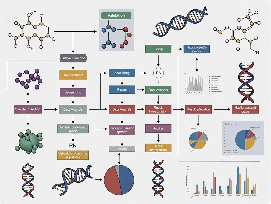
Figure 1: Experimental workflow for ecogenomic signature validation, showing the process from sample collection to signature identification.
The experimental protocol involves:
Sample Collection and Preparation: Metagenomic datasets are compiled from target (human gut) and non-target (other body sites, animal guts, environmental) habitats. These include both viral-like particle (VLP)-enriched viromes and whole-community metagenomes [1].
Sequence Data Processing: Quality control, assembly, and annotation of metagenomic sequences are performed using standard bioinformatic pipelines [18].
Reference-Based Profiling: The complete genome sequence of ϕB124-14 serves as a reference. Translated open reading frames (ORFs) from the phage genome are used as queries against metagenomic datasets [1].
Quantitative Assessment: The cumulative relative abundance of sequences with significant similarity to ϕB124-14 ORFs is calculated for each habitat. This metric represents the density of the phage's genetic signature across different environments [1].
Comparative Analysis: The representation of ϕB124-14 sequences is compared against control phages from non-gut habitats (e.g., marine Cyanophage SYN5, rhizosphere-associated Burkholderia prophage KS10) to establish habitat specificity [1].
Key Findings: Signature Validation Across Habitats
The ecogenomic profiling of ϕB124-14 consistently demonstrated a strong human gut-associated signal, as detailed in the table below.
Table 1: Ecogenomic profiling of ϕB124-14 across different habitat types based on metagenomic analysis [1].
| Habitat Type | Sample Type | Relative Representation of ϕB124-14 Signature | Statistical Significance |
|---|---|---|---|
| Human Gut | Viral Metagenomes (Viromes) | Significantly enriched | p < 0.05 vs. environmental habitats |
| Human Gut | Whole Community Metagenomes | Enriched (vs. other body sites) | p < 0.05 vs. other human body sites |
| Other Mammalian Guts (Porcine, Bovine) | Viral Metagenomes | Moderate representation | Not significant vs. human gut |
| Environmental (Marine, Freshwater) | Viral Metagenomes | Low representation | p < 0.05 vs. human gut |
| Other Human Body Sites | Whole Community Metagenomes | Low representation | p < 0.05 vs. human gut |
Analysis of viral metagenomes revealed a significantly greater mean relative abundance of ϕB124-14-encoded ORFs in human gut viromes compared to environmental datasets [1]. When compared to control phages, ϕB124-14 displayed a distinct gut-associated enrichment pattern not observed in phages from other habitats. Cyanophage SYN5, for instance, showed significantly greater representation in marine environments, while ϕB124-14 showed significantly greater representation in human-derived datasets [1].
This ecogenomic signature proved sufficiently discriminatory to distinguish 'contaminated' environmental metagenomes (subject to simulated in silico human faecal pollution) from uncontaminated datasets, highlighting its potential application in source tracking [1].
Technological Applications: From Signature to Tools
Development of Quantitative Molecular Assays
The validated ecogenomic signature of ϕB124-14 provided the foundation for developing culture-independent microbial source tracking (MST) tools. Researchers employed a "biased genome shotgun strategy" to identify human sewage-associated genetic regions within the ϕB124-14 genome [17]. This process involved:
- Target Identification: Screening 12,026 bp (25.6%) of the ϕB124-14 genome to identify genetic regions with strong human faecal association [17].
- Primer Design: Designing PCR primers for regions amenable to amplification while meeting standard design parameters [17].
- Assay Validation: Testing candidate assays against a panel of 100 individual faecal samples from diverse animal species to evaluate specificity and sensitivity [17].
Table 2: Performance comparison of ϕB124-14 bacteriophage-like qPCR assays against other human-associated fecal source identification methods [17].
| Methodology | Target Type | Specificity | Sensitivity | Notes |
|---|---|---|---|---|
| ϕB124-14 Bacteriophage-like qPCR | Viral | Superior | High | Developed from ecogenomic signature |
| HF183/BacR287 qPCR | Bacterial (Bacteroides) | Lower than ϕB124-14 | Comparable | Widely used but less specific |
| HumM2 qPCR | Bacterial (Bacteroides) | Lower than ϕB124-14 | Comparable | – |
| crAssphage CPQ056 & CPQ064 | Viral | Lower than ϕB124-14 | High | More recently discovered target |
| Culture-Based GB-124 | Bacteriophage (Host) | High | Variable | Requires cultivation, longer turnaround |
The resulting ϕB124-14 bacteriophage-like qPCR assays demonstrated superior specificity compared to top-performing DNA-based bacterial and viral human-associated methods, with strong correlation to culture-based GB-124 measurements in sewage influent [17].
Genome Signature-Based Sequence Recovery
Beyond targeted PCR assays, the ϕB124-14 ecogenomic signature enabled the development of advanced bioinformatic approaches for mining metagenomic data. The Phage Genome Signature-Based Recovery (PGSR) strategy exploits similarities in global nucleotide usage patterns (tetranucleotide usage profiles) between phage infecting related host species [18].
This approach allowed researchers to interrogate conventional whole-community metagenomes and recover "subliminal" phage sequences with high fidelity—sequences that were poorly represented in VLP-derived viral metagenomes and often missed by conventional alignment-driven methods [18]. When applied to human gut metagenomes, this strategy successfully recovered 85 metagenomic fragments classified as phage, with sizes ranging from 10-63.7 kb, 16 of which represented near full-length or complete phage genomes [18].
The Scientist's Toolkit: Essential Research Reagents and Materials
Table 3: Key research reagent solutions for ecogenomic signature research, as derived from the ϕB124-14 case study.
| Reagent/Material | Function/Application | Specific Examples from Literature |
|---|---|---|
| Reference Phage Genomes | Baseline for comparative genomics and metagenomic profiling | ϕB124-14 (complete genome), ϕB40-8, Cyanophage SYN5, Burkholderia prophage KS10 [15] [1] |
| Host Bacterial Strains | Phage propagation, host range determination, assay development | Bacteroides fragilis GB-124, other B. fragilis strains for host specificity testing [15] [19] |
| Metagenomic Datasets | Ecological profiling, signature validation across habitats | Human gut viromes, whole community gut metagenomes, environmental viromes [1] [18] |
| Bioinformatic Pipelines | Sequence analysis, signature identification, phylogenetic placement | Tetranucleotide usage profiling, ORF similarity analysis, genome signature-based recovery tools [1] [18] |
| PCR/Primer Design Tools | Development of culture-independent detection assays | "Biased genome shotgun" approach for primer design targeting human-associated genetic regions [17] |
The case study of phage ϕB124-14 provides a comprehensive framework for validating ecogenomic signatures across habitats. Through a multi-faceted approach combining comparative genomics, metagenomic profiling, and experimental validation, researchers established that individual phage can encode discernible habitat-associated signals diagnostic of underlying microbiomes [1]. The successful translation of this ecogenomic signature into specific, sensitive molecular tools for microbial source tracking [17] demonstrates the practical utility of such fundamental ecological research. Furthermore, the genome signature-based approaches developed through this work [18] offer powerful methods for accessing the considerable "biological dark matter" within microbial viromes, promising new insights into the structure and function of complex microbial ecosystems across diverse habitats.
The genus Novosphingobium, a member of the Sphingomonadaceae family, represents a group of metabolically versatile Alphaproteobacteria with a remarkable ability to inhabit diverse ecological niches. These microorganisms have been isolated from environments ranging from rhizosphere soil and plant surfaces to heavily contaminated soils, and marine and freshwater ecosystems [20]. Their metabolic plasticity and adaptability make them key players in nutrient cycling and bioremediation. A growing body of genomic evidence suggests that the phylogenetic relationships between Novosphingobium strains are less indicative of functional genotype similarity than the selective pressures of their specific habitats [20]. This article examines the habitat-specific gene enrichment patterns in Novosphingobium strains, validating the concept that ecogenomic signatures are profoundly shaped by environmental parameters, with significant implications for microbial ecology and applied biotechnology.
Comparative Genomic Analysis of Habitat-Specific Traits
Comparative genomic studies of diverse Novosphingobium strains have revealed that genome size, coding potential, and functional gene content vary significantly across different habitats, underscoring a high degree of genomic plasticity [20]. The table below summarizes key genomic features and habitat-specific metabolic traits identified in Novosphingobium.
Table 1: Genomic Features and Habitat-Specific Metabolic Traits of Novosphingobium
| Habitat | Representative Strains | Genome Size (bp) | Key Habitat-Specific Genes/Features | Bioremediation Capabilities |
|---|---|---|---|---|
| Rhizosphere | Novosphingobium sp. P6W, N. rosa NBRC 15208 | 6,537,300 - 6,952,763 | Alkane sulfonate (ssuABCD) assimilation [20] | Plant growth promotion [21] |
| Contaminated Soil | N. barchamii LL02, N. lindaniclasticum LE124 | 4,857,928 - 5,307,348 | Ectoine biosynthesis genes; wide variety of mono- and dioxygenases [20] | Degradation of hexachlorocyclohexane (HCH), polycyclic aromatic hydrocarbons (PAHs), and 2,4-dichlorophenoxyacetic acid (2,4-D) [20] [22] [23] |
| Marine Water | Novosphingobium sp. PP1Y, N. pentaromaticivorans US6-1 | 5,313,905 - 5,457,578 | Habitat-specific β-barrel outer membrane protein hubs (e.g., PP1Y_AT17644) [20] | Degradation of aromatic compounds at oil-water interfaces [20] [21] |
| Freshwater | Novosphingobium sp. THN1, AAP1 | 4,232,088 - 4,750,579 | Habitat-specific β-barrel outer membrane protein hubs (e.g., Saro_1868) [20] | Microcystin degradation (mlr gene cluster) [21] |
Analysis of core genomes has demonstrated that the enrichment of specific gene sets is a response to microenvironmental conditions. For instance, while certain traits like ectoine biosynthesis were initially assumed to be marine-specific, their presence in isolates from contaminated soil reveals a broader relevance in osmolytic regulation [20]. Furthermore, sulfur acquisition and metabolism are the only core genomic traits that differ significantly in proportion between ecological groups, with alkane sulfonate assimilation being exclusive to rhizospheric isolates [20].
Table 2: Key Enzymes and Metabolic Pathways in Novosphingobium Bioremediation
| Enzyme Class | Specific Enzymes | Target Substrates | Relevance |
|---|---|---|---|
| Mono- and Dioxygenases | PAH hydroxylating dioxygenase (PahAB) [23] | Polycyclic Aromatic Hydrocarbons (PAHs) such as phenanthrene, pyrene [23] | Initial oxidation of aromatic rings, broad substrate specificity [20] [23] |
| Dehydrohalogenases | LinA variants [24] | Hexachlorocyclohexane (HCH) isomers [24] | Dechlorination of persistent organic pollutants [24] |
| Haloalkane Dehalogenases | LinB [24] | β-hexachlorocyclohexane (β-HCH) [24] | Hydrolytic dehalogenation in HCH degradation pathway [24] |
| Microcystinase | MlrA [21] | Microcystin-LR (MC-LR) [21] | Hydrolyzes cyclic microcystin to a linear intermediate [21] |
Experimental Protocols for Uncovering Habitat-Specific Signatures
The identification of habitat-specific genes and regulatory hubs in Novosphingobium relies on a suite of advanced genomic and metagenomic techniques. The following workflow visualizes a generalized protocol for such ecogenomic studies.
Diagram Title: Workflow for Identifying Habitat-Specific Genomic Signatures
Detailed Methodological Breakdown
Strain Selection and Genome Sequencing: The process begins with the selection of multiple Novosphingobium strains isolated from well-defined habitats such as rhizosphere soil, contaminated sites, and marine and freshwater environments [20]. High-quality genomic DNA is extracted from pure cultures. Sequencing can be performed using platforms like the Pacific Biosciences (PacBio) RSII system for long-read sequencing, which is advantageous for assembling complete genomes and plasmids [21]. For a culture-independent approach, shotgun metagenomic sequencing of environmental samples on an Illumina NextSeq 550 system is employed [25].
Genome Assembly and Annotation: For pure cultures, sequence assembly is conducted using specialized pipelines such as the SMRT Analysis pipeline in conjunction with the HGAP assembler [21]. For complex environmental samples, metaSPAdes is used for de novo assembly of metagenomic sequences [25]. The assembled contigs are then binned to reconstruct Metagenome-Assembled Genomes (MAGs). Gene prediction is performed using tools like Prodigal, followed by functional annotation against databases including NCBI NR, COG, KEGG, and Swiss-Prot to assign putative functions to coding sequences [21].
Pangenome and Comparative Analysis: The pangenome, comprising the core genome (genes shared by all strains) and the flexible genome (genes present in a subset), is constructed [20] [21]. Orthologous protein analysis helps identify the core gene set. Comparative genomic analysis then focuses on identifying genes that are uniquely enriched or present in strains from a particular habitat. This includes analyzing genomic islands, which are often associated with niche adaptation, and profiling specific metabolic pathways, such as those for sulfur acquisition or aromatic compound degradation [20].
Identification of Regulatory Hubs and Validation: Protein-protein interaction (PPI) analysis can be conducted in silico to identify key proteins that act as hubs within metabolic networks. In Novosphingobium, this approach has revealed habitat-specific β-barrel outer membrane proteins as potential key hubs in different environments [20]. For catabolic pathways, regulator genes (e.g., pahR for PAH degradation) can be identified, and their function validated through the construction of reporter gene-based biosensors to confirm inducibility by specific substrates [23].
The Scientist's Toolkit: Key Research Reagents and Solutions
The following table details essential materials and tools derived from the cited research, which are crucial for conducting ecogenomic and functional studies on Novosphingobium.
Table 3: Research Reagent Solutions for Novosphingobium Studies
| Reagent/Resource | Function/Application | Example Use Case |
|---|---|---|
| M9 Minimal Medium | Defined medium for bacterial growth with specific carbon sources. | Culturing Novosphingobium with PAHs or other xenobiotics as sole carbon source to study degradation pathways [23]. |
| Qiagen DNeasy PowerSoil Pro Kit | Extraction of high-quality environmental DNA (eDNA) from complex samples like soil and sediments. | Preparing DNA for shotgun metagenomic sequencing of mangrove or contaminated soil ecosystems [25] [26]. |
| KAPA HyperPrep Kit (Roche) | Library preparation for next-generation sequencing. | Constructing sequencing libraries from eDNA for Illumina platforms [25]. |
| metaSPAdes Assembler | De novo assembly of metagenomic sequencing reads. | Reconstructing microbial genomes directly from environmental samples without cultivation [25]. |
| Prodigal Software | Prediction of protein-coding genes in genomic and metagenomic sequences. | Annotating open reading frames in assembled Novosphingobium genomes [21]. |
| pKSPA-R Plasmid | Biosensor construct with a reporter gene under the control of a PAH-inducible promoter. | Detecting low concentrations (as low as 4 ppb) of PAHs in water samples [23]. |
| LinA & LinB Enzymes | Recombinant dehydrohalogenase and haloalkane dehalogenase. | Enzymatic bioremediation studies for degrading HCH isomers, including refolding protocols for inclusion bodies [24]. |
The compelling evidence from comparative genomic studies firmly establishes that Novosphingobium strains possess highly plastic genomes, which are dynamically shaped by their environmental niches. The enrichment of specific metabolic traits—such as sulfur assimilation in rhizosphere isolates, diverse oxygenases in contaminated soil strains, and unique outer membrane protein hubs across all habitats—provides a robust validation of ecogenomic signature theory. These habitat-specific genetic repertoires not only determine the organism's functional role in its ecosystem but also present a treasure trove of biocatalytic potential. For researchers in drug development and environmental biotechnology, understanding these patterns is pivotal for harnessing Novosphingobium and similar microbes for targeted applications, from designing precise bioremediation strategies to discovering novel, stable enzymes for industrial processes.
The genomic architecture of prokaryotes is characterized by a mosaic of core and accessory elements, each playing a distinct role in evolutionary adaptation and ecological resilience. The core genome, comprising genes shared by all strains of a species, encodes essential housekeeping functions central to basic cellular processes [27] [28]. In contrast, the accessory genome consists of genes present in only a subset of strains, providing niche-specific adaptations that enable survival in diverse environments [29] [30]. Understanding the relative stability and evolutionary dynamics of these genomic components across different ecosystems is fundamental to deciphering microbial evolution, pathogenesis, and environmental adaptation. This comparison guide objectively analyzes the stability characteristics of core genomic features versus accessory genes, synthesizing experimental data from recent genomic studies to validate ecogenomic signatures across habitats.
Fundamental Concepts and Definitions
Core Genome Characteristics
The core genome represents the genetic backbone of a species, maintained across all members through evolutionary time. These genes typically include essential metabolic pathways, transcription and translation machinery, and DNA replication mechanisms [28] [31]. Core genes are generally subject to strong purifying selection that removes deleterious mutations, maintaining functional integrity across generations [27] [32]. In Enterococcus faecium, for instance, functional analysis of core genes revealed predominant roles in growth, DNA replication, transcription, translation, carbohydrate and amino acid metabolism, stress response, and transporters [31]. The stability of these genes provides phylogenetic signals for reconstructing evolutionary relationships among bacterial strains [30].
Accessory Genome Characteristics
The accessory genome, sometimes called the "flexible" genome, consists of genes acquired through horizontal gene transfer or lost through deletion events [29] [30]. This genomic compartment includes plasmids, phages, genomic islands, and other mobile genetic elements that confer context-dependent advantages [30] [31]. Accessory genes often encode functions related to environmental sensing, nutrient acquisition, stress tolerance, and host interaction [33] [10]. In the Bacillus cereus group, accessory genes contribute significantly to ecological divergence between clades, with different gene functions enriched in different clades [32].
Table 1: Fundamental Characteristics of Core and Accessory Genomes
| Feature | Core Genome | Accessory Genome |
|---|---|---|
| Definition | Genes shared by all strains of a species | Genes present in a subset of strains |
| Evolutionary Rate | Generally slower due to purifying selection | Generally faster due to diversifying selection |
| Primary Maintenance Mechanism | Vertical inheritance | Horizontal gene transfer |
| Typical Functions | Essential housekeeping functions | Niche-specific adaptations |
| Impact of Mutation | Often deleterious | Potentially adaptive |
Quantitative Comparative Analysis of Genomic Stability
Mutation and Recombination Rates
Comparative genomic studies across multiple bacterial species reveal distinct patterns of mutation and recombination between core and accessory genomes. Research on Streptococcus pneumoniae employing the mcorr method—a coalescent-based population genetics approach that analyzes correlated substitutions—found that core genes often have higher recombination rates than accessory genes [27]. This finding challenges conventional assumptions that accessory genomes experience more frequent recombination. The same study reported that while recombination rates were higher in core genomes, mutational divergence was lower, suggesting that divergence-based homologous recombination barriers could contribute to differences in recombination rates between genomic compartments [27].
Selection Pressures Across Ecosystems
Different selection pressures act on core and accessory genomes, driving distinct evolutionary patterns:
Purifying selection is prevalent in core genomes, removing deleterious alleles that impair essential cellular functions [32]. This conservative pressure maintains functional integrity across environments.
Diversifying selection more frequently affects accessory genes, promoting genetic variation that facilitates adaptation to specific ecological niches [32]. In the Bacillus cereus group, genes under diversifying selection show signs of frequent horizontal gene transfer, promoting diversification between clades [32].
Ecological determinants shape accessory genome content, with different bacterial clades maintaining distinct repertoires of accessory genes optimized for their specific habitats [32] [10]. For instance, Blastococcus species isolated from extreme environments possess accessory genes enhancing heavy metal resistance and pollutant degradation [10].
Table 2: Selection Pressures on Core vs. Accessory Genomes in Different Ecosystems
| Ecosystem Type | Core Genome Selection | Accessory Genome Selection |
|---|---|---|
| Host-associated (e.g., E. coli ST131) | Strong purifying selection maintaining basic cellular functions | Diversifying selection for host-specific adaptations (e.g., virulence factors) |
| Extreme environments (e.g., Blastococcus in stone niches) | Conservation of essential metabolic pathways | Positive selection for stress response genes (e.g., heavy metal resistance) |
| Generalist species (e.g., Bacillus cereus group) | Stable across clades with minimal functional variation | Clade-specific enrichment for niche adaptation |
Experimental Approaches for Assessing Genomic Stability
Genomic Stability Assessment Protocols
Research into core and accessory genome stability employs several established methodological frameworks:
Backbone Stability (BS) Analysis quantifies the conservation of core gene order between genomes. The BS coefficient between genome i and genome j is defined as: BSij = Nijcn / Nijtot, where Nijcn is the number of conserved edges and Nijtot is the total edges (conserved + non-conserved) in the comparison [29]. This approach measures how conserved the core gene order is between strains, with values approaching 1 indicating highly similar organization.
Genome Organization Stability (GOS) Analysis integrates both genome rearrangements and the effect of gene insertions/deletions: GOSij = Nijcn / (Nijcc + Nijac/2), where Nijcc represents edges connecting core genes and Nij_ac represents edges between core and accessory genes [29]. This method accounts for neighborhoods broken by insertion/deletion of accessory genes, providing a more comprehensive stability assessment.
Pan-genome Analysis involves clustering all genes from multiple genomes into orthology groups, typically using tools like Roary or Panaroo with sequence identity thresholds (e.g., ≥90-95% identity and coverage) [28] [10]. This approach discriminates between core genes (shared by all isolates) and accessory genes (subset-specific), enabling quantitative comparison of their evolutionary dynamics.
Figure 1: Experimental workflow for comparative analysis of core and accessory genome stability, integrating multiple bioinformatics approaches.
Key Research Reagents and Computational Tools
Table 3: Essential Research Toolkit for Genomic Stability Analysis
| Tool/Resource | Function | Application in Stability Research |
|---|---|---|
| Roary [33] [31] | Pan-genome pipeline | Rapid large-scale pan-genome analysis, identifies core and accessory genes |
| Panaroo [10] | Pan-genome analysis with graph-based approach | Identifies core/accessory genes, handles assembly errors |
| OrthoFinder [10] | Orthogroup inference | Identifies groups of orthologous genes across multiple genomes |
| IQ-TREE [33] [10] | Phylogenetic inference | Constructs maximum likelihood trees from core gene alignments |
| CheckM [28] [10] | Genome quality assessment | Evaluates completeness and contamination of genomes pre-analysis |
| mcorr [27] | Correlated substitution analysis | Infers homologous recombination parameters without phylogenetic reconstruction |
| fastANI [10] | Average Nucleotide Identity | Calculates genomic similarity for species demarcation |
Ecological Determinants of Genomic Stability
Habitat-Specific Evolutionary Pressures
Environmental characteristics exert distinct selective pressures that shape the stability of core and accessory genomes differently:
In extreme environments characterized by desiccation, salinity, alkalinity, and heavy metal contamination (e.g., stone-dwelling Blastococcus habitats), microorganisms exhibit highly dynamic genetic composition with a small core genome and large accessory genome, indicating significant genomic plasticity [10]. This configuration enables rapid adaptation to fluctuating conditions while maintaining essential functions.
For host-associated bacteria like E. coli ST131, which moves frequently between human, avian, and domesticated animal hosts, the core genome remains stable across hosts, while the accessory genome differentiates into distinct clusters associated with specific resistance genes (e.g., CTX-M type) [30]. This pattern demonstrates how conserved core genomes facilitate host generality, while flexible accessory genomes enable host-specific adaptations.
In pathogenic species like Streptococcus pneumoniae, core genes experience higher recombination rates than accessory genes, potentially increasing the efficiency of selection in conserved genomic regions [27]. This contrasts with traditional models suggesting that accessory genomes evolve more rapidly through recombination.
Ecogenomic Signatures Across Taxonomic Groups
Different bacterial taxa exhibit distinctive patterns of core and accessory genome evolution:
Enterobacteriaceae (E. coli, Salmonella): These species typically display open pan-genomes where the accessory genome continuously expands as new strains are sequenced [28]. The core genome represents a relatively small fraction (often <50%) of the total gene pool, with substantial accessory genomes facilitating ecological flexibility.
Actinobacteria (Blastococcus, Modestobacter): Species from extreme environments show remarkable genomic plasticity, with accessory genomes enriched in genes for substrate degradation, nutrient transport, and stress tolerance [10]. The core genome remains highly conserved, maintaining essential functions across niches.
Firmicutes (Bacillus cereus group, Enterococcus faecium): These groups demonstrate clade-specific selection patterns, where different gene functions are enriched in different clades for both core and accessory genomes [31] [32]. This facilitates ecological divergence while maintaining phylogenetic coherence.
Figure 2: Ecological determinants shaping core and accessory genome evolution across different habitat types.
The comparative analysis of core genomic features and accessory genes reveals a fundamental paradigm in microbial evolution: core genomes provide phylogenetic stability through strong purifying selection and conserved gene order, while accessory genomes facilitate ecological adaptability through dynamic gene content and higher evolutionary plasticity. Quantitative assessments across diverse ecosystems demonstrate that core genes can experience substantial recombination—sometimes at higher rates than accessory genes—but maintain lower mutational divergence overall [27].
These patterns of genomic stability have profound implications for ecogenomic signature validation. The conservation of core genes across environments provides reliable markers for phylogenetic reconstruction and species demarcation, while the flexible nature of accessory genomes enables fine-scale adaptation to specific ecological niches [32] [10]. This dual evolutionary strategy—maintaining a stable functional core while permitting peripheral innovation—represents a highly successful evolutionary strategy across microbial taxa.
For researchers investigating bacterial pathogenesis, environmental adaptation, or evolutionary dynamics, these findings underscore the necessity of integrated approaches that examine both core and accessory genomic components [30]. Future studies leveraging expanding genomic datasets and refined analytical methods will continue to enhance our understanding of how genomic stability patterns shape microbial diversity across Earth's ecosystems.
Advanced Computational Methods for Ecogenomic Signature Detection and Application
Chaos Game Representations (CGR) and k-mer Frequency Analysis
Within the field of ecogenomics, the validation of signatures across diverse habitats depends on robust methods to compare genetic sequences efficiently. Alignment-free sequence comparison techniques have become essential for processing the vast volumes of data generated by modern sequencing technologies, avoiding the computational bottlenecks of traditional alignment-based methods [34] [35]. Among these, Chaos Game Representation (CGR) and k-mer Frequency Analysis are two pivotal approaches. CGR translates sequences into a graphical, coordinate-based format, capturing complex patterns in a visual and mathematical form [36] [37]. In parallel, k-mer analysis breaks down sequences into short substrings of length k, using their statistical distribution to infer sequence characteristics and relationships [34]. This guide provides a comparative analysis of these two methodologies, detailing their principles, experimental protocols, and performance to inform their application in ecogenomic signature research.
Comparative Analysis: Core Principles and Ecogenomic Applications
The table below summarizes the fundamental characteristics and habitat-related applications of CGR and k-mer analysis.
Table 1: Core Principles and Ecogenomic Applications of CGR and K-mer Analysis
| Feature | Chaos Game Representation (CGR) | K-mer Frequency Analysis |
|---|---|---|
| Core Principle | Iterative mapping of sequences into a coordinate space (e.g., 2D or 3D square) [36] [38]. | Enumeration of all contiguous subsequences of length k within a sequence [34]. |
| Primary Output | An image or a trajectory of points in a fractal pattern [37]. | A frequency vector of all possible k-mers [34] [39]. |
| Sequence Information Encoded | Markov Properties: Reveals higher-order statistical patterns and dependencies between nucleotides [36]. | Compositional Features: Captures the frequency and, in some advanced methods, the position distribution of k-mers [39] [35]. |
| Key Ecogenomic Applications | - Phylogenetic Analysis: Unique genomic signatures aid in constructing evolutionary trees [36] [38].- Pattern Visualization: Identifying and visualizing repetitive elements and compositional biases in metagenomic sequences [37]. | - Taxonomic Classification & Metagenomics: Rapid assignment of sequences to taxonomic groups in diverse environmental samples [34].- Biomarker Discovery: Identifying unique k-mers (e.g., nullomers) as signatures for specific organisms or environmental adaptations [34] [40]. |
Performance and Experimental Data
When deployed for specific bioinformatics tasks, the two methods exhibit distinct performance characteristics, strengths, and limitations, as detailed in the following table.
Table 2: Experimental Performance and Practical Considerations
| Aspect | Chaos Game Representation (CGR) | K-mer Frequency Analysis |
|---|---|---|
| Handling of Unequal Sequence Lengths | Frequency Chaos Game Representation (FCGR) transforms sequences into equal-sized matrices by counting k-mer frequencies in a grid, facilitating comparison [36]. | Inherently handles sequences of different lengths by working with frequency vectors, though longer k-values can lead to sparse data [34]. |
| Computational Efficiency | The CGR algorithm itself is iterative and computationally light. However, global similarity comparison of entire trajectories can be complex [38]. | Highly efficient and scalable for large datasets, with numerous optimized algorithms available for k-mer counting [34] [35]. |
| Sensitivity to Mutations | Substitutions: Alter the trajectory path, changing point positions [38].Insertions/Deletions: Change the total number of points in the trajectory, affecting the overall shape [38]. | Substitutions: Directly alter the k-mers at the mutation site.Insertions/Deletions: Cause a frame-shift in all subsequent k-mers, leading to a larger change in the k-mer profile [34]. |
| Reported Performance | A 3D CGR method using shape signatures was shown to produce phylogenetic trees comparable to alignment-based methods and was robust to different mutation types [38]. | Methods like IEPWRMkmer, which combine k-mer frequency and position information, have demonstrated high efficiency and reliability in phylogenetic tree reconstruction, as validated by Robinson-Foulds distance metrics [39]. |
| Key Limitations | Traditional 2D CGR point-by-point distance can be biased by the spatial arrangement of nucleotides, which may not reflect true biological divergence [38]. | The choice of k is critical; short k can lack specificity, while long k leads to computational overhead and data sparsity (overfitting) [34]. |
Experimental Protocols
Workflow for K-mer Frequency Analysis in Metagenomic Classification
A typical workflow for using k-mer analysis in an ecogenomic context, such as classifying sequences in a metagenomic sample, involves the following steps. This protocol is widely used in tools for taxonomic classification and signature discovery [34] [40].
Title: K-mer Analysis Workflow
Step-by-Step Protocol:
- Input Sequence Data: Gather whole genome or metagenomic sequencing reads from environmental samples (e.g., soil, water) [35].
- Sequence Fragmentation: If working with whole genomes, they may be computationally fragmented into reads to standardize analysis.
- K-mer Generation: Select a value for k (e.g., k=9 to k=31 is common). Slide a window of length k across each sequence, extracting every possible k-mer [34]. The choice of k is a critical parameter that balances specificity and computational load.
- K-mer Counting & Frequency Vector Creation: For each sequence, count the occurrence of each possible k-mer. This generates a high-dimensional frequency vector representing that sequence [34] [39].
- Dimensionality Reduction (Optional but common): Use techniques like Principal Component Analysis (PCA) to project the high-dimensional k-mer vectors into a 2D or 3D space to visualize sequence relationships and identify clusters [35].
- Dissimilarity Matrix Calculation: Calculate pairwise distances between sequences based on their k-mer frequency vectors. Common distance measures include Manhattan or Euclidean distance [39].
- Downstream Analysis: Use the dissimilarity matrix for tasks such as phylogenetic tree construction, taxonomic classification of metagenomic reads, or identifying signature k-mers that define a specific habitat [34] [40].
Workflow for Chaos Game Representation (CGR)
The following protocol outlines the generation of a standard 2D CGR and its extension to a Frequency Matrix (FCGR), which is often used for machine learning applications [36] [37].
Title: CGR and FCGR Workflow
Step-by-Step Protocol:
- Define Coordinate Space: Define a 2D unit square [0,1] x [0,1]. For a 3D CGR, a cube would be used [38].
- Initialize Plot and Assign Nucleotides: Assign each nucleotide to a corner of the square (e.g., A=(0,0), C=(0,1), G=(1,1), T=(1,0)). The starting point, x₀, is typically the center of the square (0.5, 0.5) [37].
- Iterative Mapping: For each nucleotide in the sequence from first to last, calculate the next point using the iterative function: xᵢ = xᵢ₋₁ + 0.5 * (yᵢ - xᵢ₋₁) where xᵢ₋₁ is the previous point and yᵢ is the corner coordinate of the current nucleotide [37]. This formula places each new point halfway between the previous point and the current nucleotide's corner.
- Final CGR Plot: After processing the entire sequence, the resulting plot of points is the CGR, a unique fractal representation of the sequence.
- Generate FCGR Matrix (For Comparison): To compare sequences quantitatively, the CGR plot is divided into a 2k x 2k grid. The number of points falling into each cell of the grid is counted, producing a Frequency Chaos Game Representation (FCGR) matrix. This matrix is a normalized k-mer frequency table that can be used as an input for machine learning models or direct sequence comparison [36].
- Use in Comparison/ML: The FCGR matrices, which are equal-sized for all sequences, can be compared using distance metrics or used to train classifiers for ecological source tracking [36].
The Scientist's Toolkit: Essential Research Reagents and Computational Solutions
The table below lists key computational tools and concepts that serve as essential "reagents" in experiments involving CGR and k-mer analysis.
Table 3: Key Research Reagent Solutions for Alignment-Free Analysis
| Tool/Concept | Type | Function in Analysis |
|---|---|---|
| FCGR Matrix [36] | Computational Representation | Transforms a DNA sequence of any length into a fixed-size, numerical matrix representing k-mer frequencies, enabling machine learning. |
| Nullomers / Nullpeptides [34] | Biological Concept & Computational Target | K-mers absent from a genome or proteome. They can serve as highly specific biomarkers for pathogen detection or cancer diagnostics in ecogenomic studies. |
| IEPWRMkmer Method [39] | Computational Algorithm | An alignment-free distance measure that combines k-mer frequency and positional information via information entropy, improving phylogenetic accuracy. |
| Seqwin [40] | Software Tool | An open-source framework that uses weighted pan-genome minimizer graphs to efficiently identify signature sequences unique to a target group of microbes. |
| Digital Filter (FIR) [35] | Signal Processing Technique | A filter applied to k-mer signature signals to calculate k-mer density along a sequence, aiding in the detection of regions with distinctive word frequencies. |
| Minimizers [34] | Computational Algorithm | A heuristic to select a representative subset of k-mers from a longer sequence, significantly reducing memory consumption and computational runtime. |
Machine Learning and Neural Networks for Signature Classification
The accurate classification of signatures—whether genomic or image-based—is a cornerstone of modern scientific research, enabling everything from the diagnosis of disease to the understanding of ecological adaptations. This guide objectively compares the performance of various machine learning (ML) and deep learning (DL) models in signature classification tasks. The context is a broader thesis on validating ecogenomic signatures across diverse habitats, a field that relies on robust classification to link genetic markers to environmental functions. For researchers, scientists, and drug development professionals, selecting the optimal model is not merely a technical choice but a critical step in ensuring that research findings are both reliable and translatable to real-world applications. This guide provides a structured comparison of leading algorithms, supported by experimental data and detailed methodologies, to inform these decisions.
Methodological Framework: A Comparative Approach
The performance evaluation of ML and DL models for signature classification follows a structured, comparative methodology. This section outlines the core experimental protocols common to the studies cited in this guide, ensuring a consistent basis for comparing the results presented in subsequent sections.
Model Selection and Training
A suite of standard and advanced models is typically evaluated. Commonly assessed machine learning models include Support Vector Machine (SVM), K-Nearest Neighbors (KNN), Random Forest (RF), and Decision Tree (DT). For deep learning, models often include Artificial Neural Network (ANN), Long Short-Term Memory (LSTM) networks, and specialized Convolutional Neural Networks (CNNs) [41] [42]. Models are trained on labelled datasets where the "signature" (e.g., gene expression profile, image features) is the input and a class (e.g., diseased/healthy, bacterial/viral, signature author) is the output.
Performance Metrics
Model performance is quantified using standard metrics to ensure objectivity. Key metrics include:
- Accuracy: The proportion of total correct predictions (both positive and negative) among the total number of cases examined.
- Balanced Accuracy: Useful for imbalanced datasets, it is the average accuracy obtained from each class.
- Area Under the Receiver Operating Characteristic Curve (AUROC): A measure of the model's ability to distinguish between classes, where 1.0 represents perfect discrimination and 0.5 represents a random guess [43] [42].
- Sensitivity and Specificity: The true positive rate and true negative rate, respectively [43].
Validation and Testing
To prevent overfitting and ensure generalizability, models are validated on held-out test data not used during training. In some studies, an independent validation cohort is used to test the final model, providing a robust assessment of its real-world performance [43]. For genomic signatures, validation may also involve demonstrating clinical utility by comparing the signature's performance against established biomarkers like C-reactive protein (CRP) [43].
Table 1: Key Experimental Protocols in Signature Classification Studies
| Protocol Aspect | Description | Example from Literature |
|---|---|---|
| Feature Selection | Identifying the most informative variables (e.g., genes, pixels) for classification. | Highly Variable Genes (HVG), Principal Component Analysis (PCA) [42]. |
| Model Training | The process of feeding data to an algorithm to learn the classification function. | Training Neural Networks with cross-entropy loss [44]. |
| Cross-Validation | Assessing how the model will generalize to an independent dataset. | Held-out test sets; independent validation cohorts [43] [42]. |
| Performance Benchmarking | Comparing new models against established baselines or state-of-the-art models. | Comparing a novel three-gene signature against CRP and leukocyte count [43]. |
Comparative Performance of Classification Models
The efficacy of ML and DL models varies significantly depending on the data type and classification task. The following data, synthesized from multiple research efforts, provides a quantitative comparison.
Performance in Network Intrusion Detection
In a study on signature-based intrusion detection, which classifies network traffic as normal or intrusive, several models were evaluated. The results demonstrate that while traditional ML models perform well, DL models excel in detecting complex patterns [41].
Table 2: Model Performance in Intrusion Detection [41]
| Model | Model Type | Key Performance Findings |
|---|---|---|
| Support Vector Machine (SVM) | Machine Learning | Effective results; considered a promising solution for real-world IDS due to versatility and explainability. |
| Random Forest (RF) | Machine Learning | Effective results; promising for real-world applications due to versatility and explainability. |
| K-Nearest Neighbors (KNN) | Machine Learning | Showed effective results in classification. |
| Decision Tree (DT) | Machine Learning | Showed effective results in classification. |
| Long Short-Term Memory (LSTM) | Deep Learning | Rapidly finds long-term and complex patterns; high precision, accuracy, and recall; suitable for nuanced, evolving threats. |
| Artificial Neural Network (ANN) | Deep Learning | Rapidly finds complex patterns; highly effective with high precision, accuracy, and recall. |
Performance in Genomic Signature Classification
For biological applications, such as distinguishing between viral and bacterial infections or classifying diseased cells, the performance requirements for models are extreme due to the direct implications for diagnosis and treatment.
In one study, a minimal three-gene signature (HERC6, IGF1R, NAGK) derived from host blood transcriptomes was used to discriminate between viral and bacterial infections. The classification performance, likely powered by a logistic regression model, was exceptional. It achieved an AUROC of 0.976 (95% CI 0.919–1.000), with a sensitivity of 97.3% and specificity of 100% in one validation cohort. In a second cohort that included SARS-CoV-2 patients, it maintained an AUROC of 0.953, sensitivity of 88.6%, and specificity of 94.1%. This significantly outperformed traditional biomarkers like CRP (AUROC 0.833) [43].
In Parkinson's disease research, a Neural Network (NN) classifier applied to single-nuclei RNA sequencing data achieved a mean balanced accuracy of 0.984 in distinguishing diseased from healthy cells, outperforming logistic regression, random forest, and support vector machines. This high accuracy was crucial for downstream interpretation and gene discovery [42].
Performance in Handwritten Signature Image Classification
A novel CNN architecture (Si-CNN+NC) designed for offline handwritten signature classification demonstrated superior performance compared to several well-known pre-trained models. Its superior speed and accuracy make it suitable for applications requiring rapid processing, such as in criminal detection and forgery prevention [44].
Table 3: Model Performance in Handwritten Signature Image Classification [44]
| Model | Model Type | Key Performance Findings |
|---|---|---|
| Si-CNN+NC | Deep Learning (Novel CNN) | Outperformed others in both accuracy and speed; superior performance on benchmark datasets. |
| Si-CNN | Deep Learning (Novel CNN) | Achieved higher accuracy than benchmark models; fast and lightweight. |
| GoogleNet | Deep Learning (Pre-trained) | Outperformed by the proposed Si-CNN+NC model. |
| DenseNet201 | Deep Learning (Pre-trained) | Outperformed by the proposed Si-CNN+NC model. |
| Inceptionv3 | Deep Learning (Pre-trained) | Outperformed by the proposed Si-CNN+NC model. |
| ResNet50 | Deep Learning (Pre-trained) | Outperformed by the proposed Si-CNN+NC model. |
Experimental Protocols in Detail
To ensure reproducibility and provide a deeper understanding of the comparative data, this section elaborates on the experimental protocols for two key studies.
Protocol 1: Validating a Three-Gene Viral Infection Signature
This study aimed to derive and validate a blood transcriptional signature for detecting viral infections, including COVID-19, in emergency department settings [43].
- Sample Collection: Whole-blood RNA was prospectively collected from adults (≥18 years) presenting with suspected infection to a major UK hospital. Participants were recruited into discovery and validation cohorts.
- Discovery Cohort: RNA sequencing was performed on samples from 56 participants with confirmed bacterial infections and 27 with viral infections. Differential gene expression analysis identified host genes that were significantly over- or under-expressed.
- Signature Derivation: A feature selection method (Forward Selection-Partial Least Squares, FS-PLS) was used to identify the most parsimonious set of discriminating genes, resulting in a three-gene signature (HERC6, IGF1R, NAGK). A logistic regression model was developed using this signature.
- Validation: The signature was translated into an RT-qPCR assay and validated on two independent prospective cohorts: one with undifferentiated fever and another with PCR-confirmed COVID-19 or bacterial infection. Performance was assessed by calculating AUROC, sensitivity, and specificity.
Protocol 2: Neural Networks for Parkinson's Disease Gene Signatures
This research introduced an explainable ML framework to identify molecular markers of Parkinson's disease (PD) from single-nuclei transcriptomes [42].
- Data Preparation: Four publicly available snRNAseq datasets from post-mortem midbrains of PD patients and controls were used. Cells were annotated into broad types (e.g., astrocytes, microglia, dopaminergic neurons).
- Feature Selection and Model Training: Highly Variable Genes (HVGs) were identified for each cell type. Neural Network (NN) classifiers were then trained using these HVGs to predict disease status at the single-cell level.
- Model Interpretation: The Local Interpretable Model-agnostic Explanations (LIME) method was applied to the trained NNs. LIME approximates the local decision boundary for each prediction, assigning an importance score (Z-score) to each gene, thereby revealing the transcriptional markers that most strongly influenced the classification of a cell as "diseased."
- Validation: The generalizability of the LIME-identified gene signatures was tested by comparing their importance across different datasets and against genes identified by traditional differential expression analysis.
The Scientist's Toolkit: Essential Research Reagents and Materials
The following table details key reagents and computational tools essential for conducting research in signature classification, particularly in the ecogenomic domain.
Table 4: Key Research Reagent Solutions for Signature Classification
| Item | Function/Application | Specific Example |
|---|---|---|
| Whole-blood RNA Collection Kits | Standardized collection and stabilization of RNA from blood samples for transcriptomic studies. | Used in the discovery and validation of the three-gene viral infection signature [43]. |
| RT-qPCR Assays | Translating a discovered genomic signature into a rapid, clinically applicable diagnostic test. | Used to validate the three-gene signature in emergency department cohorts [43]. |
| Single-nuclei/cell RNA Sequencing Kits | Profiling gene expression at the level of individual cells to identify cell-type-specific signatures. | Used to generate snRNAseq data from post-mortem midbrains for Parkinson's disease research [42]. |
| High-Performance Computing (HPC) Cluster | Providing the computational power required for training complex deep learning models on large genomic or image datasets. | Essential for running neural networks, CNNs, and other resource-intensive algorithms [41] [42] [44]. |
| Curated Reference Genomes | Serving as a baseline for genomic taxonomy, assembly, and functional annotation in ecogenomic studies. | Used in cyanobacteria ecogenomics to establish a phylogenomic framework and link taxa to ecological niches [45]. |
Integrated Workflow: From Data to Ecological Validation
The process of signature classification and validation, especially within an ecogenomic context, follows a logical sequence that integrates computational modeling with biological and ecological inquiry. The diagram below illustrates this overarching workflow.
The comparative analysis presented in this guide reveals a clear landscape for signature classification. Traditional machine learning models like SVM and Random Forest offer strong, interpretable performance for many tasks and are well-suited for real-world applications where explainability is key [41]. However, for the most challenging problems involving complex, high-dimensional data such as single-cell transcriptomes or nuanced image patterns, deep learning models—particularly Neural Networks and specialized CNNs—consistently achieve superior accuracy and robustness [42] [44]. The choice of model is therefore contingent on the specific data type, the required performance benchmarks, and the need for interpretability. As the field of ecogenomics moves toward standardizing methods to meet stakeholder needs in conservation and medicine [46], the adoption of these high-performing, validated classification frameworks will be crucial for generating reliable, comparable, and actionable biological insights.
The validation of ecogenomic signatures across diverse habitats represents a frontier in biodiversity research. This guide compares emerging approaches for constructing composite DNA signatures that integrate information from nuclear and organellar genomes. Where nuclear genomes provide comprehensive genetic blueprints subject to recombination and biparental inheritance, organellar genomes (from chloroplasts and mitochondria) offer haploid, non-recombining markers with different evolutionary trajectories [47] [48]. The integration of these complementary systems enables researchers to address fundamental questions in species delimitation, adaptive potential, and evolutionary history with unprecedented resolution [48] [46].
The strategic value of composite signatures lies in their capacity to reveal different aspects of evolutionary history. While nuclear genomes reflect complex genealogies influenced by biparental inheritance and recombination, organellar genomes provide maternally inherited markers useful for tracing lineage-specific patterns [48]. This multi-compartment approach has transformed applications ranging from phylogenetic reconstruction to conservation prioritization, allowing scientists to circumvent limitations inherent in single-marker systems [47] [48].
Comparative Analysis of Genomic Integration Approaches
Performance Metrics Across Methodologies
Table 1: Comparison of genomic approaches for constructing composite DNA signatures
| Methodology | Genomic Compartments Accessed | Species Discrimination Power | Technical Challenges | Best Application Context |
|---|---|---|---|---|
| Short-read sequencing | Nuclear, plastid, mitochondrial | Moderate | Assembly difficulties for complex regions | Biodiversity monitoring, phylogenomics [49] |
| Long-read sequencing (HiFi) | Nuclear, plastid, mitochondrial | High | Higher input DNA requirements, cost | Complete organelle genome assembly, NUMT/NUPT detection [50] |
| Deep genome skimming (DGS) | Plastid, mitochondrial, single-copy nuclear genes | High to Very High | Computational resource demands | Species circumscription, resolving complex taxa [48] |
| Target enrichment (Hyb-Seq) | Targeted nuclear loci, organellar genomes | High | Custom probe design required, cost | Phylogenetics of closely related species [48] |
Quantitative Assessment of Organellar-Nuclear DNA Transfer
Table 2: Documented organellar DNA integration events in plant genomes
| Species | NUPTs Detected | NUMTs Detected | Plastome Coverage by NUPTs | Mitogenome Coverage by NUMTs | Reference |
|---|---|---|---|---|---|
| Cicuta virosa | 6,686 | 6,237 | 99.93% | 77.04% | [50] |
| Triticum urartu | Highest number reported | Not specified | Not specified | Not specified | [47] |
| Arabidopsis thaliana | Not specified | 620 kb largest NUMT | Not specified | Not specified | [47] |
| Oryza sativa | 131 kb largest NUPT | Not specified | Not specified | Not specified | [47] |
| Gossypium hirsutum | 135 kb largest NUPT | Not specified | Not specified | Not specified | [47] |
Experimental Protocols for Signature Validation
Integrated Workflow for Composite DNA Signature Analysis
The following diagram illustrates the comprehensive workflow for developing and validating composite DNA signatures, integrating experimental and computational approaches:
Workflow for Composite DNA Signature Analysis - This diagram outlines the integrated experimental and computational pipeline for developing composite DNA signatures, from sample collection to ecogenomic application.
Detailed Methodological Protocols
Genome Assembly and Quality Assessment
High-fidelity genome assembly forms the foundation for reliable composite signature development. The hybrid assembly approach combining long-read and short-read technologies has demonstrated superior performance for recovering both nuclear and organellar genomes [50] [51]. For the Cicuta virosa genome, researchers employed PacBio HiFi sequencing yielding 79 Gb of data with average read lengths of 16,471 bp, followed by assembly using Hifiasm, producing a draft nuclear genome of 1,265.91 Mb with N50 contig size of 19.63 Mb [50]. Quality assessment should include BUSCO analysis (98.8% completeness for eudicot genes in C. virosa), contig alignment to organellar genomes to identify potential contamination, and mapping rate evaluation [50].
For mitochondrial genome-specific assembly, MitoHiFi v3.2.2 has proven effective, as demonstrated in the Indrella ampulla mitogenome assembly of 13,887 bp [51]. Annotation refinement should incorporate multiple tools: GeSeq and ARWEN for tRNA prediction, with manual curation to remove false-positive tRNAs within protein-coding regions [51].
Detection and Validation of Organellar-Nuclear DNA Integration
The detection of NUPTs and NUMTs requires specialized bioinformatic pipelines. The standard approach involves:
- Reference-based BLASTN analysis: Nuclear contigs are aligned against complete plastome and mitogenome references using BLASTN with careful parameter optimization [47] [50].
- Hit filtering and characterization: Detected fragments are filtered based on sequence identity (typically 80-100%) and length parameters, with detailed annotation of their genomic locations and structural arrangements [47].
- Cluster identification: NUPTs/NUMTs are analyzed for non-random distribution patterns, including tight clusters, loose clusters, and mosaic structures containing both plastid and mitochondrial sequences [47].
- Functional transfer validation: Putative functional organellar-derived genes in the nucleus require additional evidence including expression data and protein targeting prediction [50].
In the C. virosa study, this approach identified 6,686 NUPTs covering 99.93% of the plastome and 6,237 NUMTs covering 77.04% of the mitogenome, with sequence identities ranging from 80-100%, indicating multiple transfer events across evolutionary timescales [50].
Multi-locus Species Circumscription Protocol
The Multilayer Precision Species Circumscription Approach (MPSCA) integrates data from multiple genomic compartments [48]:
- Plastome analysis: Assemble complete chloroplast genomes and identify hypervariable regions (8 identified in Epimedium)
- Single-copy nuclear gene recovery: Utilize deep genome skimming to recover hundreds of single-copy nuclear genes
- Micro-morphological correlation: Combine with stable morphological characteristics (e.g., leaf epidermal features in Epimedium)
- Concordance analysis: Assess congruence between different marker systems to resolve taxonomic ambiguities
This approach successfully discriminated Epimedium species that remained unresolved using single-marker systems, with single-copy nuclear genes proving particularly valuable due to their higher evolutionary rates and biparental inheritance [48].
The Scientist's Toolkit: Essential Research Reagents and Platforms
Table 3: Key research reagents and platforms for composite signature analysis
| Category | Specific Tools/Platforms | Primary Function | Performance Considerations |
|---|---|---|---|
| Sequencing Platforms | PacBio HiFi, Oxford Nanopore, Illumina | DNA sequencing | Long-read technologies essential for complex regions; short-read for accuracy |
| Assembly Software | Hifiasm, Flye, MaSuRCA, QuickMerge | Genome assembly and merging | Hybrid approaches show superior contiguity metrics [50] [51] |
| Annotation Tools | Prokka, GALBA, GeSeq, ARWEN | Structural annotation | Multi-tool approaches improve accuracy, especially for tRNA genes [10] [51] |
| Specialized Analysis | MitoHiFi, RepeatMasker, BLAST+ | Organelle genome assembly, repeat masking, homology search | Specialized pipelines needed for organellar genomes [51] |
| Quality Assessment | BUSCO, Merqury, CheckM, BlobToolKit | Assembly and annotation quality | Multiple assessment tools recommended for comprehensive evaluation [10] [51] |
Data Interpretation and Ecological Validation
Analytical Frameworks for Signature Implementation
The ecological validation of composite DNA signatures requires specialized analytical approaches:
Phylosymbiosis assessment: Evaluate how host evolutionary history structures microbial communities across taxonomic scales, with host phylogeny dominating at broader scales and ecological factors modifying patterns among closely related species [52]. Statistical frameworks should quantify the congruence between host phylogeny and microbiome composition, accounting for confounding factors like diet and geography.
Demographic history reconstruction: Implement Pairwise Sequentially Markovian Coalescent (PSMC) analysis to infer historical population dynamics from genomic data [51]. Critical parameters include depth filtering thresholds (minimum 6X, maximum 48X for I. ampulla), generation time estimates, and mutation rate assumptions, with 100 bootstrap replicates recommended for confidence assessment [51].
Co-occurrence network analysis: Identify metabolically interdependent taxa through correlation-based network inference, particularly focusing on organisms with streamlined genomes and potential auxotrophies [53]. This approach revealed co-occurrent cohorts of freshwater prokaryotes with complementary biosynthetic capabilities, where streamlined genomes showed higher prevalence and relative abundance [53].
Standards and Reporting Frameworks
The harmonization of biodiversity genomics research practices is essential for comparative analyses and stakeholder adoption [46]. Key reporting standards include:
- Minimum information standards: Complete documentation of sequencing depth, assembly metrics, and annotation methods
- Genetic diversity metrics: Standardized measurements of genome-wide heterozygosity, population structure, and adaptive variation
- Functional annotation consistency: Orthologous gene calling using established databases and cutoffs
- Data accessibility: Public archiving of raw data, assembled genomes, and annotation files
Initiatives like the European Reference Genome Atlas (ERGA) are developing standardized metrics for measuring and reporting genetic diversity to enhance interpretability and comparability across studies [46].
Fecal pollution poses a significant threat to water security and public health, contributing to the spread of waterborne diseases and environmental degradation [54]. Traditional culture-based methods for detecting fecal contamination, while useful for confirming its presence, suffer from critical limitations: they are time-consuming, cannot determine the pollution source, and often show weak correlation with pathogenic factors [54] [55]. Microbial Source Tracking (MST) has emerged as a sophisticated suite of analytical protocols that overcome these limitations by using host-associated characteristics of various microorganisms to identify the specific origins of fecal contamination in water systems [56] [57]. The development and validation of these methodologies within the broader context of ecogenomic signatures represent a significant advancement in environmental microbiology, enabling more targeted remediation strategies and improved risk assessments [2].
Methodological Approaches in Microbial Source Tracking
MST methodologies can be broadly categorized into two distinct approaches, each with unique operational frameworks, advantages, and limitations.
Library-Dependent Methods
Library-dependent methods are culture-based techniques that rely on isolate-by-isolate typing of bacteria cultured from various fecal sources and water samples [56] [58]. These methods involve creating extensive libraries of biochemical or genotypic "fingerprints" from bacterial strains of known fecal sources, which are then compared to isolates from environmental samples for classification [57].
These methods include phenotypic approaches such as antibiotic resistance analysis and carbon source utilization patterns, as well as genotypic techniques like ribotyping, pulse-field gel electrophoresis (PFGE), and repetitive DNA sequence PCR (Rep-PCR) [56] [58]. While library-dependent methods can provide valuable data, they present significant limitations including high cost, extended processing time, requirement for experienced personnel, and geographical specificity of libraries that often limits their broad application [56].
Library-Independent Methods
Library-independent methods represent a more recent advancement in MST and are primarily based on detecting specific host-associated genetic markers in DNA extracted directly from water samples without culturing [56] [58]. This approach typically utilizes polymerase chain reaction (PCR) methods to amplify gene targets that are specifically associated with particular host populations [56].
The most common library-independent methods include:
- Host-specific bacterial PCR/qPCR: Targeting host-associated genetic markers from bacterial groups such as Bacteroidetes [54] [58]
- Host-specific viral PCR: Utilizing viruses known to infect specific hosts [54]
- Mitochondrial DNA detection: Targeting host-derived mitochondrial sequences [59]
- eDNA metabarcoding: Using next-generation sequencing to comprehensively characterize diverse fecal sources [59]
Key advantages of library-independent methods include reduced processing time, elimination of geographical library limitations, and the ability to provide absolute quantification of pollution indicators through quantitative real-time PCR (qPCR) [54] [56].
Table 1: Comparison of Major MST Methodological Approaches
| Feature | Library-Dependent Methods | Library-Independent Methods |
|---|---|---|
| Basis | Culture-based identification of isolates [56] | Detection of host-associated genetic markers [56] |
| Analysis Time | Days to weeks | Hours to days |
| Geographical Application | Limited to specific regions [56] | Broad application possible |
| Expertise Required | High [56] | Moderate to High |
| Cost | High (library development) [56] | Moderate to High |
| Primary Targets | E. coli, Enterococcus, other fecal indicators [58] | Bacteroidetes markers, host-specific viruses, mitochondrial DNA [54] [59] |
Performance Comparison of MST Markers and Methodologies
Rigorous evaluation of MST method performance is essential for selecting appropriate protocols for water quality investigations. Performance is typically measured through sensitivity (ability to correctly identify true positives) and specificity (ability to correctly identify true negatives) across different host categories [58].
Performance of Library-Dependent Methods
Comprehensive studies have revealed variable performance across different library-dependent methods. Antibiotic resistance analysis (ARA) of E. coli demonstrates relatively low sensitivity (24-27%) for human sources but higher specificity (83-86%) for non-human sources [58]. Ribotype analysis shows improved sensitivity (85%) and specificity (79%) for human sources when using E. coli isolates from reference feces [58]. Carbon source utilization patterns exhibit particularly low sensitivity (12%) for human sources but very high specificity (98%) for non-human sources [58].
Performance of Library-Independent Methods
Library-independent methods generally demonstrate superior and more consistent performance metrics. The human-associated Bacteroidetes marker HF183 shows sensitivity ranging from 70-100% and specificity of 100% in blind samples [58]. Ruminant-associated markers (CF128) demonstrate excellent sensitivity (97-100%) and specificity (93-100%) across various studies [58]. A recent meta-analysis further confirmed that PCR/qPCR-based methods significantly enhance the diagnostic odds ratio compared to conventional approaches, with dye-based (SYBR) and probe-based (TaqMan) methods showing particularly high performance for human-associated markers [54].
Table 2: Performance Characteristics of Selected Library-Independent MST Markers
| Target Marker | Host Category | Sensitivity (%) | Specificity (%) | Reference |
|---|---|---|---|---|
| HF183 | Human | 70-100 | 100 | [58] |
| CF128 | Ruminants | 97-100 | 93-100 | [58] |
| Bacteroides thetaiotaomicron | Human | 78-92 | 76-98 | [58] |
| Dog-associated (DF475) | Canine | 40 | 86 | [58] |
| Avian | Birds | Varies by marker | Varies by marker | [59] |
Experimental Protocols and Workflows
Implementing MST investigations requires careful consideration of sampling strategies, laboratory methodologies, and analytical approaches.
Sampling Design and Sample Processing
Effective MST begins with appropriate sampling strategies that consider temporal and spatial variability of fecal contamination. Samples should be collected in sterile containers and processed promptly to preserve nucleic acid integrity [59] [57]. For molecular methods, typical protocols involve filtering 100-500 mL of water through 0.22-μm filters to capture microbial biomass [59]. DNA extraction is then performed using commercial kits, often with modifications such as increased bead-beating time or alternative bead types to improve cell disruption [59].
Molecular Detection Methods
PCR/qPCR Protocols: Quantitative PCR assays typically involve reaction mixtures containing hot start PCR master mix, specific forward and reverse primers (10 μM), template DNA, and nuclease-free water [59]. Thermal cycling conditions generally include initial denaturation (95°C for 10 min), 35-45 cycles of denaturation (95°C for 30 s), primer-specific annealing (50-60°C for 1 min), and extension (72°C for 40 s), followed by final extension (72°C for 5 min) [54] [59].
eDNA Metabarcoding Workflow: This advanced approach involves a two-step PCR process: (1) initial amplification of target regions (e.g., mitochondrial 16S rRNA) with limited cycles (10 cycles) to reduce amplification bias, and (2) nested PCR using Illumina linker-attached primers (35 cycles) to prepare sequencing libraries [59]. Sequencing is performed on next-generation platforms, followed by bioinformatic analysis to assign taxonomic classifications.
Figure 1: Microbial Source Tracking Decision Workflow. This diagram illustrates key decision points in the MST process, from initial study design through data interpretation, highlighting the divergent paths for library-dependent and library-independent approaches [57].
Ecogenomic Signatures and Advanced Applications
The emerging field of ecogenomics has significantly advanced MST capabilities by revealing habitat-associated signatures in microbial and viral genomes that serve as diagnostic markers for fecal pollution sources [2].
Bacteriophage Ecogenomic Signatures
Recent research has demonstrated that individual bacteriophages encode clear habitat-related 'ecogenomic signatures' based on the relative representation of phage-encoded gene homologues in metagenomic datasets [2]. These signatures can segregate metagenomes according to environmental origin and distinguish contaminated environmental metagenomes from uncontaminated datasets [2]. The ɸB124-14 phage, for instance, encodes an ecogenomic signature that can identify human fecal pollution in environmental waters, demonstrating the potential for phage-based MST tools [2].
eDNA Metabarcoding for Comprehensive Profiling
eDNA metabarcoding represents a powerful expansion of MST capabilities, enabling comprehensive characterization of diverse fecal sources through amplification and sequencing of universal marker genes such as mitochondrial 16S rRNA [59]. This approach facilitates detection of multiple potential fecal contributors simultaneously, providing a more complete picture of contamination sources than single-marker MST methods [59]. Applications in urban freshwater beaches have revealed diverse wildlife contributions, including mallard duck, muskrat, beaver, raccoon, gull, and numerous other species that traditional methods might miss [59].
Figure 2: Complementary Relationship Between eDNA Metabarcoding and Microbial Source Tracking. Integration of these approaches provides more comprehensive fecal pollution profiling than either method alone [59].
Research Reagent Solutions and Essential Materials
Successful implementation of MST methodologies requires specific research reagents and materials optimized for environmental sample processing and analysis.
Table 3: Essential Research Reagents and Materials for MST Investigations
| Reagent/Material | Specific Function | Application Examples |
|---|---|---|
| Norgen Soil Plus DNA Extraction Kit | Environmental DNA extraction from filters | eDNA metabarcoding studies [59] |
| Hot Start PCR Master Mix | High-specificity amplification of target sequences | qPCR detection of host-specific markers [59] |
| Host-Specific Primers (e.g., HF183) | Selective amplification of host-associated genetic markers | Human fecal contamination tracking [54] [58] |
| 0.22-μm Nitrocellulose Membrane Filters | Concentration of microbial biomass from water samples | Sample processing for molecular detection [59] |
| SYBR Green/TaqMan Probes | Detection and quantification of amplified DNA | Real-time PCR for marker quantification [54] |
| Zirconium Beads | Enhanced cell disruption during DNA extraction | Improved DNA yield from environmental samples [59] |
| Illumina Sequencing Reagents | Next-generation sequencing of amplified gene regions | eDNA metabarcoding for comprehensive source profiling [59] |
Microbial Source Tracking represents a critical advancement in environmental water quality assessment, transitioning from simple detection of fecal contamination to sophisticated identification of pollution sources. The integration of ecogenomic principles has further enhanced MST capabilities, revealing habitat-associated signatures in microbial and viral genomes that serve as robust diagnostic markers [2]. While library-independent molecular methods, particularly PCR/qPCR-based approaches, have demonstrated superior performance for most applications [54] [58], the emerging integration of eDNA metabarcoding with traditional MST markers promises even more comprehensive fecal source profiling [59]. Continued refinement of MST methodologies, standardized protocols, and expanded validation across diverse geographical regions will further strengthen the application of these tools for protecting water resources and public health. The validation of ecogenomic signatures across habitats represents a promising frontier for developing next-generation MST tools with enhanced discriminatory power and accuracy [2].
The field of microbial diagnostics is undergoing a revolutionary shift with the integration of ecogenomic signatures—distinct, habitat-specific patterns of nucleic acid sequences that serve as fingerprints for microbial communities and individual pathogens [60] [1]. This paradigm leverages the fundamental principle that microbial genomes evolve distinct oligonucleotide usage patterns influenced by their environmental niches, creating identifiable signatures that transcend mere taxonomic classification [60]. These signatures provide a powerful framework for comparing microbial communities across diverse habitats, enabling breakthroughs in pathogen identification, microbiome diagnostics, and microbial source tracking [1].
The validation of these ecogenomic signatures across various habitats forms the critical foundation for modern metagenomic analysis techniques. By quantifying habitat-specific patterns in tetra-nucleotide usage or identifying the over-representation of phage-encoded gene homologues in specific environments, researchers can develop highly sensitive diagnostic tools that detect subtle microbial community perturbations associated with disease states [60] [1]. This approach has demonstrated particular utility in clinical settings where rapid, accurate pathogen identification directly impacts patient outcomes, especially in complex cases involving immunocompromised individuals or mixed infections [61] [62].
Comparative Analysis of Metagenomic Technologies
The advancement of ecogenomic signature research has been propelled by several next-generation sequencing (NGS) technologies, each with distinct operational characteristics and performance metrics. The table below provides a systematic comparison of the primary metagenomic pathogen detection platforms:
Table 1: Performance Comparison of Major Metagenomic Pathogen Detection Technologies
| Technology | Key Principle | Turnaround Time | Cost (USD) | Sensitivity | Specificity | Key Advantages |
|---|---|---|---|---|---|---|
| Metagenomic NGS (mNGS) | Untargeted sequencing of all nucleic acids | ~20 hours [61] | $840 [61] | 79.05% [62] | Varies by pathogen | Detects rare/novel pathogens; hypothesis-free [61] |
| Amplification-based tNGS | Ultra-multiplex PCR amplification of target pathogens | ~8 hours [61] | Lower than mNGS [61] | 40.23% (gram-positive bacteria); 71.74% (gram-negative bacteria) [61] | 98.25% (DNA viruses) [61] | Rapid results; cost-effective [61] |
| Capture-based tNGS | Probe-based enrichment of target sequences | Intermediate [61] | Intermediate [61] | 99.43% [61] | 74.78% (DNA viruses) [61] | High sensitivity; detects AMR genes [61] [63] |
| Conventional Culture | Microbial growth on selective media | 24-48 hours [64] | Lower | 16.03% [62] | High | Gold standard; provides viability data [62] |
Table 2: Analytical Performance Across Pathogen Types in Lower Respiratory Infections
| Pathogen Category | mNGS Detection | Amplification-based tNGS | Capture-based tNGS | Conventional Culture |
|---|---|---|---|---|
| Total Species Identified | 80 species [61] | 65 species [61] | 71 species [61] | 28 species [64] |
| Gram-positive Bacteria | 89.7% [62] | 40.23% [61] | High [61] | 22.2% [65] |
| Gram-negative Bacteria | 89.7% [62] | 71.74% [61] | High [61] | 79.2% [65] |
| Fungi | 89.7% [62] | 55.6% [65] | Moderate [61] | 55.6% [65] |
| Viruses | 89.7% [62] | High [61] | 74.78% (DNA viruses) [61] | Not detected |
| Atypical Pathogens | High [62] [65] | Limited [65] | High [61] | Limited [65] |
The quantitative comparison reveals a clear trade-off between the breadth of detection (mNGS) and analytical sensitivity for targeted pathogens (tNGS). While mNGS identifies the highest number of species (80 species compared to 71 for capture-based tNGS and 65 for amplification-based tNGS), capture-based tNGS demonstrates superior overall sensitivity (99.43%) particularly for challenging bacterial pathogens [61]. Amplification-based tNGS shows notable limitations for gram-positive bacteria (40.23% sensitivity) but excels in specificity for DNA viruses (98.25%) [61].
Experimental Methodologies for Ecogenomic Signature Validation
Habitat Signature Identification (HabiSign Algorithm)
The HabiSign algorithm represents a novel alignment-free approach for comparing microbial communities and identifying habitat-specific sequences based on tetra-nucleotide usage patterns [60]. The methodology employs the following rigorous workflow:
Reference Point Identification:
- Genome Selection: 237 completely sequenced microbial genomes (one representative from each genus) are downloaded from NCBI to comprehensively represent diverse oligonucleotide usage patterns [60].
- Fragment Processing: Each genome is split into non-overlapping 1000 bp fragments, with a 128-dimensional vector computed for each fragment containing frequencies of all possible tetra-nucleotides (complementary tetra-nucleotides are counted together) [60].
- Clustering: Fragments are clustered using k-means clustering with Manhattan distance, generating 631 centroid vectors that serve as reference points (RPs) in the feature vector space [60].
Metagenomic Signature Generation:
- For each sequence in a metagenome, tetra-nucleotide frequencies are calculated and transformed into a 128-dimensional vector [60].
- The distance of this vector to each of the 631 RPs is computed, identifying the closest RP and those within 1.01 times the minimum distance ("hit profile") [60].
- The habitat signature is calculated as the propensity (Hij) of each RP to be mapped by sequences from metagenome j, normalized by the original frequency of genomic fragments in the RP cluster [60].
Figure 1: HabiSign Workflow for Ecogenomic Signature Identification
Phage Ecogenomic Signature Profiling
Beyond tetra-nucleotide signatures, phage-encoded gene patterns serve as powerful ecogenomic indicators:
Principle: Individual bacteriophages associated with specific microbial ecosystems encode discernible habitat-associated signals derived from co-evolution with their hosts [1].
Methodology:
- ORF Analysis: Calculate cumulative relative abundance of sequences similar to translated phage open reading frames (ORFs) across diverse metagenomic datasets [1].
- Habitat Specificity Assessment: Compare representation of phage ORFs across habitats (e.g., human gut, porcine gut, bovine gut, aquatic environments) using statistical tests [1].
- Signal Validation: Demonstrate that habitat-associated enrichment patterns are specific to phage from that environment (e.g., gut-associated φB124-14 shows significantly greater representation in human gut viromes versus environmental datasets) [1].
Clinical Validation Study Designs
Respiratory Infection Studies:
- Sample Collection: Bronchoalveolar lavage fluid (BALF) samples collected from patients with suspected lower respiratory tract infections [61] [62].
- Comparative Testing: Parallel analysis using mNGS, targeted NGS (both amplification-based and capture-based), and conventional microbiological tests (culture, immunological tests, PCR) [61].
- Clinical Correlation: Pathogen detection results correlated with comprehensive clinical diagnosis established by clinicians based on symptoms, radiological findings, and test results [61].
Immunocompromised Patient Studies:
- Cohort Design: Prospective or retrospective enrollment of immunocompromised and immunocompetent patients with community-acquired pneumonia [62].
- Outcome Measures: Sensitivity/specificity calculations, pathogen spectrum identification, and assessment of clinical impact through documentation of treatment adjustments and patient outcomes [62].
The Scientist's Toolkit: Essential Research Reagents and Platforms
Table 3: Essential Research Reagents and Platforms for Ecogenomic Signature Studies
| Category | Specific Product/Platform | Application in Ecogenomic Research |
|---|---|---|
| DNA Extraction Kits | QIAamp UCP Pathogen DNA Kit [62] | Optimal recovery of microbial DNA from clinical samples |
| Host Depletion Reagents | Benzonase [62] | Digestion of host nucleic acids to improve microbial sequencing depth |
| Library Preparation | Ovation Ultralow System V2 [62] | Construction of sequencing libraries from low-input samples |
| Target Enrichment | Respiratory Pathogen ID Panel (Illumina) [63] | Probe-based capture of pathogen sequences for tNGS |
| Sequencing Platforms | Illumina NextSeq 550 [62] | High-throughput short-read sequencing for mNGS |
| Oxford Nanopore Technologies [64] | Long-read sequencing for full-length 16S rRNA analysis | |
| Bioinformatics Tools | Fastp [62] | Quality control and adapter trimming of raw sequencing data |
| Bowtie2 [62] | Removal of host-derived sequences by alignment to reference genome | |
| Kraken [62] | Taxonomic classification of metagenomic sequences | |
| MicroTrait [10] | Prediction of ecological traits from genomic data | |
| Reference Databases | PATRIC [62] | Curated database of bacterial genomic information |
| PLaBAse-PGPT-db [10] | Specialized database for plant growth-promoting traits |
Technological Workflows in Clinical Practice
The application of ecogenomic signatures in clinical diagnostics follows standardized workflows that ensure reproducibility and accuracy:
Figure 2: Clinical Metagenomics Workflow
Sample Preparation Considerations:
- Sample Type Selection: Bronchoalveolar lavage fluid (BALF) demonstrates superior sensitivity for respiratory pathogens compared to blood or sputum [62].
- Volume Requirements: Minimum 5-10 mL BALF recommended for comprehensive pathogen detection [61].
- Control Implementation: Inclusion of positive controls (known pathogens), negative controls (sterile water), and internal controls (spiked calibrators) in each sequencing run [62].
- Host Depletion: Critical step to increase microbial sequencing depth; achieved through benzonase treatment [62] or centrifugation-based methods [65].
Clinical Validation and Diagnostic Performance
Respiratory Infection Diagnostics
In a comprehensive study of 205 patients with suspected lower respiratory tract infections, metagenomic approaches demonstrated significant advantages over conventional methods [61]. The application of ecogenomic signatures enabled precise differentiation between colonization and infection through abundance thresholds and phylogenetic context. When benchmarked against comprehensive clinical diagnosis, capture-based tNGS demonstrated superior accuracy (93.17%) compared to mNGS and amplification-based tNGS [61]. Notably, mNGS identified the broadest spectrum of pathogens (80 species) but with longer turnaround time (20 hours) and higher cost ($840) compared to tNGS approaches [61].
For immunocompromised patients with community-acquired pneumonia, mNGS demonstrated significantly enhanced sensitivity (79.05%) compared to culture methods (16.03%), with particular value in detecting polymicrobial infections [62]. Treatment adjustments guided by mNGS results occurred in 73.21% of patients, with 50.60% experiencing beneficial clinical effects [62].
Microbiome Dysbiosis Indices
Beyond pathogen detection, ecogenomic signatures enable quantification of microbial community disturbances:
Diversity Metrics:
- Species Richness: Significant decreases observed in Crohn's disease, COVID-19, pulmonary tuberculosis, and hypertension compared to healthy controls [66].
- Shannon Diversity Index: Reduced in 12 of 40 disease-control comparisons across multiple studies, indicating consistent dysbiosis patterns [66].
Machine Learning Applications: Random forest classifiers trained on gut microbial signatures achieved high accuracy in distinguishing diseased individuals from controls (AUC = 0.776) and high-risk patients from controls (AUC = 0.825) [66]. These models successfully classified cases and controls in 28 of 40 disease comparisons with an average AUC of 0.759, demonstrating the robust diagnostic potential of ecogenomic signatures [66].
The validation of ecogenomic signatures across diverse habitats represents a transformative advancement in biomedical diagnostics. Technologies leveraging these signatures—whether through untargeted mNGS or hypothesis-driven tNGS—provide powerful tools for pathogen identification and microbiome analysis. The experimental data clearly indicates that capture-based tNGS offers optimal performance for routine diagnostic testing with its superior sensitivity and accuracy, while mNGS remains invaluable for detecting rare pathogens and hypothesis-free exploration [61]. Amplification-based tNGS serves as a cost-effective alternative for resource-limited settings requiring rapid results [61].
Future directions in ecogenomic signature research include the development of standardized reference databases, refinement of abundance thresholds for clinical significance, and integration of machine learning algorithms for pattern recognition. As these technologies become more accessible and cost-effective, they hold the promise of revolutionizing personalized medicine through precise microbiome-based diagnostics and targeted therapeutic interventions.
Overcoming Technical Challenges in Ecogenomic Signature Discrimination
Limitations of Nuclear DNA Signatures for Closely Related Taxa
In the field of ecogenomics, the validation of robust molecular signatures across diverse habitats is a cornerstone of reliable species identification, biodiversity assessment, and phylogenetic reconstruction. For decades, nuclear DNA (nDNA) signatures have been instrumental in these endeavors, providing a wealth of information from the biparentially inherited genome. Their use spans from DNA barcoding to population genetics and phylogenetic studies. However, within the context of a broader thesis on validating ecogenomic signatures, a critical and often underappreciated challenge emerges: the limited resolution of nDNA signatures for distinguishing closely related taxa. This limitation arises from a complex interplay of biological and evolutionary factors that can obscure the very genetic boundaries researchers seek to define. This guide objectively compares the performance of nDNA signatures with alternative genomic approaches, presenting experimental data that delineates their constraints in delimiting species, particularly in recently diverged lineages or complex evolutionary scenarios. By examining the underlying causes and presenting viable solutions, this analysis aims to equip researchers with the knowledge to select appropriate methodologies for validating ecogenomic signatures across varied habitats.
Theoretical Framework: Why Nuclear DNA Signatures Fail for Close Taxa
The inability of nDNA signatures to reliably distinguish closely related species stems from fundamental genetic and evolutionary processes. Understanding these mechanisms is crucial for interpreting experimental results and selecting appropriate analytical methods.
Incomplete Lineage Sorting (ILS): When species diverge over short evolutionary timescales, ancestral genetic polymorphisms may not have had sufficient time to become fixed in the descendant species. This means that allelic variation from the common ancestor can be randomly sorted into the new species, leading to paraphyletic or polyphyletic patterns at individual nuclear loci. Consequently, gene trees constructed from single nDNA markers often conflict with the true species tree, reducing the reliability of nDNA signatures for species delimitation [67] [68].
Ongoing Gene Flow and Hybridization: Closely related taxa often remain reproductively compatible, allowing for gene flow across species boundaries. This introgression homogenizes nDNA sequences, as nuclear markers are inherited from both parents. This process can erase the very genetic distinctions that nDNA signatures aim to detect, even when taxa are morphologically and ecologically distinct [68].
Intragenomic Variation and Concerted Evolution: The nuclear genome contains multi-gene families, such as ribosomal DNA (rDNA), where intragenomic variation exists among copies. The internal transcribed spacer (ITS) region, a common barcoding marker for fungi, is particularly prone to this issue. While concerted evolution works to homogenize these sequences, the process is often incomplete, leading to varying patterns of variation within and between species that can confound simple identification and barcoding efforts [69].
Contrast with Organellar DNA: Unlike nDNA, mitochondrial (mtDNA) and chloroplast (cpDNA) genomes are typically uniparentially inherited and do not undergo recombination. This often results in a faster coalescence time, meaning that mtDNA or cpDNA can achieve fixation of mutations more rapidly than nDNA after a speciation event, sometimes providing higher resolution for distinguishing recently diverged taxa [70] [68].
Experimental Evidence and Comparative Data
Empirical studies across diverse life forms—from plants and animals to fungi and prokaryotes—consistently demonstrate the challenges of using nDNA signatures for closely related taxa. The following table summarizes key experimental findings that highlight these limitations.
Table 1: Experimental Evidence of Limitations in Nuclear DNA Signatures
| Study System | Methodology | Key Finding on nDNA Limitations | Reference |
|---|---|---|---|
| Pine Taxa (Pinus mugo complex) | 79 nuclear gene fragments (1212 SNPs) & mtDNA | Majority of nuclear loci showed homogenous patterns and low net divergence between taxa due to shared polymorphisms, despite clear ecological and phenotypic differences. | [68] |
| Tree Genus Milicia | Nuclear SNPs, SSRs, and sequences (nDNA At103) vs. plastid DNA | Plastid sequences (psbA-trnH, trnC-ycf6) failed to provide distinct clades, whereas nuclear SSR and sequence data revealed hidden species diversity. |
[67] |
| Prokaryotes & Microeukaryotes | Genome signature comparison (δ* difference) |
Dinucleotide relative abundance signatures (a form of nDNA signature) showed that closely related bacterial species like E. coli and E. fergusonii could not be reliably differentiated. | [71] [70] |
| Fungi | Analysis of rDNA intragenomic variation | The intragenomic variation of the ITS region, the universal fungal barcode, can be problematic for species delimitation and identification. | [69] |
| H. sapiens vs. P. troglodytes | Composite DNA signature (nDNA + mtDNA) | Conventional nDNA signatures alone failed to achieve separation between these closely related primates, a limitation overcome by composite signatures. | [70] |
Detailed Experimental Protocol: A Representative Case
The study on the Pinus mugo complex provides an exemplary protocol for investigating the resolution of nDNA signatures in closely related taxa [68].
Sampling and DNA Extraction: Researchers collected 153 individuals from 16 natural populations of P. mugo, P. uncinata, and P. uliginosa, focusing on allopatric stands to avoid hybrid zones. Haploid DNA was extracted from megagametophytes of germinated seeds, ensuring clear haplotype identification without the ambiguity of heterozygosity.
Locus Selection and Amplification: A total of 79 nuclear gene fragments associated with adaptive traits (e.g., stress response, photoperiodism) were targeted. PCR amplification was performed using primers designed from P. taeda cDNA. Additionally, three mtDNA fragments were amplified to compare with nuclear data.
Sequencing and Data Analysis: Sanger sequencing of PCR products was conducted. Data analysis included measures of nucleotide diversity (π), population mutation parameter (θW), and tests for signatures of selection (e.g., using coalescent and outlier detection methods). The population structure inferred from biparentially inherited nuclear markers was compared to that from maternally inherited mtDNA.
This experimental design directly contrasts the phylogenetic signal from multiple nDNA loci with that from mtDNA, allowing for a robust assessment of their relative power to delimit closely related conifer taxa.
Table 2: Performance Comparison of Genomic Regions for Delimiting Closely Related Taxa
| Genomic Region | Inheritance | Key Advantages | Key Limitations for Close Taxa | Recommended Use |
|---|---|---|---|---|
| Nuclear DNA (single loci, e.g., ITS) | Biparental | Biparental history, high information content for deeper phylogenies | Incomplete Lineage Sorting, gene flow, intragenomic variation | Use with caution; require multiple unlinked loci. |
| Nuclear SNPs/SSRs (multi-locus) | Biparental | High-throughput, genome-wide coverage | Development cost, ascertainment bias, data complexity | Powerful for population structure and recent gene flow. |
| Mitochondrial DNA (mtDNA) | Usually Maternal | Fast coalescence, no recombination, haploid | Sensitive to bottlenecks, can be decoupled from species history due to selective sweeps | Primary marker for initial species screening in animals. |
| Chloroplast DNA (cpDNA) | Usually Maternal | Fast coalescence, no recombination, haploid | Lower mutation rate than mtDNA in plants, slow evolution | Phylogeography and species delimitation in plants. |
| Composite Signatures (nDNA + mtDNA) | Combined | Leverages strengths of both genomes; higher discrimination power | Requires sequencing and analysis of multiple genomic compartments | Recommended solution for overcoming limitations of single-genome approaches. |
Proposed Solutions and Alternative Approaches
To overcome the inherent limitations of nDNA signatures, researchers have developed several innovative methodologies that enhance taxonomic resolution.
Composite Genomic Signatures
This approach involves combining information from the nuclear genome with that from organellar genomes (mitochondrial or chloroplast) to create a composite DNA signature. Research has demonstrated that while conventional nDNA signatures failed to separate H. sapiens and P. troglodytes or E. coli and E. fergusonii, their composite signatures were successfully differentiated in all tested cases. This method is particularly powerful because it integrates the biparental evolutionary history of the nucleus with the typically faster-evolving and uniparentially inherited organellar genomes, creating a more comprehensive taxonomic identifier [70].
Low-Coverage Genome Skimming with Machine Learning (varKoding)
A novel method known as varKoding uses exceptionally low-coverage genome skim data (less than 10 Mbp) to create two-dimensional images representing the genomic signature of a species based on k-mer frequencies. These images are then classified using neural networks (e.g., transformer architectures). This method bypasses the need for genome assembly and alignment, and has demonstrated high precision (>91%) in species identification across eukaryotes and prokaryotes, exceeding the performance of alternative methods. Its robustness to sequencing platforms and minimal data requirements make it highly scalable for biodiversity science [72].
Multi-Locus Species Delimitation with Co-dominant Markers
Employing multiple, unlinked nuclear markers, such as Single Nucleotide Polymorphisms (SNPs) or Simple Sequence Repeats (SSRs), can provide the necessary resolution. For instance, in the Milicia study, both SNPs and SSRs were able to reveal cryptic genetic clusters that corresponded to reproductively isolated species, a finding confirmed by the "Fields For Recombination" method applied to nuclear sequence data. This multi-locus approach mitigates the stochastic effects of ILS at any single locus [67].
The following diagram illustrates the logical decision process for selecting the appropriate method based on the research goal and taxonomic group.
The Scientist's Toolkit: Essential Research Reagents and Materials
The following table details key reagents, materials, and computational tools essential for conducting research in this field.
Table 3: Research Reagent Solutions for Ecogenomic Signature Studies
| Item Name | Function/Application | Specific Example/Note |
|---|---|---|
| DNeasy Plant Mini Kit (Qiagen) | High-quality DNA extraction from complex plant tissues. | Used for extracting DNA from haploid megagametophytes in pine studies [68]. |
| SbfI Restriction Enzyme | Key reagent for RADseq library preparation for SNP discovery. | Used in developing SNP markers for population structure analysis in Milicia [67]. |
| BigDye Terminator v3.1 Kit | Sanger sequencing of PCR-amplified gene fragments. | Standard for sequencing nuclear and mitochondrial loci in population genetics [68]. |
| Illumina HiSeq Platform | High-throughput sequencing for genome skimming and RADseq. | Generates data for varKoding and SNP calling [72] [67]. |
| PyTorch Framework | Open-source machine learning library for training neural networks. | Used in varKoding for taxonomic identification from genomic signature images [72]. |
| CodonCode Aligner | Software for editing, assembling, and aligning Sanger sequencing data. | Essential for processing sequence data from multiple individuals and loci [68]. |
The validation of ecogenomic signatures across diverse habitats is a fundamental challenge that requires a critical understanding of the tools at our disposal. While nuclear DNA signatures provide invaluable insights, this comparison guide has delineated their pronounced limitations in resolving closely related taxa, evidenced by experimental data from plants, animals, and microbes. These limitations, rooted in incomplete lineage sorting, gene flow, and intragenomic variation, are not merely theoretical but have practical consequences for species delimitation and biodiversity assessment. The promising solutions outlined—composite signatures, machine learning-aided genomic skimming, and multi-locus approaches—offer a path forward, enabling researchers to overcome these constraints. As ecogenomics continues to evolve, the selection of a robust, methodologically sound approach will be paramount for generating reliable data that accurately reflects the complex tapestry of life across our planet's ecosystems.
Within the expanding field of genomic research, the validation of ecogenomic signatures across diverse habitats presents a significant challenge, particularly when dealing with closely related species or complex environmental samples. Genomic signatures are quantitative characteristics of a DNA sequence that are pervasive throughout a genome but dissimilar between organisms of different species [73]. Chaos Game Representation (CGR), an alignment-free method for genomic sequence comparison, has shown promise as a potential genomic signature [73] [74]. However, conventional CGR signatures of nuclear DNA (nDNA) alone often lack sufficient discriminatory power for distinguishing closely related species, such as H. sapiens and P. troglodytes or E. coli and E. fergusonii [73] [75].
To address this limitation, additive signature methods have emerged as a transformative approach, enhancing discrimination power by combining multiple sources of genomic information. These methods are particularly valuable for ecological studies where species identification, classification, and relationship mapping depend on robust, high-resolution signatures. Additive methods open new possibilities for analyzing raw unassembled next-generation sequencing (NGS) data, genomes of extinct organisms, synthetic genomes, and environmental samples where high-quality assembled data may be unavailable [73].
Understanding Additive Genomic Signatures
The Foundation: Chaos Game Representation
The Chaos Game Representation is a graphical representation method that converts DNA sequences into two-dimensional images, where patterns correspond to the frequencies of k-mers (substrings of length k) within the sequence [73]. This alignment-free approach enables computationally efficient genome comparisons even between sequences with little common evolutionary history [73]. CGR has evolved from qualitative image analysis to a quantitative tool using various distance measures, including approximated information distance (AID), Structural Dissimilarity Index (DSSIM), Euclidean distance, and Pearson correlation distance [73].
The fundamental hypothesis behind using CGR as a genomic signature is that each species possesses unique k-mer frequencies that are consistent throughout its genome but differ from other species. While this hypothesis was validated for mitochondrial DNA (mtDNA) across all sequenced mitochondrial genomes available in NCBI GenBank [73] [74], comprehensive analysis revealed limitations in nuclear DNA signatures, especially for closely related taxa.
The Additive Approach: Conceptual Framework
Additive genomic signatures represent a paradigm shift from single-source to multi-source genomic characterization. The core concept involves combining information from multiple DNA sequences to create a composite signature with enhanced discriminatory power [73]. This approach recognizes that different genomic elements (nuclear, mitochondrial, chloroplast, or plasmid DNA) contribute complementary information to the overall genomic identity of an organism.
Two specific implementations of additive signatures have been developed:
Composite DNA Signatures: Combine conventional nDNA signatures with organellar DNA signatures (mtDNA, chloroplast DNA-cpDNA, or plasmid DNA-pDNA) from the same organism [73]. This approach addresses the observed phenomenon that CGR patterns of nuclear and organellar DNA sequences from the same organism can be completely different [73], yet together provide a more complete genomic profile.
Assembled DNA Signatures: Combine information from many short DNA subfragments (e.g., 100 basepairs) of a given DNA fragment to produce a recognizable signature [73] [75]. This method maintains distinguishing power while using shorter contiguous sequences, making it particularly valuable for working with raw NGS read data or degraded environmental samples.
Comparative Analysis of Signature Methods
Performance Comparison
Extensive computational experiments comparing conventional and additive signature methods have been conducted using a dataset totaling 1.45 gigabase pairs of nuclear/nucleoid genomic sequences from 42 different organisms spanning all major kingdoms of life [73]. The performance assessment involved a three-step process: (1) random sampling of nDNA fragments and signature construction, (2) pairwise distance calculation between signatures using multiple metrics, and (3) separation assessment via Multi-Dimensional Scaling (MDS) and clustering algorithms [73].
Table 1: Discrimination Performance of Signature Types
| Signature Type | Data Requirements | Discrimination Power | Best Use Cases |
|---|---|---|---|
| Conventional nDNA | Long contiguous sequences (thousands to hundreds of thousands of bp) | Fails for closely related species | High-quality assembled genomes from distantly related taxa |
| Composite DNA | nDNA + organellar DNA (mtDNA, cpDNA, or pDNA) | Successful differentiation in all tested cases, including closely related species | Species identification, classification, phylogenetic studies |
| Assembled DNA | Multiple short fragments (e.g., 100 bp) | Equivalent to conventional signatures with less sequence information | NGS read data, degraded samples, metagenomic studies |
The experimental results demonstrated that while conventional nDNA signatures failed to differentiate closely related species such as H. sapiens and P. troglodytes or E. coli and E. fergusonii, composite DNA signatures successfully achieved separation in all tested cases [73]. Similarly, assembled DNA signatures maintained discrimination power while operating on significantly shorter DNA fragments, reducing the requirement for long contiguous sequences [73] [75].
Quantitative Distance Metrics
The effectiveness of genomic signature methods depends heavily on the distance metrics used to compare signatures. Research has evaluated multiple distance functions for their ability to differentiate signatures in computational experiments:
Table 2: Distance Metrics for Signature Comparison
| Distance Metric | Computational Efficiency | Discrimination Sensitivity | Applications |
|---|---|---|---|
| Approximated Information Distance (AID) | High | Moderate | Initial screening, large datasets |
| Structural Dissimilarity Index (DSSIM) | Medium | High | Image-based CGR comparison |
| Euclidean Distance | High | Moderate | General-purpose CGR comparison |
| Pearson Correlation Distance | Medium | High | Pattern similarity assessment |
| Manhattan Distance | High | Moderate | High-dimensional CGR data |
| Descriptor Distance | Medium | High | Complex pattern recognition |
The choice of distance metric depends on the specific application, with AID offering computational simplicity for initial analysis [73], while DSSIM and other more complex metrics may provide enhanced sensitivity for challenging discrimination tasks [73].
Experimental Protocols for Additive Signatures
Composite Signature Generation Protocol
The methodology for creating composite DNA signatures involves systematic data collection and integration from multiple genomic sources:
Sequence Acquisition:
- Obtain nuclear/nucleoid DNA sequences from the target organisms
- Obtain organellar DNA sequences (mtDNA, cpDNA, or pDNA) from the same organisms
- For ecological studies, ensure representative sampling across habitats
Fragment Sampling:
- Randomly sample 150 kbp nDNA fragments from every chromosome (20 per chromosome, or all fragments if fewer) [73]
- Similarly sample representative fragments from organellar genomes
- For fragmented environmental samples, use all available contiguous sequences
Signature Construction:
- Generate CGR for each DNA fragment using standard algorithms [73]
- For composite signatures, integrate nDNA and organellar DNA CGRs using additive methods
- Apply appropriate normalization to account for different sequence lengths and compositions
Distance Calculation:
- Compute pairwise distances between all signatures using selected metrics (AID, DSSIM, Euclidean, etc.)
- Construct distance matrices for subsequent analysis
Separation Assessment:
- Apply Multi-Dimensional Scaling (MDS) to produce 3D Molecular Distance Maps [73]
- Assess separation using k-means clustering or separating plane algorithms
- Validate discrimination power through statistical significance testing
Assembled Signature Generation Protocol
The assembled DNA signature approach modifies the conventional method to work with shorter DNA fragments:
Fragment Preparation:
- Divide available DNA sequences into short subfragments (e.g., 100 basepairs)
- For environmental samples, use raw NGS reads without assembly
Signature Generation:
- Generate individual CGRs for each short subfragment
- Apply additive methods to combine multiple subfragment CGRs into a single assembled signature
Validation:
- Compare discrimination power of assembled signatures against conventional signatures
- Optimize subfragment length and quantity for specific applications
Figure 1: Assembled Signature Generation Workflow
Research Reagent Solutions
Implementing additive signature methods requires specific computational tools and resources. The following table details essential research reagents for conducting these analyses:
Table 3: Essential Research Reagents and Computational Tools
| Resource Type | Specific Tool/Format | Function in Analysis |
|---|---|---|
| Sequence Data | NCBI GenBank, SRA | Source of genomic sequences for signature generation |
| CGR Algorithms | Custom implementations in Python/R | Conversion of DNA sequences to CGR images |
| Distance Metrics | AID, DSSIM, Euclidean | Quantification of differences between signatures |
| Visualization Tools | MDS, Phylogenetic Trees | Representation of signature relationships |
| Statistical Packages | R, Python scikit-learn | Clustering and separation assessment |
| Specialized Software | KAMERIS, CGRclust | Alignment-free subtyping and clustering |
The methodology for genomic signature analysis is available in R language implementations [76], providing researchers with accessible tools for applying these methods to ecological and biomedical research questions.
Applications in Ecogenomic Research
Habitat-Specific Signature Validation
Additive signature methods offer particular value for validating ecogenomic signatures across different habitats. The enhanced discrimination power enables researchers to:
- Track species distribution across ecological gradients
- Identify cryptic species with high morphological similarity
- Monitor population dynamics in response to environmental changes
- Detect invasive species in complex environmental samples
- Reconstruct phylogenetic relationships from fragmented ancient DNA
The composite signature approach is especially valuable for ecological studies where organellar DNA (often mtDNA) may be more readily amplified from environmental samples than nuclear markers. Similarly, the assembled signature method facilitates working with metagenomic data where complete genomes are unavailable.
Integration with Other Omics Approaches
Additive genomic signatures can be integrated with other molecular characterization methods to create comprehensive ecological assessment tools:
Figure 2: Multi-Omics Integration for Ecological Assessment
Gene expression signatures, identified through microarray analysis and supervised machine learning [76], can complement genomic signatures to provide insights into functional adaptations across habitats. This integrated approach offers a more complete understanding of how genomic composition and expression patterns interact with environmental factors.
Additive signature methods represent a significant advancement in genomic analysis, effectively addressing the limitation of conventional CGR signatures in discriminating closely related species. By combining information from multiple genomic sources—whether different genomic compartments (composite signatures) or multiple short fragments (assembled signatures)—these methods enhance discrimination power while increasing flexibility for working with various data types.
For ecological research focused on validating signatures across habitats, additive methods offer practical solutions for analyzing complex environmental samples, degraded DNA, and unassembled NGS data. The robust performance of composite signatures, successfully differentiating even closely related species in all tested cases [73], provides ecological researchers with a powerful tool for species identification, distribution mapping, and relationship studies.
As genomic technologies continue to evolve and ecological datasets expand, additive signature methods will play an increasingly important role in deciphering patterns of biodiversity, ecosystem function, and evolutionary relationships across diverse habitats and taxonomic groups.
Addressing DNA Damage and Contamination in Low-Quality Samples
In the field of ecogenomics, where research aims to validate genomic signatures across diverse habitats, the integrity of DNA starting material presents a fundamental challenge. The successful extraction of meaningful biological information from environmental samples is critically dependent on the quality of the isolated nucleic acids. However, ecogenomic samples—ranging from soil and water to degraded tissue and museum specimens—are frequently characterized by low DNA quantity, fragmentation, and various forms of molecular damage. These limitations significantly hinder the reliability of downstream analyses, including next-generation sequencing (NGS) and polymerase chain reaction (PCR), potentially compromising the validation of habitat-specific genomic signatures.
This guide objectively compares current methodologies for handling challenging genomic samples, with a specific focus on practical solutions for researchers engaged in ecogenomic studies. We evaluate performance metrics across different platforms and provide detailed experimental protocols to assist scientists and drug development professionals in selecting appropriate strategies for their specific sample types, ensuring that data integrity is maintained from sample collection through final analysis.
Understanding DNA Degradation in Environmental Samples
DNA degradation in environmental and archival samples occurs through multiple biochemical pathways that collectively compromise nucleic acid integrity. Understanding these mechanisms is essential for developing effective countermeasures in ecogenomic research.
The primary mechanisms include:
- Oxidative Damage: Caused by exposure to environmental stressors like heat and UV radiation, leading to base modifications and strand breaks that interfere with replication and sequencing [77].
- Hydrolytic Damage: Involves the breakdown of DNA backbone bonds by water molecules, resulting in depurination and fragmentation, particularly in aqueous environmental samples [77].
- Enzymatic Breakdown: Nucleases present in biological samples rapidly degrade DNA if not properly inactivated during collection or storage [77].
- Formalin-Induced Damage: In FFPE samples, formaldehyde causes DNA-protein crosslinks, cytosine deamination (leading to C>T artifacts), and oxidative base lesions that compromise sequencing accuracy [78].
These degradation processes result in fragmented DNA with non-uniform ends that complicate library preparation and introduce sequencing artifacts. In the context of ecogenomic signature validation, such damage can manifest as reduced library yields, shifts in variant allele frequencies, and biases in GC-rich sequence retention, ultimately threatening the reliability of cross-habitat comparisons [78].
Comparative Analysis of DNA Extraction and Library Preparation Methods
DNA Extraction Performance for Challenging Samples
The initial extraction step is crucial for determining the success of all subsequent analyses. We compared two specialized DNA extraction methods optimized for degraded samples using museum specimen lysates, measuring DNA yield and fragment size distribution.
Table 1: Performance Comparison of DNA Extraction Methods for Challenging Samples
| Extraction Method | Principle | Average DNA Yield | Optimal Fragment Size Recovery | Cost per Sample | Throughput Potential |
|---|---|---|---|---|---|
| Rohland et al. Method [79] | Silica bead-based binding with Buffer D | High | 35-300 bp | Low | High |
| Patzold et al. Method [79] | Modified commercial column-based kit | High | 50-500 bp | Medium | Medium |
The data indicates that while both methods recover similar DNA yields, the Rohland method offers advantages in cost-effectiveness and throughput potential, making it particularly suitable for large-scale ecogenomic studies where numerous samples must be processed simultaneously [79].
Library Preparation Method Comparison for Degraded DNA
Following extraction, library preparation methods must accommodate fragmented DNA while minimizing artifacts. We evaluated three approaches using DNA from museum specimens, with performance measured by library complexity and adapter-dimer formation.
Table 2: Library Preparation Method Performance for Degraded DNA
| Library Method | Principle | Input DNA Flexibility | Adapter-Dimer Formation | Cost per Sample | Best Application in Ecogenomics |
|---|---|---|---|---|---|
| NEB Next Ultra II [79] | Commercial kit with uracil-tolerant polymerase | Moderate (1ng-100ng) | Moderate (requires bead clean-up) | High | High-quality extracts with moderate damage |
| IDT xGen ssDNA & Low-Input [79] | Single-stranded DNA adaptation | High (sub-nanogram input) | Low | Very High | Precious, extremely low-input samples |
| Santa Cruz Reaction (SCR) [79] | DIY method with modular indexing | Very High (wide input range) | Very Low | Very Low | Large-scale projects with degraded samples |
The Santa Cruz Reaction (SCR) method emerges as a particularly efficient solution for ecogenomic studies, demonstrating robust performance with severely degraded DNA while offering significant cost advantages—a critical consideration for projects processing hundreds or thousands of environmental samples [79]. The SCR protocol incorporates uracil-tolerant polymerases and optimized cycling conditions to handle deaminated cytosines common in aged specimens, with indexing PCR cycles calibrated based on DNA input to prevent overamplification.
Advanced Quality Control Frameworks for Damaged DNA
Integrated QC Framework for FFPE and Archival Samples
A nanoscale quality control framework that integrates multiple assessment techniques provides a comprehensive solution for evaluating DNA integrity in challenging samples. This approach combines gel electrophoresis, quantitative PCR (qPCR), and next-generation sequencing to establish correlations between fragmentation levels and amplification efficiency [78].
Key components of this framework include:
- Gel Electrophoresis: Provides visual assessment of DNA fragmentation patterns and approximate size distribution through band intensity analysis [78].
- qPCR Amplification Efficiency: Measures the ability of DNA samples to amplify across targets of varying lengths, with reduced efficiency in longer amplicons indicating fragmentation [78].
- Targeted NGS Validation: Enables precise quantification of damage-induced artifacts and evaluation of enzymatic repair efficacy at specific genomic loci [78].
Research demonstrates a quantifiable inverse correlation between DNA fragmentation and amplification efficiency in FFPE samples. This relationship enables effective sample stratification, guiding researchers to direct high-integrity specimens toward whole-exome sequencing while reserving heavily degraded samples for targeted short-amplicon assays [78].
Mitochondrial DNA Analysis Algorithm for Low-Quality Material
For ancient or highly degraded samples where nuclear DNA is largely inaccessible, mitochondrial analysis offers a valuable alternative. A recently developed variant calling algorithm specifically addresses challenges in mtDNA analysis from low-quality materials [80].
This algorithm incorporates:
- Modified Analysis Parameters: Customized settings for Converge software and IGV viewer that account for damage patterns in degraded DNA [80].
- EMPOP Database Integration: Leverages phylogenetic alignment and fine-tuned haplogrouping for accurate haplotype classification [80].
- Low Coverage Compensation: Additional analytical steps for samples with insufficient regional coverage to maintain analytical sensitivity [80].
This approach has proven particularly valuable in forensic ecogenomics, where it reduces manual interpretation labor while improving variant calling accuracy in 70-80 year old bone samples, demonstrating its efficacy with severely compromised material [80].
Experimental Protocols for Challenging Sample Types
Optimized DNA Extraction from Difficult Matrices
Protocol for Bone Demineralization and DNA Extraction Bone presents particular challenges due to its mineralized structure, requiring a combination of chemical and mechanical disruption [77].
Reagents Required:
- 0.5 M EDTA, pH 8.0 (for demineralization)
- Lysis Buffer C: 200mM Tris pH8, 25mM EDTA pH8, 0.05% Tween-20, 0.4 mg/ml Proteinase K [79]
- Binding Buffer D [79]
- Silica beads or magnetic beads [79]
- 80% ethanol (molecular grade)
Procedure:
- Mechanical Disruption: Crush 50-100mg of bone using a mixer mill or similar device. For difficult samples, the Bead Ruptor Elite system provides precise control over homogenization parameters to minimize DNA shearing while effectively disrupting the mineralized matrix [77].
- Demineralization: Incubate bone powder in 1mL 0.5M EDTA for 24-48 hours at 4°C with gentle agitation. Note that while EDTA is essential for demineralization, it is a known PCR inhibitor, requiring careful optimization of concentration and thorough removal in subsequent steps [77].
- Lysis: Centrifuge demineralized material and resuspend in 500μL Lysis Buffer C. Incubate overnight at 56°C with constant agitation.
- DNA Purification: Transfer lysate to a new tube and add 5 volumes of Binding Buffer D with silica beads. Incubate for 3 hours with rotation.
- Wash and Elution: Pellet beads and wash twice with 80% ethanol. Dry beads for approximately 5 minutes until visibly dry, then elute DNA in 50μL elution buffer [79].
Santa Cruz Reaction (SCR) Library Preparation Protocol
The SCR method provides an effective, low-cost solution for building sequencing libraries from degraded DNA [79].
Reagents Required:
- AmpliTaq Gold Master Mix (uracil-tolerant)
- Custom adapters with unique dual indexes
- QuantBio SparQ beads or similar SPRI beads
- SCR assembly reagents [79]
Procedure:
- Library Assembly: Set up SCR reaction according to published protocol [81]. Use half reaction volumes to conserve precious samples.
- Indexing PCR: Amplify based on DNA input without qPCR quantification:
- 2-4.9ng: 10 cycles
- 5-19.9ng: 8 cycles
- 20-29.9ng: 6 cycles
- 30-41ng: 4 cycles [79]
- Clean-up: Purify with 1.2x SPRI beads to retain small fragments characteristic of degraded DNA.
- Quality Control: Assess library size distribution using Agilent Tapestation with D1000 tapes.
This protocol maximizes the recovery of informative sequences from damaged templates while minimizing the formation of adapter dimers that can compromise sequencing efficiency.
Visualization of Experimental Workflows
DNA Integrity Assessment Workflow
Integrated Damage Mitigation Strategy
The Scientist's Toolkit: Essential Research Reagents and Materials
Table 3: Essential Research Reagents for Challenging DNA Samples
| Reagent/Material | Function | Application Notes |
|---|---|---|
| EDTA (0.5M, pH 8.0) [77] | Demineralization agent for bony samples | Chelates calcium; requires optimization to avoid PCR inhibition |
| Binding Buffer D [79] | Silica-binding solution for DNA extraction | Compatible with both column and bead-based purification |
| Proteinase K [79] | Protein digestion during lysis | Essential for accessing DNA in cross-linked samples |
| PreCR Repair Mix [78] | Enzymatic repair of DNA damage | Addresses base damage and strand breaks; improves amplification |
| Silica Beads/Magnetic Beads [79] | DNA binding and purification | Enable high-throughput processing of multiple samples |
| AmpliTaq Gold Master Mix [79] | Uracil-tolerant PCR amplification | Bypasses C>T artifacts from cytosine deamination |
| SPRI Beads [79] | Size-selective cleanup | 1.2x ratio preserves small fragments in degraded samples |
The validation of ecogenomic signatures across habitats demands rigorous approaches to address the inherent challenges of DNA damage and contamination in environmental samples. Our comparison demonstrates that method selection should be guided by both sample characteristics and research objectives. The Rohland extraction method combined with the Santa Cruz Reaction library preparation provides a cost-effective, high-throughput solution for large-scale ecogenomic studies, while the integrated QC framework enables appropriate sample stratification and methodological alignment.
Looking forward, the development of damage-aware bioinformatics algorithms—such as the mtDNA variant calling protocol for challenging material—will further enhance our ability to extract meaningful biological signals from compromised samples [80]. By implementing these optimized wet-lab and computational approaches, researchers can significantly improve the reliability of ecogenomic signature validation, ultimately strengthening our understanding of genomic adaptations across diverse habitats and environmental conditions.
Multi-Label Classification for Handling Signature Uncertainty
The validation of ecogenomic signatures—distinct, habitat-associated genetic patterns diagnostic of specific microbial environments—represents a critical frontier in microbial ecology and diagnostic microbiology. Within this research domain, multi-label classification (MLC) has emerged as an essential computational framework for addressing the complex uncertainty inherent in signature identification and application. Unlike traditional single-label classification where an instance is assigned to only one category, MLC enables researchers to assign multiple relevant labels simultaneously to a single genomic sample, thereby better capturing the biological complexity of environmental microbial communities [82]. This capability is particularly valuable when analyzing metagenomic data where microbial signatures may overlap across different habitats or when a single sample contains genetic material from multiple sources.
The integration of MLC methodologies into ecogenomic research directly addresses several fundamental challenges in signature validation: quantifying prediction confidence across multiple potential habitats, modeling interdependent relationships between signature components, and handling the inherent uncertainty when signatures are not perfectly discrete. As Stachler et al. demonstrated in their investigation of bacteriophage ϕB124-14, individual phage can encode clear habitat-related 'ecogenomic signatures' based on the relative representation of phage-encoded gene homologues in metagenomic datasets [1] [2]. However, the application of these signatures for microbial source tracking requires sophisticated computational approaches to resolve uncertainty when signatures suggest multiple possible habitats or when environmental contamination creates ambiguous classification scenarios.
Table 1: Key Challenges in Ecogenomic Signature Validation Addressed by Multi-Label Classification
| Challenge | Traditional Approach Limitations | MLC-Enhanced Solutions |
|---|---|---|
| Signature Ambiguity | Single-label classifiers force assignment to one habitat | Probabilistic assignment to multiple potential habitats |
| Habitat Overlap | Unable to represent shared genetic features | Explicit modeling of label correlations and co-occurrence |
| Uncertainty Quantification | Limited to overall prediction confidence | Label-wise uncertainty decomposition for each signature component |
| Partial Signature Matching | Binary classification decisions | Graded relevance scores for multiple habitats |
| Contamination Detection | Difficulty identifying mixed sources | Simultaneous assignment of primary and secondary habitat labels |
This comparative guide examines the current landscape of MLC methodologies as applied to signature uncertainty handling, with particular emphasis on their performance characteristics, experimental requirements, and applicability to ecogenomic research. By objectively comparing the capabilities of different MLC approaches, we provide researchers with evidence-based guidance for selecting appropriate classification strategies based on their specific signature validation objectives and experimental constraints.
Comparative Analysis of Multi-Label Classification Methods
The selection of an appropriate MLC method represents a critical decision point in designing ecogenomic signature validation pipelines. Different approaches offer distinct advantages and limitations in handling signature uncertainty, computational demands, and data requirements. Based on current research, we have identified and compared several prominent MLC methodologies with particular relevance to genomic signature applications.
Method Categories and Performance Metrics
Uncertainty-Based Batch Selection, as proposed by Zhou et al., introduces a novel approach that assesses uncertainty for each label by considering differences between successive predictions and the confidence of current outputs [83] [84]. This method further leverages dynamic uncertainty-based label correlations to emphasize instances whose uncertainty is synergistically expressed across multiple labels. Empirical studies demonstrate this approach improves performance and accelerates convergence of various multi-label deep learning models, achieving superior results compared to five competing methods [83].
Label-Wise Uncertainty Decomposition represents another significant advancement, building a hierarchical Bayesian methodology for multi-label classification that leverages Type II likelihood and Empirical Bayes [85]. This approach estimates and decomposes label-wise uncertainties by the law of the total variance, providing intuitively interpretable uncertainty measures that combine model variance, model bias, and noise components. When applied to out-of-distribution detection tasks, this method achieves a approximately 6.88% lower FPR95 score than the second-best method on the NUS-WIDE dataset [85].
Zero-Shot Learning (ZSL) for Multi-Label Classification offers particular promise for ecogenomic applications where labeled training data may be limited. As investigated by Abdeen et al., ZSL requires no training data and can effectively address classification challenges when dealing with numerous classes at varying levels of abstraction [86]. In evaluations using industrial datasets with 377 requirements and 1968 labels from 6 output spaces, the top-performing model (T5-xl) achieved maximum Fβ = 0.78 and a novel distance metric Dn = 0.04 across 5 out of 6 output spaces [86].
Table 2: Performance Comparison of Multi-Label Classification Methods
| Method | Core Approach | Uncertainty Handling | Reported Performance Advantages | Computational Demand |
|---|---|---|---|---|
| Uncertainty-Based Batch Selection [83] [84] | Dynamic assessment of prediction fluctuations and confidence | Label-wise uncertainty with correlation modeling | Superior to 5 competitors; accelerated convergence | Moderate |
| Label-Wise Uncertainty Decomposition [85] | Hierarchical Bayesian with Type II likelihood | Decomposes uncertainty into variance, bias, and noise | ~6.88% lower FPR95 vs. second-best method | High |
| Zero-Shot Learning [86] | Pre-trained language models without fine-tuning | Implicit through model architecture | Fβ = 0.78, Dn = 0.04 on industrial dataset | Low to Moderate |
| Traditional Deep MLC [82] | End-to-end neural network training | Limited to output probabilities | Strong performance with sufficient data | High |
| Data Augmentation + Transfer Learning [82] | Enhanced training data with pre-trained models | Varies with base architecture | Effective in few-shot scenarios | Moderate |
Experimental Protocols and Validation Frameworks
Robust experimental design is essential for meaningful comparison of MLC methods in ecogenomic applications. The N-way K-shot paradigm has emerged as a standard framework for evaluating few-shot learning scenarios, where training data is divided into a support set (providing K examples per label for model learning) and a query set (used to evaluate generalization to unseen instances) [82]. This approach closely mirrors the practical constraints faced in ecogenomic research where reference signatures for certain habitats may be limited.
For uncertainty quantification, the protocol established in label-wise decomposition methodologies involves hierarchical Bayesian modeling with the following key steps: (1) specification of prior distributions over model parameters, (2) Type II likelihood maximization to ensure higher data likelihood, (3) application of the law of total variance to decompose uncertainty into constituent components, and (4) empirical validation through out-of-distribution detection tasks [85]. This structured approach provides a comprehensive framework for assessing not just whether a classification is correct, but how confident the model is in its predictions for each label.
In benchmarking studies, researchers should employ multiple evaluation metrics to capture different aspects of performance. Traditional metrics including precision, recall, F1, and Fβ provide overall performance measures, while novel distance metrics like Dn offer more nuanced evaluation of how far hierarchical classification results are from ground truth [86]. For ecogenomic applications specifically, habitat-specific precision and recall metrics are essential, as overall performance may mask critical variations in detection capability for signatures from different environmental sources.
Experimental Data and Performance Benchmarks
Comprehensive performance evaluation requires standardized datasets and carefully designed benchmarking protocols. Current research provides quantitative insights into the capabilities of different MLC approaches under various experimental conditions, offering guidance for method selection based on specific application requirements.
Quantitative Performance Comparisons
In direct comparative studies, uncertainty-based batch selection methods have demonstrated significant advantages over alternative approaches. Zhou et al. reported consistent performance improvements across various deep multi-label learning models and datasets from different domains, with the method maintaining superiority regardless of the underlying neural architecture [83] [84]. This suggests that the benefits of dynamic uncertainty assessment and label correlation modeling generalize well across different application contexts, an important characteristic for ecogenomic research where signature profiles may vary considerably across different habitat types.
The empirical evaluation of label-wise uncertainty decomposition revealed particularly strong performance in out-of-distribution detection tasks, achieving a FPR95 (false positive rate at 95% true positive rate) approximately 6.88% lower than the second-best method on the NUS-WIDE dataset [85]. This enhanced capability to identify when input data differs from the training distribution is particularly valuable for ecogenomic signature validation, where environmental samples may contain novel or previously uncharacterized genetic elements that fall outside established signature profiles.
In the domain of low-resource classification scenarios, zero-shot learning approaches have demonstrated remarkable effectiveness. The comprehensive evaluation by Abdeen et al. examining 9 language models with reduced parameter counts (up to 3B) and 5 large language models (up to 70B) found that smaller models could achieve competitive performance, with T5-xl reaching maximum Fβ = 0.78 and Dn = 0.04 across most output spaces [86]. This suggests that effective MLC for signature validation may not necessarily require massive computational resources, making these approaches accessible to research groups with varying infrastructure capabilities.
Table 3: Detailed Performance Metrics Across Method Categories
| Method Category | Precision | Recall | F1-Score | Domain-Specific Metrics | Data Efficiency |
|---|---|---|---|---|---|
| Uncertainty-Based Batch Selection [83] [84] | Not specified | Not specified | Superior to competitors | Improved convergence speed | Moderate |
| Label-Wise Uncertainty Decomposition [85] | Not specified | Not specified | Not specified | FPR95: ~6.88% improvement | Requires sufficient data for reliable uncertainty estimation |
| Zero-Shot Learning [86] | Varies by model | Varies by model | Varies by model | Dn = 0.04 (best performance) | High (requires no training data) |
| Few-Shot Learning with Augmentation [82] | Enhanced through data augmentation | Maintained despite limited data | Competitive in low-data regimes | Effective for long-tail distributions | High |
| Traditional Deep MLC [82] | High with sufficient data | High with sufficient data | High with sufficient data | Performance degrades with data scarcity | Low |
Domain-Specific Validation Studies
In ecogenomic applications specifically, research by Stachler et al. demonstrated that phage-encoded ecological signals possess sufficient discriminatory power for environmental classification, successfully segregating metagenomes according to environmental origin and distinguishing 'contaminated' environmental metagenomes from uncontaminated datasets [1] [2]. This foundational work established the potential for genetic signatures to serve as reliable indicators of habitat characteristics, while simultaneously highlighting the need for sophisticated classification approaches capable of handling the uncertainty inherent in environmental samples.
The critical importance of uncertainty quantification was further emphasized by research showing that individual phage can encode clear habitat-related 'ecogenomic signatures' based on relative representation of phage-encoded gene homologues in metagenomic datasets [1]. However, the effective utilization of these signatures for applications such as microbial source tracking requires careful attention to uncertainty management, as classification errors could lead to incorrect conclusions about contamination sources or habitat characteristics.
Visualization of Methodologies and Workflows
Effective implementation of MLC methods for signature uncertainty management requires clear understanding of their underlying workflows and methodological relationships. The following diagrams provide visual representations of key processes and comparative architectures.
Uncertainty-Based Batch Selection Workflow
Label-Wise Uncertainty Decomposition Architecture
Successful implementation of MLC methods for signature uncertainty requires both computational resources and domain-specific data assets. The following table summarizes key components of the research toolkit for scientists working in this interdisciplinary field.
Table 4: Essential Research Resources for MLC in Ecogenomic Signature Validation
| Resource Category | Specific Tools/Components | Function/Purpose | Implementation Considerations |
|---|---|---|---|
| Computational Frameworks | Deep neural networks (C2AE, MPVAE) [83] | Feature-label space alignment and correlation modeling | Architecture selection impacts label correlation capture |
| Uncertainty Quantification | Hierarchical Bayesian models [85] | Label-wise uncertainty decomposition and interpretation | Computational intensity vs. interpretability tradeoffs |
| Data Augmentation | Synonym replacement, back-translation [82] | Addressing data scarcity in few-shot scenarios | Critical for long-tailed label distributions |
| Pre-trained Models | BERT, T5, Llama [86] [82] | Zero-shot and few-shot learning capabilities | Model size vs. performance tradeoffs |
| Evaluation Metrics | Precision, Recall, F1, Fβ, Dn [86] | Comprehensive performance assessment | Multiple metrics needed for complete picture |
| Genomic Reference Data | Bacteriophage ϕB124-14 genome [1] [2] | Ecogenomic signature reference standard | Habitat-specific signature validation |
| Metagenomic Datasets | Human gut, porcine gut, bovine gut, aquatic viromes [1] | Method validation across diverse habitats | Critical for assessing generalization capability |
Based on our comprehensive comparison of multi-label classification methods for handling signature uncertainty, we recommend researchers consider the following evidence-based guidelines for method selection:
For high-stakes applications where uncertainty interpretation is critical, such as diagnostic tool development, label-wise uncertainty decomposition methods provide the most comprehensive framework for understanding and quantifying different sources of uncertainty [85]. The ability to distinguish between model variance, bias, and data noise offers valuable insights for method refinement and risk assessment.
In scenarios with limited labeled training data, which frequently occurs when validating new ecogenomic signatures, zero-shot learning approaches offer compelling advantages [86] [82]. The demonstrated effectiveness of language models with reduced parameter counts makes this approach computationally accessible while maintaining strong performance.
For large-scale dataset processing where computational efficiency is prioritized, uncertainty-based batch selection methods provide an optimal balance of performance and efficiency [83] [84]. The dynamic assessment of prediction stability and label correlations accelerates model convergence while maintaining classification accuracy.
As ecogenomic signature research continues to evolve, the integration of these MLC methodologies will play an increasingly important role in translating signature discoveries into reliable classification tools for environmental monitoring, public health protection, and microbial ecology research.
Optimizing k-mer Length and Sequence Coverage for Reliable Detection
In the validation of ecogenomic signatures across diverse habitats, the reliable detection of genetic signals hinges on two fundamental parameters: k-mer length and sequence coverage. The process of k-merization, which involves breaking down DNA sequences into shorter fragments of length k, forms the foundational layer for a vast array of genomic analyses, from genome assembly and metagenomic binning to functional annotation. Selecting an optimal k-mer length is not a one-size-fits-all endeavor; it represents a critical trade-off between specificity and sensitivity, profoundly influenced by the coverage uniformity of the underlying sequencing data. In ecological genomics, where researchers often deal with complex communities or poorly characterized genomes, suboptimal parameter choices can obscure true ecological signals, lead to misassemblies, or cause the failure to detect key organisms or genes.
The interplay between k-mer length and sequence coverage creates a complex optimization landscape. Longer k-mers offer higher specificity, reducing random matches in repetitive regions—a common challenge in plant genomes or microbial communities with closely related strains. However, they require higher coverage to ensure all k-mers are sufficiently sampled, making them susceptible to coverage dropouts in high-GC regions or other biased areas of the genome. Conversely, shorter k-mers provide better tolerance for lower coverage and sequencing errors but increase the risk of false-positive matches in repetitive genomes, potentially conflating distinct genomic elements. This guide synthesizes recent experimental findings to provide a structured framework for selecting these parameters to maximize detection reliability in ecogenomic research.
Comparative Performance Analysis of k-mer Strategies
Performance Metrics Across k-mer Sizes and Tokenization Strategies
Table 1: Impact of k-mer length and tokenization strategy on model performance and computational efficiency in genomic language models [87]
| k-mer Size | Tokenization Strategy | Vocabulary Size | Relative Sequence Length (Tokens) | Key Performance Characteristics |
|---|---|---|---|---|
| k=3 | Fully Overlapping | 69 | L - 2 | Captures minimal context; high token count; lower computational efficiency. |
| k=6 | Non-overlapping (AgroNT) | 4,101 | ⌈L/6⌉ + 2 | Balanced context and efficiency; used by state-of-the-art AgroNT model. |
| k=6 | Fully Overlapping | 4,101 | L - 5 | Preserves local context; generally enhances prediction performance. |
| k=8 | Fully Overlapping | 65,541 | L - 7 | Captures extensive context; can approach state-of-the-art performance. |
The choice of k-mer length directly influences the biological context a model can capture. Studies on transformer-based genomic language models (gLMs) demonstrate that a thoughtful design of the k-mer tokenizer plays a critical role in model performance, often outweighing the importance of model scale [87]. For tasks such as splice site prediction and alternative polyadenylation site prediction in plant genomics, fully overlapping k-mer tokenization generally enhances performance by preserving local sequence context. Notably, models with optimized k-mer tokenization, despite being smaller, can perform on par with larger, more resource-intensive models like AgroNT, offering efficient alternatives for researchers with limited computational resources [87].
Influence of Sequencing and Fragmentation Methods on Coverage
Table 2: Impact of DNA fragmentation method on coverage uniformity and variant detection in Whole Genome Sequencing (WGS) [88]
| Fragmentation Method | Coverage Uniformity | Variant Detection in High-GC Regions | Best Suited Sample Types | Key Advantage |
|---|---|---|---|---|
| Mechanical Fragmentation | More uniform across GC spectrum | Higher sensitivity | FFPE, Blood, Saliva | Lower SNP false-negative/positive rates at reduced sequencing depths. |
| Enzymatic Fragmentation | Pronounced imbalances, particularly in high-GC regions | Reduced sensitivity | Standard DNA extracts | Faster and more amenable to automation. |
The reliability of k-mer-based detection is fundamentally constrained by the uniformity of the sequence coverage from which k-mers are drawn. Research comparing four PCR-free WGS library preparation workflows revealed that mechanical fragmentation yields a more uniform coverage profile across different sample types and across the GC spectrum [88]. This uniformity is "pivotal" because uneven read distributions can obscure clinically—and by extension, ecologically—relevant variants. Enzymatic workflows demonstrated more pronounced coverage imbalances, particularly in high-GC regions, which directly affected the sensitivity of variant detection. For ecogenomic studies aiming to detect rare species or low-frequency variants, mechanical fragmentation maintains lower Single Nucleotide Polymorphism (SNP) false-negative and false-positive rates even at reduced sequencing depths, thereby highlighting the advantages of consistent coverage for resource-efficient WGS [88].
Experimental Protocols for k-mer Optimization
Protocol 1: Systematic k-mer Tokenization for Genomic Language Models
This protocol is adapted from studies on transformer-based genomic language models for plant genomic tasks [87].
1. Sequence Preparation and k-merization:
- Extract subsequences (e.g., 510 bp) from reference genomes with a defined stride (e.g., 255 bp for 50% overlap).
- For each sequence, generate k-mers using a sliding window. Test k values ranging from 3 to 8.
- Implement two primary tokenization strategies:
- Fully Overlapping: Slide the k-mer window by one nucleotide at a time (e.g., from "ATGCCT", extract "ATG", "TGC", "GCC", "CCT" for k=3).
- Non-overlapping: Generate k-mers that do not share nucleotides (e.g., from "ATGCCT", extract "ATG" and "CCT" for k=3).
2. Model Training and Fine-Tuning:
- Pre-train a BERT-style model using a masked language modeling objective, where 15% of random k-mers are masked and the model is trained to predict the original tokens.
- For downstream task evaluation (e.g., splice site prediction), fine-tune the pre-trained models on specifically annotated datasets.
- Use benchmarks such as the Plant Genomic Benchmarks (PGB) or a newly constructed RNA-seq splicing site dataset for Arabidopsis thaliana.
3. Performance Evaluation:
- Assess model performance using metrics like accuracy, precision, and recall for the specific genomic task.
- Evaluate computational efficiency by tracking the number of generated tokens and training time. The number of tokens
T_kfor a sequence of lengthLis calculated as:- Non-overlapping k-mers:
T_k = ⌈L/k⌉ + 2 - Fully-overlapping k-mers:
T_k = L - k + 1 + 2
- Non-overlapping k-mers:
Protocol 2: k-mer-Based Genome Size Estimation Using HiFi Reads
This protocol is designed for precise genome size estimation, a critical first step in ecogenomic studies, using HiFi reads [89].
1. k-mer Spectrum Generation:
- Use a high-performance k-mer counter like
FastKto generate k-mer frequency spectra from HiFi read datasets. - Execute this process for a continuous range of k-mer lengths (e.g., from k=15 to k=51) to observe the impact of k value on the spectra. Avoid relying on a single k-mer length.
2. Genome Size Inference with GenomeScope 2.0:
- For each k-mer histogram, run GenomeScope 2.0 to model the genome characteristics and estimate genome size (GS), heterozygosity, and repeat content.
- The core formula for the simple GS estimation is:
GS = (Total number of k-mers) / (Peak k-mer coverage).
3. Steady-Value Calculation and Closed-Loop Validation:
- Observe the variation in predicted GS across the range of k-mer lengths. The estimates will often converge in a specific region of k values.
- Implement a steady-value calculation to derive a final, robust GS estimate from this region of convergence, as done in the LVgs pipeline.
- For validation, compare the k-mer spectra of the resulting assembly back to the original read k-mer spectra in a closed-loop manner to assess assembly completeness and accuracy.
Visualization of Workflows and Logical Relationships
Decision Framework for k-mer Length Selection
The following diagram illustrates the logical workflow for selecting an optimal k-mer length based on genomic context and research goals.
Myloasm Metagenomic Assembly with Polymorphic k-mers
The diagram below outlines the innovative use of polymorphic k-mers in the myloasm assembler for resolving strain-level variation in metagenomes [90].
The Scientist's Toolkit: Essential Research Reagents and Solutions
Table 3: Key research reagents, tools, and computational solutions for k-mer-based analyses
| Tool/Reagent Name | Type | Primary Function in k-mer Analysis | Key Feature/Benefit |
|---|---|---|---|
| Covaris truCOVER Kit | Reagent/Library Prep | Mechanical DNA fragmentation for WGS | Maximizes coverage uniformity, minimizing GC-bias for reliable k-mer sampling [88]. |
| PacBio HiFi Reads | Sequencing Technology | Generation of long, accurate reads | Enables robust k-mer analysis across a wider length range, ideal for complex genomes [89]. |
| LVgs Pipeline | Computational Tool | Genome size estimation | Integrates FastK & GenomeScope 2.0 for closed-loop, precise GS estimation using multi-k analysis [89]. |
| kMermaid | Computational Tool | Functional read classification | Uses AA k-mer profiles for ultrafast, unambiguous mapping of reads to homologous protein clusters [91]. |
| Myloasm | Computational Tool | Metagenome assembler | Uses polymorphic k-mers (SNPmers) to resolve strain-level variation in long-read metagenomes [90]. |
| Hugging Face Transformers | Computational Library | Genomic Language Model training | Provides framework for pre-training BERT models with custom k-mer tokenizers [87]. |
The optimization of k-mer length and sequence coverage is not merely a technical preliminary but a foundational step that dictates the success of ecogenomic signature validation. The evidence indicates that a reflexive reliance on default parameters risks introducing substantial bias, especially in complex environmental samples. The most reliable detection framework adopts a holistic strategy: it begins with wet-lab methods that maximize coverage uniformity, such as mechanical fragmentation for critical WGS applications [88]. It then employs computational tools that either leverage a spectrum of k-mer lengths, like the LVgs pipeline for genome size estimation [89], or use biologically informed k-mer strategies, such as polymorphic k-mers in myloasm for strain resolution [90] and overlapping tokenization for genomic language models [87].
For researchers validating signatures across habitats, the key is to align k-mer strategy with the specific biological question and genomic context. When targeting broad ecological patterns, such as phylum-level distribution or overall functional potential, shorter k-mers and standard pipelines may suffice. However, for resolving fine-scale patterns—such as strain-level biogeography, horizontal gene transfer of antibiotic resistance genes [90], or the precise impact of a single-nucleotide variant on gene regulation
Validation Frameworks and Comparative Performance of Ecogenomic Signatures
The validation of habitat-specific genetic patterns, or ecogenomic signatures, is a critical step in moving from descriptive microbial surveys to predictive ecology and reliable biotechnological tools. These signatures—distinct patterns in the genetic makeup of a microbial community—hold promise for applications ranging from tracking fecal pollution in water to understanding how ecosystems respond to environmental stress. However, their predictive power depends entirely on rigorous experimental testing to confirm they are specific, reproducible, and transferable beyond a single sample. This guide objectively compares the performance of different model systems and experimental approaches used to validate ecogenomic signatures, providing a structured overview of the supporting data and methodologies that inform this burgeoning field.
Comparative Analysis of Experimental Validation Systems
The choice of experimental model is fundamental to testing signature specificity. The table below compares the performance of three established systems used in ecogenomic research.
Table 1: Performance Comparison of Model Systems for Ecogenomic Signature Validation
| Model System / Experimental Approach | Primary Application in Validation | Key Performance Metrics | Reported Specificity & Sensitivity | Major Advantages | Significant Limitations |
|---|---|---|---|---|---|
| Experimental Life Support System (ELSS) [92] | Coral reef microbial community dynamics | Physicochemical parameter stability; microbial community stabilization (evenness, taxonomic composition); host-microbe transplantation success [92] | Bacterial communities in ELSS were "similar to those observed at shallow coral reef sites"; transplantation "significantly altered" specific host-associated communities, demonstrating system responsiveness [92] | Controlled, multi-factorial design; enables hypothesis testing for causation; includes multiple reef biotopes (sediment, water, hosts) [92] | Potential for "microbial adaptation to altered environmental conditions"; initial recovery period required for stabilization [92] |
| ΦB124-14 Phage Ecogenomic Signature [93] [17] | Microbial Source Tracking (MST) for human fecal pollution | Cumulative relative abundance of phage ORFs in metagenomes; qPCR assay specificity/sensitivity [93] [17] | Signature "segregate[d] metagenomes according to environmental origin"; novel qPCR assays showed 95% specificity for human sewage, outperforming some established methods [93] [17] | Clear human-gut associated signal; demonstrated discriminatory power for biotechnological application (MST) [93] | Specificity can vary with assay design; not all phage genomes encode strong habitat signals [93] |
| In Silico Sequence Signature Analysis [94] | Broad comparison of microbial community samples | Accuracy in clustering samples into pre-defined groups; recovery of environmental gradients [94] | The d2S dissimilarity measure achieved "superior performance" in clustering samples and recovering gradients like diet and temperature [94] | Does not require assembly or reference databases; applicable to any NGS dataset; fast and computationally efficient [94] | Performance depends on sequencing depth and choice of k-tuple size; provides correlation, not causation [94] |
Detailed Experimental Protocols & Data
Controlled Microcosm Experiment: Coral Reef ELSS
The following protocol was used to establish and validate a complex coral reef model system [92].
1. System Design:
- The ELSS consisted of multiple glass aquaria (microcosms), each connected to a reservoir.
- A hydraulic pump maintained a constant water flow (approx. 8.64 mL/s) between the microcosm and reservoir.
- Each microcosm contained a layer of sediment and synthetic seawater [92].
2. Environmental Control:
- Temperature: Maintained at 28°C using water bath tanks and heaters to simulate Pacific reef conditions.
- Lighting: Programmable luminaires simulated a 12-hour diurnal cycle, providing specific intensities of photosynthetically active radiation (PAR), UVA, and UVB.
- Water Renewal: 1 L of synthetic seawater (35 ppt) was replaced daily in each reservoir.
- Aeration: Constant and equal aeration was provided to all microcosms [92].
3. Biological Community Assembly:
- Sediment: A mixture of sterilized commercial aragonite sand and natural coral reef sediment was added to each microcosm.
- Organisms: After an 8-day stabilization period, five reef species were introduced: the hard corals Montipora digitata and Montipora capricornis, the soft coral Sarcophyton glaucum, a Zoanthus sp. zoanthid, and a Chondrilla sp. sponge [92].
4. Validation Measurements:
- Physicochemical: Daily monitoring of temperature, pH, dissolved oxygen, and salinity. Weekly measurements of dissolved inorganic nutrients (NO₃⁻, NO₂⁻, NH₄⁺, PO₄³⁻) in the water column.
- Biological: Chlorophyll fluorescence of corals/zoanthid was measured via Pulse Amplitude Modulation (PAM) at days 0 and 34.
- Microbial: Bacterial community composition in sediment, water, and host organisms was analyzed via sequencing to compare with natural reefs and assess stability over time [92].
Ecogenomic Signature Validation via Metagenomic Profiling
This protocol outlines the in silico method for identifying and validating a habitat-specific signature in a bacteriophage genome [93].
1. Signature Identification:
- Target Genome: A phage genome (ɸB124-14) known to be associated with a specific habitat (human gut) is selected.
- Metagenomic Data Collection: Publicly available viral and whole-community metagenomic datasets from various habitats (e.g., human gut, porcine gut, bovine gut, marine, freshwater) are compiled.
- Sequence Similarity Search: Each open reading frame (ORF) from the target phage genome is used as a query against the metagenomic datasets using tools like BLAST.
- Abundance Calculation: The cumulative relative abundance of sequences similar to the target phage's ORFs is calculated for each metagenomic dataset [93].
2. Signature Specificity Testing:
- Statistical Comparison: The relative abundance of the signature is compared across different habitat types (e.g., human gut vs. environmental viromes) using statistical tests. A true ecogenomic signature will show significantly higher abundance in its habitat of origin.
- Control Comparisons: The process is repeated for control phages from other habitats (e.g., a marine cyanophage) to confirm that the observed signal is specific to the target phage and not a general feature of all phages or the dataset [93].
3. Functional Application (qPCR Assay Development):
- Target Region Selection: As demonstrated with ΦB124-14, bioinformatic analysis identifies human-sewage-associated genetic regions within the phage genome [17].
- Primer/Probe Design: Specific primers and probes are designed for these target regions.
- Wet-Lab Validation: The qPCR assays are run against a panel of fecal samples from various animals and human sewage to determine specificity (percentage of non-human samples that test negative) and sensitivity (percentage of human sewage samples that test positive) [17].
Visualization of Experimental Workflows
The following diagram illustrates the logical flow and key decision points in the process of ecogenomic signature validation, integrating both in silico and experimental approaches.
Diagram 1: Ecogenomic Signature Validation Workflow. This flowchart outlines the iterative process of testing signature specificity, from initial computational discovery to final validation in a controlled model system.
The Scientist's Toolkit: Key Research Reagents & Materials
The following table catalogs essential materials and tools used in the featured experiments for ecogenomic signature validation.
Table 2: Essential Research Reagents and Solutions for Ecogenomic Validation
| Reagent / Material / Tool | Function in Validation Experiment | Specific Example from Literature |
|---|---|---|
| Synthetic Seawater | Provides a consistent, controllable chemical environment for aquatic microcosms, free from unknown biological contaminants. | Coral Reef Salt (e.g., CORAL PRO SALT, Red Sea) mixed to 35 ppt with deionized water [92]. |
| Programmable Luminaire System | Precisely controls light intensity and photoperiod to simulate natural conditions or test the effect of light-related stressors (e.g., UVB). | Systems like "Reef—SET" with UV and full-spectrum fluorescent tubes to simulate a 12h tropical diurnal cycle [92]. |
| Hydraulic Pump & Flow System | Maintains water circulation and exchange between microcosms and reservoirs, preventing stagnation and ensuring homogeneous conditions. | CompactON 300 pumps (EHEIM) used to maintain a flow rate of ~8.64 mL/s [92]. |
| Pulse Amplitude Modulation (PAM) Fluorometer | Measures chlorophyll fluorescence in vivo to assess the photosynthetic efficiency and health of symbiotic organisms like corals. | Used to monitor coral and zoanthid health in the ELSS at days 0 and 34 [92]. |
| Metagenomic Datasets | Serve as reference backgrounds for in silico tests of signature specificity across different habitats (e.g., gut, ocean, soil). | Publicly available viromes and whole-community metagenomes from human gut, porcine gut, marine environments, etc. [93]. |
| Specialized Bioinformatics Tools | Software for sequence assembly, binning, host prediction, and calculating signature dissimilarity. | Tools like MetaBAT2 (binning), VirSorter (viral sequence identification), d2S (dissimilarity measure), and tetranucleotide frequency analysis (host prediction) [95] [5] [94]. |
| qPCR Assay Reagents | Enable the translation of an in silico signature into a rapid, sensitive, and specific tool for environmental detection. | Primers and probes designed from human-associated regions of the ΦB124-14 phage genome for detecting human sewage pollution [17]. |
In the field of microbial ecology, ecogenomic signatures—characteristic genetic patterns that distinguish microbial communities from different habitats—have emerged as powerful tools for environmental monitoring, public health protection, and ecosystem studies. The core premise underlying these signatures is that microorganisms, including bacteria and their viruses (bacteriophages), encode genetic markers that are diagnostic of their habitat of origin. However, a critical challenge remains: demonstrating that these signatures maintain their predictive power when transferred across different environmental contexts or geographical locations, a property known as transferability.
The validation of signature transferability represents a fundamental step in moving from observational studies to robust biotechnological applications. Without rigorous cross-habitat verification, ecogenomic signatures risk being context-dependent observations with limited practical utility. This comparison guide examines the experimental approaches, performance metrics, and methodological considerations for assessing ecogenomic signature transferability across multiple research contexts, providing researchers with a framework for evaluating and comparing different signature types.
Comparative Performance of Ecogenomic Signatures
Habitat Discrimination Capabilities
Table 1: Performance Comparison of Ecogenomic Signatures Across Habitats
| Signature Type | Target Organism | Source Habitat | Transfer Habitats Tested | Discrimination Accuracy | Key Limiting Factors |
|---|---|---|---|---|---|
| ΦB124-14 phage ecogenomic signature [93] | Bacteroides fragilis phage | Human gut | Porcine gut, bovine gut, aquatic environments | Successfully distinguished human gut viromes from other mammalian guts and environmental data sets [93] | Specificity to human gut microbiome, representation in metagenomes |
| Blastococcus genomic traits [10] | Blastococcus species | Stone/arid environments | Archaeological sites, heavy metal-contaminated soils | No direct correlation found between ecological traits and isolation source [10] | Genetic plasticity, small core genome, large accessory genome |
| CPR bacterial genomic features [96] | Patescibacteria | Groundwater | Freshwater lakes (epilimnion vs. hypolimnion) | Lifestyle strategies varied from free-living to host-associated across lineages [96] | Metabolic reduction, dependency relationships, local environmental conditions |
| Bacterial human-associated genetic markers [17] | Bacteroides species | Human gut | Wastewater, surface waters | HF183/BacR287: 92.6% sensitivity, 93.0% specificity; HumM2: 91.5% sensitivity, 98.0% specificity [17] | Host specificity, environmental persistence, assay optimization |
Quantitative Detection Performance
Table 2: Analytical Performance Metrics for Sewage-Associated Detection Methods
| Methodology | Target | Sensitivity in Sewage | Specificity Against Non-Human Sources | Comparative Performance Notes |
|---|---|---|---|---|
| ΦB124-14_1752 qPCR assay [17] | Bacteroides phage ΦB124-14 | 93.8% (45/48 sewage samples) | 98.9% (1/92 non-human samples) | Similar to viral markers CPQ056 and CPQ064, superior to bacterial markers HF183/BacR287 and HumM2 in specificity [17] |
| ΦB124-14_2156 qPCR assay [17] | Bacteroides phage ΦB124-14 | 89.6% (43/48 sewage samples) | 98.9% (1/92 non-human samples) | Statistically similar performance to ΦB124-14_1752 assay [17] |
| Viral marker CPQ_056 [17] | crAssphage | 97.9% (47/48 sewage samples) | 94.6% (5/92 non-human samples) | Higher sensitivity but lower specificity than ΦB124-14 assays [17] |
| Viral marker CPQ_064 [17] | crAssphage | 95.8% (46/48 sewage samples) | 95.7% (4/92 non-human samples) | Intermediate performance profile [17] |
| Bacterial marker HF183/BacR287 [17] | Bacteroides 16S rRNA | 97.9% (47/48 sewage samples) | 93.0% (6/92 non-human samples) | High sensitivity but cross-reacts with some non-human sources [17] |
Experimental Protocols for Signature Validation
Metagenomic Analysis of Habitat Association
The foundational protocol for establishing ecogenomic signatures involves comparative metagenomic analysis across habitats [93]:
Reference Genome Selection: Identify candidate signature organisms with suspected habitat specificity (e.g., ΦB124-14 phage for human gut)
Metagenomic Dataset Curation: Compile viral and whole-community metagenomes from multiple habitat types (human gut, other mammalian guts, various aquatic environments)
Homology Analysis: Calculate cumulative relative abundance of sequences similar to signature organism open reading frames (ORFs) in each metagenome
Statistical Discrimination Testing: Use statistical methods to determine if signature organism ORFs show significantly greater representation in source habitat versus non-source habitats
Control Comparisons: Include non-target phage genomes (e.g., cyanophage SYN5 from marine environments) to verify habitat-specificity of observed patterns
This approach successfully demonstrated that ΦB124-14 phage encodes a discernible human gut-associated signature, with significantly greater representation of its gene homologues in human gut viromes compared to environmental datasets [93].
qPCR Assay Development and Validation
For applied environmental monitoring, qPCR assays provide a practical implementation of ecogenomic signatures [17]:
Target Region Identification: Screen signature genome for human-associated genetic regions using a "biased genome shotgun strategy"
Primer and Probe Design: Design candidate qPCR assays against identified target regions
Specificity Testing: Evaluate assays against comprehensive fecal panels (human and non-human sources)
Sensitivity Determination: Quantify detection limits and performance across diluted sewage samples
Comparative Performance Assessment: Benchmark new assays against established bacterial and viral human-associated markers
This protocol yielded two novel ΦB124-14 bacteriophage-like qPCR assays (ΦB124-141752 and ΦB124-142156) that exhibited superior specificity (98.9%) compared to top-performing bacterial methods [17].
Cross-Habitat Model Transferability Assessment
For broader ecological applications, model transferability testing follows this protocol [97]:
Model Training: Develop species distribution models using presence records and environmental variables in a training area
Grain Size Optimization: Test multiple spatial resolutions of predictor variables to identify optimal grain size
Model Transfer: Apply trained models to geographically distinct transfer areas with independent occurrence records
Performance Validation: Compare model predictions against observed distribution patterns in transfer areas
Variable Importance Analysis: Identify which environmental variables maintain predictive power across habitats
This approach revealed that model transferability is highly dependent on grain size selection, with finer grain sizes generally improving transfer accuracy but requiring optimization for specific applications [97].
Visualization of Experimental Workflows
Ecogenomic Signature Development and Validation
Figure 1: Ecogenomic signature development and application workflow illustrating the process from initial discovery to applied environmental monitoring.
Model Transferability Assessment Framework
Figure 2: Cross-habitat model transferability assessment framework showing the process from model development to transfer validation.
The Scientist's Toolkit: Essential Research Reagents and Materials
Table 3: Key Research Reagents and Materials for Ecogenomic Signature Studies
| Category | Specific Reagents/Materials | Research Function | Application Examples |
|---|---|---|---|
| Reference Genomes | ΦB124-14 phage genome [93], Blastococcus genomes [10], CPR bacterial genomes [96] | Provide basis for homology searches and signature identification | Habitat association studies through metagenomic analysis [93] |
| Metagenomic Datasets | Human gut viromes, environmental viromes, whole community metagenomes [93] | Enable assessment of signature distribution across habitats | Calculating cumulative relative abundance of signature genes [93] |
| qPCR Assay Components | ΦB124-141752 and ΦB124-142156 primer/probe sets [17] | Facilitate specific detection of signature targets in environmental samples | Sewage pollution measurement in wastewater and surface waters [17] |
| Bioinformatics Tools | CheckM [10] [96], Prokka [10], MicroTrait [10], PGPg_finder [10] | Assess genome quality, perform annotation, and extract ecological traits | Pangenome analysis, functional trait prediction [10] |
| Modeling Algorithms | MAXENT [97], T-LoCoH [98] | Predict species distribution and delineate home ranges | Modeling invasive plant distribution [97], animal movement analysis [98] |
| Experimental Systems | Experimental Life Support System (ELSS) [92] | Provide controlled environments for studying microbial communities | Investigation of coral reef associated bacterial communities [92] |
Critical Factors Influencing Signature Transferability
Biological Constraints on Transferability
The transferability of ecogenomic signatures across habitats is constrained by several biological factors. Host dependency relationships significantly impact signature stability, as demonstrated by Candidate Phyla Radiation (CPR) bacteria that exhibit a spectrum of lifestyles from putative free-living to host-associated [96]. The genomic plasticity of target organisms, illustrated by Blastococcus species' small core genome and large accessory genome, can decouple genetic features from specific habitats [10]. Furthermore, metabolic capabilities determine environmental persistence, with reduced genomes in CPR bacteria limiting their independent survival across habitat types [96].
The specificity of host relationships plays a crucial role, as evidenced by the superior transferability of ΦB124-14 phage signatures compared to broader taxonomic markers. This phage infects a restricted set of human-associated Bacteroides fragilis strains, creating a tight host association that translates to high habitat specificity [93] [17]. Similarly, differential environmental persistence affects transferability, with phage signatures generally demonstrating longer environmental persistence than their bacterial hosts, making them more reliable for environmental monitoring applications [93].
Methodological Considerations for Optimization
Spatial and temporal resolution profoundly impact transferability outcomes. In species distribution modeling, grain size selection directly affects model performance, with finer grain sizes (50m vs. 1km) generally improving transferability by better capturing critical habitat features [97]. Parameter optimization through cross-validation approaches significantly enhances methodological robustness, as demonstrated in T-LoCoH home range estimation where automated parameter selection outperformed subjective guidelines [98].
The selection of appropriate control comparisons validates transferability claims. Research on ΦB124-14 included comparisons with cyanophage SYN5 (marine environment) and Burkholderia prophage KS10 (plant rhizosphere), providing critical context for evaluating the human gut specificity of the observed signature [93]. Additionally, standardized validation frameworks using consistent performance metrics (sensitivity, specificity, accuracy) across multiple habitat types enable meaningful comparison of different ecogenomic signatures and their transferability potential [17].
Cross-habitat verification represents a critical validation step for ecogenomic signatures, separating context-dependent observations from robust biomarkers with practical utility. The comparative analysis presented in this guide demonstrates that signature transferability varies considerably across target organisms, habitat types, and methodological approaches. Phage-based signatures like ΦB124-14 show particular promise for environmental monitoring applications due to their high specificity and environmental persistence, while broader taxonomic signatures may be more appropriate for ecological studies where habitat flexibility is expected.
Future research directions should prioritize standardized transferability assessment protocols, expanded reference databases across habitat types, and multi-method approaches that combine metagenomic discovery with targeted molecular assays. Additionally, investigation into the mechanisms underlying signature preservation across habitats—whether evolutionary constraint, functional necessity, or host dependency—will enhance our ability to predict signature transferability a priori. As the field advances, rigorous cross-habitat verification will ensure that ecogenomic signatures fulfill their potential as reliable tools for understanding and monitoring ecosystem dynamics across environmental gradients.
Within the broader thesis of validating ecogenomic signatures across habitats, accurate taxonomic classification forms the foundational step that enables researchers to correlate genomic signatures with environmental adaptations. Metagenomic sequencing has revolutionized microbial ecology by allowing direct, unbiased interrogation of community composition, moving beyond culture-dependent approaches that dominated early microbiology [99]. The term "signature" in this context carries dual significance: it references both the genomic signatures imprinted in microbial DNA through environmental adaptation and specialized computational tools like the Signature web server that identify unique taxonomic markers [100] [101]. As environmental sequencing projects generate increasingly complex datasets, the performance of taxonomic classification methods across different taxonomic levels (species, genus, family, etc.) becomes critical for drawing accurate ecological inferences about habitat-specific adaptations [45].
The fundamental challenge in taxonomic classification lies in balancing competing demands of accuracy, computational efficiency, and sensitivity across diverse biological samples. Classification tools employ distinct algorithmic approaches—including DNA-to-DNA alignment, DNA-to-protein translation, and marker-based methods—each with inherent strengths and limitations that manifest differently across taxonomic ranks [99]. This comparative analysis examines the performance characteristics of leading taxonomic classification methods across multiple taxonomic levels, providing researchers with evidence-based guidance for selecting appropriate tools based on their specific research contexts within ecogenomic signature validation.
Methodological Framework for Performance Evaluation
Benchmarking Datasets and Experimental Design
Rigorous evaluation of taxonomic classifiers requires well-characterized datasets with known composition. Benchmarking studies typically employ two approaches: simulated datasets with predetermined taxonomic profiles and experimental mock communities comprising defined microbial mixtures. Mock communities provide particularly valuable assessment platforms as they replicate the complexities of actual metagenomic samples while maintaining ground truth knowledge of constituent taxa [102]. Recent evaluations have utilized several standardized mock communities, including:
- ATCC MSA-1003: 20 bacterial species across staggered abundance levels (18% to 0.02%)
- ZymoBIOMICS Gut Microbiome Standard D6331: 17 species (14 bacteria, 1 archaea, 2 yeasts) with abundances ranging from 14% to 0.0001%
- ZymoBIOMICS D6300: 10 species (8 bacteria, 2 yeasts) in even abundances [102]
These controlled datasets enable precise measurement of classification accuracy, sensitivity, and abundance estimation across taxonomic levels. For comprehensive evaluation, studies typically employ both short-read (Illumina) and long-read (PacBio HiFi, Oxford Nanopore) sequencing technologies to assess performance across different data types [102].
Key Performance Metrics
Classification performance is quantified using standardized metrics that capture different aspects of accuracy:
- Precision: The proportion of correctly identified taxa among all taxa reported by the classifier (measuring false positives)
- Recall: The proportion of true positive taxa correctly identified by the classifier (measuring false negatives)
- F1 Score: The harmonic mean of precision and recall, providing a balanced assessment
- Area Under Precision-Recall Curve (AUPR): Overall performance across all abundance thresholds
- Read Utilization: The percentage of input reads successfully classified [99] [102]
These metrics are calculated at each taxonomic level (species, genus, family, etc.) to reveal level-specific performance patterns. For abundance estimation, correlation between predicted and actual abundances provides additional important performance characterization [102].
Table 1: Standard Performance Metrics for Taxonomic Classification Evaluation
| Metric | Calculation | Interpretation | Ideal Value |
|---|---|---|---|
| Precision | True Positives / (True Positives + False Positives) | Measures false positive rate | Closer to 1.0 |
| Recall | True Positives / (True Positives + False Negatives) | Measures false negative rate | Closer to 1.0 |
| F1 Score | 2 × (Precision × Recall) / (Precision + Recall) | Balanced performance measure | Closer to 1.0 |
| AUPR | Area under precision-recall curve | Overall performance across thresholds | Closer to 1.0 |
| Read Utilization | Classified Reads / Total Reads | Efficiency of classification | Context-dependent |
Performance Analysis Across Taxonomic Levels
Species-Level Classification
Species-level classification represents the most challenging task in taxonomic profiling due to high genetic similarity between closely-related species. Performance varies significantly between methods, with long-read classifiers generally achieving superior accuracy. In evaluations using PacBio HiFi datasets of mock communities, top-performing methods including BugSeq, MEGAN-LR with DIAMOND, and sourmash demonstrated exceptional species-level detection, correctly identifying all species down to 0.1% abundance with high precision [102]. The implementation of refinement tools such as Taxometer has shown remarkable potential for improving species-level annotations, increasing the share of correct species-level contig annotations of MMseqs2 from 66.6% to 86.2% in benchmark tests [103].
The choice of reference database profoundly influences species-level performance. Methods utilizing comprehensive databases like RefSeq or GTDB generally outperform those relying on marker genes alone, particularly for rare or novel species. However, this advantage comes with increased computational requirements, creating practical trade-offs for large-scale studies [99] [103]. DNA-to-protein methods (BLASTx-like) often provide superior species-level discrimination for divergent taxa despite their computational intensity, as amino acid sequences evolve more slowly than nucleotide sequences [99].
Genus-Level Classification
Genus-level classification typically shows higher accuracy than species-level assignment across most methods. Tools leveraging k-mer-based approaches (Kraken2, Centrifuge) demonstrate particularly strong genus-level performance, with studies reporting correct annotation rates exceeding 94.8% for well-characterized human microbiome samples [102] [103]. Tetra-nucleotide frequencies (TNFs) have proven highly effective for genus-level discrimination, with models using TNFs alone able to reproduce up to 98% of Taxometer annotations at genus level [103].
Interestingly, methods tend to show more consistent performance at genus level compared to species level, with reduced variance between algorithmic approaches. This consistency reflects the stronger phylogenetic signal preserved at this taxonomic rank. Genus-level classifications also demonstrate greater resilience to database imperfections, as the core genomic features defining genera are better represented in reference databases than species-specific variations [99] [103].
Family-Level and Higher Taxonomic Ranks
Classification accuracy generally increases at higher taxonomic levels, with family-level and above typically achieving the highest performance metrics across most methods. The stronger phylogenetic conservation at these ranks makes discrimination more straightforward, and even simpler algorithms can achieve satisfactory performance. Marker-based methods like MetaPhlAn2 demonstrate excellent precision at family level and above, though they may miss taxa whose marker genes are absent from the reference database [99].
The high accuracy at these levels provides reliable phylogenetic anchoring for community analyses, enabling robust habitat comparisons in ecogenomic studies. However, the coarser resolution limits functional inferences, as significant metabolic diversity can exist within families. For profiling overall community structure across habitats, family-level classifications often provide the optimal balance between accuracy and coverage [45].
Table 2: Characteristic Performance Patterns Across Taxonomic Levels
| Taxonomic Level | Typical Precision Range | Typical Recall Range | Dominant Classification Approach | Primary Challenges |
|---|---|---|---|---|
| Species | 0.70-0.95 (varies by method) | 0.65-0.90 (varies by method) | DNA-to-DNA, DNA-to-protein | Genetic similarity, database gaps |
| Genus | 0.85-0.98 | 0.80-0.96 | k-mer-based, TNFs | Rare genera, genomic plasticity |
| Family | 0.92-0.99 | 0.90-0.98 | Marker-based, k-mer-based | Family-level phylogenetic consistency |
| Phylum | 0.98-1.00 | 0.95-0.99 | Any method with comprehensive database | Primarily database coverage |
Experimental Protocols for Method Evaluation
Standardized Benchmarking Workflow
Reproducible evaluation of taxonomic classifiers follows a systematic workflow ensuring fair comparison between methods. The protocol begins with dataset selection, prioritizing mock communities with validated composition. Sequencing data undergoes uniform quality control using tools like FastQC and MultiQC, followed by adapter removal and quality trimming with utilities such as Trimmomatic or Cutadapt [102].
Classifiers are then installed following developer recommendations and configured with standardized databases where possible to isolate algorithmic performance from database effects. Some studies employ a uniform database across all methods to specifically evaluate classification algorithms independent of database composition [99]. Each classifier processes the standardized dataset using recommended parameters, with computational performance (runtime, memory usage) monitored throughout.
The critical analysis phase involves comparing classifier outputs against ground truth compositions using dedicated benchmarking frameworks. The resulting performance metrics are calculated at each taxonomic level, with statistical tests identifying significant differences between methods. Abundance correlation analyses provide additional dimension to the evaluation, particularly for profiling methods designed specifically for relative abundance estimation [102].
Ecogenomic Signature Validation Protocol
Validation of ecogenomic signatures across habitats requires specialized protocols that extend beyond standard taxonomic benchmarking. This process begins with sample collection from contrasting environments, selected to represent the ecological gradients of interest (e.g., temperature, pH, salinity) [45] [101].
DNA extraction follows standardized protocols across all samples to minimize technical variation, with extraction efficiency verified through fluorometric quantification. Library preparation and sequencing are performed using consistent protocols, ideally with randomized sample processing to avoid batch effects. For habitat association studies, environmental parameters (temperature, pH, nutrient concentrations) are meticulously recorded for correlation with genomic signatures [101].
Bioinformatic analysis entails quality-controlled metagenomic sequencing data processed through multiple taxonomic classifiers to generate consensus community profiles. Simultaneously, genomic signatures are extracted from sequence data, typically as k-mer frequency vectors that capture pervasive sequence composition patterns [101]. Statistical analyses then correlate signature patterns with both taxonomy and environmental parameters, with validation through cross-habitat prediction models that test whether signatures from one habitat can predict taxonomy in another [45] [101].
Advanced Approaches: Integrating Classification with Ecological Validation
Machine Learning-Enhanced Classification
Machine learning approaches are increasingly enhancing traditional taxonomic classification methods, particularly for challenging samples with high microbial diversity or novel taxa. The Taxometer algorithm exemplifies this trend, employing neural networks that integrate tetra-nucleotide frequencies (TNFs) and abundance profiles with initial taxonomic annotations to refine classifications [103]. This approach demonstrates the value of incorporating multiple data types, with models combining TNFs and abundance profiles correctly predicting 18-35% more species labels than models using either feature alone [103].
Supervised machine learning has proven particularly effective for identifying environmental components in genomic signatures, achieving medium to high classification accuracies for environment categories (temperature, pH extremes) across k-mer sizes of 3-6 [101]. These methods successfully discriminate between taxonomically related organisms inhabiting contrasting environments, revealing the pervasive genomic imprint of environmental adaptation. Unsupervised approaches complement these analyses by identifying convergent genomic signatures across diverse taxonomic groups sharing similar habitats, such as the striking similarity between hyperthermophile bacteria and archaea despite their vast phylogenetic distance [101].
Ecogenomic Integration Frameworks
The integration of taxonomic classification with environmental metadata enables powerful ecogenomic frameworks that link phylogenetic identity with habitat specificity. These approaches have revealed distinct ecological preferences across cyanobacterial lineages, identifying three major ecogenomic groups: Low Temperature, Low Temperature Copiotroph, and High Temperature Oligotroph clades [45]. Such classifications demonstrate the robust connection between genomic taxonomy and environmental adaptation, validating the use of genomic signatures for habitat prediction.
Advanced frameworks further incorporate functional profiling to mechanistically link taxonomy with ecological role. By analyzing co-occurrence networks of freshwater microorganisms, researchers have documented coordinated patterns of genome streamlining and metabolic dependency across taxonomic groups [53]. These analyses reveal how microbial communities organize through complementary genomic signatures, with streamlined, high-abundance taxa frequently depending on larger-genomed neighbors for essential metabolites—a pattern consistent with the Black Queen Hypothesis of metabolic interdependence [53].
Table 3: Research Reagent Solutions for Taxonomic Classification Studies
| Reagent/Resource | Type | Primary Function | Example Implementations |
|---|---|---|---|
| Reference Databases | Data Resource | Provides reference sequences for taxonomic assignment | RefSeq, GTDB, SILVA, BLAST nt/nr [99] |
| Mock Communities | Control Material | Benchmarking classifier performance | ATCC MSA-1003, ZymoBIOMICS Standards [102] |
| Taxonomic Classifiers | Software Tools | Assign taxonomic labels to sequences | Kraken2, Centrifuge, MMseqs2, MetaPhlAn2 [99] [102] |
| Benchmarking Frameworks | Analysis Pipeline | Standardized performance evaluation | CAMI benchmarking tools, Taxometer [102] [103] |
| Sequence Processing | Quality Control | Data preprocessing and cleanup | FastQC, MultiQC, Trimmomatic, Cutadapt [102] |
This comparative analysis demonstrates that taxonomic classification performance exhibits strong dependence on taxonomic level, with generally increasing accuracy at higher taxonomic ranks. Species-level classification remains challenging, particularly for low-abundance taxa and poorly represented groups, though emerging long-read technologies and refined algorithms show promising improvements. Genus-level identification currently offers the optimal balance between resolution and reliability for most ecogenomic applications, while family-level and higher classifications provide robust phylogenetic framing for community-level analyses.
The integration of machine learning approaches with traditional similarity-based methods represents the most promising direction for enhancing classification performance across taxonomic levels. Tools like Taxometer demonstrate how pattern recognition in tetra-nucleotide frequencies and abundance profiles can compensate for limitations in sequence similarity approaches, particularly for incomplete reference databases [103]. Similarly, ecogenomic frameworks that directly incorporate environmental parameters into classification models show exceptional potential for validating habitat-associated genomic signatures [45] [101].
As genomic surveys expand across diverse ecosystems, future classification methods will need to balance increasing database comprehensiveness with computational efficiency. The integration of taxonomic signatures with functional profiling will further enhance our ability to predict ecological dynamics from sequence data. For researchers validating ecogenomic signatures across habitats, employing complementary classification approaches—combining established similarity-based methods with emerging machine learning techniques—will provide the most robust foundation for drawing ecological inferences about habitat-specific adaptations in microbial communities.
Vibrio cholerae, the bacterium responsible for cholera, demonstrates a remarkable capacity for genomic evolution, resulting in lineages with enhanced virulence, transmission capability, and resistance to antimicrobials [104]. Discriminating between strains is therefore critical for understanding epidemiology, tracking outbreaks, and developing effective public health interventions. This case study explores how genomic signatures—unique patterns in the genetic code—are used to differentiate V. cholerae strains, with a specific focus on validating these signatures across clinical and environmental habitats. The integration of ecogenomic principles allows researchers to trace transmission pathways, identify the origins of outbreaks, and monitor the emergence of potentially more successful lineages [105] [106].
Key Genomic Signatures for Strain Discrimination
The genomic landscape of V. cholerae is shaped by a dynamic interplay between its core genome and accessory genetic elements acquired through horizontal gene transfer. The table below summarizes the primary categories of genomic signatures used for strain discrimination.
Table 1: Key Genomic Signatures for Vibrio cholerae Strain Discrimination
| Signature Category | Specific Genetic Elements | Function and Discriminatory Power |
|---|---|---|
| Virulence Factors | Cholera toxin genes (ctxA, ctxB), Toxin co-regulated pilus (tcpA), Zonula occludens toxin (zot) [105] |
Determines pathogenic potential; variations in ctxB allele help distinguish biotypes and lineages [104]. |
| Mobile Genetic Elements (MGEs) | Vibrio Pathogenicity Islands (VPI-1, VPI-2), Vibrio Seventh Pandemic Islands (VSP-I, VSP-II), SXT/R391 Integrative and Conjugative Elements (ICEs) [104] [106] | Hallmarks of pandemic strains (7PET); specific profiles define transmission waves and lineages (e.g., WASA lineage) [107]. |
| Antimicrobial Resistance (AMR) Determinants | Genes like blaPER-7, plasmid-borne resistance genes, mutations in gyrA/parC [105] [108] |
Provides a fingerprint for antibiotic-resistant outbreaks and indicates acquisition of MGEs carrying resistance [105]. |
| Phage Defense Systems | Phage-Inducible Chromosomal Island-like Elements (PLEs), WonAB abortive-infection system [107] | Confers resistance to predatory vibriophages; can contribute to the success of specific epidemic lineages [107]. |
| Single Nucleotide Polymorphisms (SNPs) | Variations in core genome and intergenic regions [104] | High-resolution tracking of transmission chains and evolutionary relationships between closely related isolates. |
Experimental Protocols for Genomic Discrimination
A multi-faceted approach combining classical microbiology with advanced genomics is required to comprehensively characterize V. cholerae strains.
Sample Collection and Bacterial Isolation
- Clinical Samples: Stool samples are collected from suspected cholera patients (presenting with acute watery diarrhea with or without vomiting) using Cary Blair transport media for preservation during transport to the laboratory [105].
- Environmental Samples: Water samples (drinking water, wastewater, household effluent) are collected from relevant sites. For example, 121 environmental samples were collected from households and open drains in a study in Nairobi [105]. Samples are transported in sterile Whirl-Pak bags on ice and processed within 6 hours.
- Culture and Identification: Samples are cultured on selective media, and suspect V. cholerae colonies are confirmed using biochemical tests (e.g., API 20E system) and/or species-specific PCR [108]. Serogrouping (O1 or O139) and serotyping (Ogawa or Inaba) are performed using specific antisera [108].
Whole-Genome Sequencing (WGS) and Bioinformatics
WGS has become the gold standard for high-resolution strain discrimination.
- DNA Extraction & Library Preparation: Genomic DNA is extracted using commercial kits (e.g., QIAamp DNA Mini Kit). Sequencing libraries are prepared with kits such as the Novogene NGS DNA Library Prep Set or Nextera XT [108].
- Sequencing: Sequencing is typically performed on Illumina platforms (NovaSeq 6000, HiSeq 2500) to generate high-coverage (e.g., 328-fold mean coverage), paired-end reads [108].
- Bioinformatic Analysis:
- Quality Control: Raw reads are filtered to remove adapters and low-quality sequences using tools like FqCleanER [108].
- De Novo Assembly: Filtered reads are assembled into contigs using assemblers such as SPAdes [108].
- Annotation: Assembled genomes are annotated with tools like Prokka to identify genes and other genomic features [105].
- Signature Identification:
- Phylogenetic Analysis: A core genome alignment is used to construct a phylogenetic tree (e.g., using Maximum Likelihood methods) to visualize strain relatedness [105] [104].
- SNP Calling: The number of SNP differences between isolates is calculated pairwise. Clusters of related isolates are often defined as those with fewer than 15 core genome SNPs [104].
- Mobile Genetic Elements & Resistance Genes: MGEs are identified based on genomic location and comparison to known databases. Antibiotic resistance genes are detected using tools like ResFinder [105] [108].
The following diagram illustrates the core workflow for genomic analysis of V. cholerae.
Comparative Genomic Data: Clinical vs. Environmental Strains
Genomic analysis of strains from different habitats reveals critical differences in their virulence, resistance, and evolutionary history. Data from a 2022-2023 outbreak in Nairobi provides a clear example.
Table 2: Comparative Genomic Analysis of Clinical and Environmental V. cholerae Isolates from a 2022-2023 Outbreak, Nairobi [105]
| Characteristic | Clinical Isolates (n = 70) | Environmental Isolates (n = 17) |
|---|---|---|
| Key Virulence Genes | 100% carried ctxA, ctxB7, zot, hlyA |
Lacked ctxB; harbored toxR, als, hlyA |
| Antibiotic Resistance Profile | 100% resistant to ampicillin, cefotaxime, ceftriaxone, cefpodoxime | Variable resistance: 59% to ampicillin, 41% to trimethoprim-sulfamethoxazole, 47% to nalidixic acid |
| Susceptibility | Susceptible to gentamicin and chloramphenicol | Susceptible to gentamicin and chloramphenicol |
| Mobile Genetic Elements | All harbored IncA/C2 plasmids and AMR genes (e.g., blaPER-7) |
Not specified |
| Phylogenetic Analysis | Highly clonal, closely related to 2016 Kenyan outbreak genomes (15 SNPs, T13 lineage) | High genetic diversity, clustered outside the 7th pandemic El Tor lineage |
Ecogenomic Interpretation of Data
The data in Table 2 highlights several key ecogenomic principles:
- Habitat-Specific Selection: The presence of a full suite of virulence genes (
ctxA,ctxB,zot) exclusively in clinical isolates underscores strong selection for pathogenicity in the human host. Conversely, environmental strains lack key toxins but retain other genes that may be advantageous in aquatic ecosystems [105]. - Transmission Insights: The high clonality of clinical isolates and their close genetic relationship to a prior outbreak (15 SNPs) suggests the 2022-2023 outbreak resulted from the re-emergence of a previously circulating strain rather than a new introduction. The diverse environmental strains are not the immediate source of the human outbreak, but they may act as a reservoir for resistance and virulence genes [105].
- Antibiotic Resistance Dissemination: The uniform multi-drug resistance profile in clinical isolates, linked to specific plasmids and genes (
blaPER-7), points to the successful spread of a resistant clone in the human population. The variable resistance in environmental strains indicates a different selective pressure or a more diverse pool of resistance determinants [105].
The Scientist's Toolkit: Essential Research Reagents and Materials
The following table details key reagents, tools, and resources essential for conducting genomic discrimination studies of V. cholerae.
Table 3: Research Reagent Solutions for V. cholerae Genomic Studies
| Item | Function/Application | Specific Examples / Notes |
|---|---|---|
| DNA Extraction Kits | High-quality genomic DNA extraction for sequencing. | QIAamp DNA Mini Kit (Qiagen), FavorPrep Tissue Genomic DNA Extraction Kit [108]. |
| Library Prep Kits | Preparation of sequencing-ready libraries from DNA. | Novogene NGS DNA Library Prep Set, Nextera XT Kit (Illumina) [108]. |
| Selective Culture Media | Isolation and enrichment of V. cholerae from complex samples. | Thiosulfate-citrate-bile salts-sucrose (TCBS) agar, Cary Blair transport media [105]. |
| Serotyping Reagents | Determination of O1/O139 serogroup and Ogawa/Inaba serotype. | Polyclonal O1 and monospecific Ogawa/Inaba antisera (e.g., from Mast Diagnostics) [108]. |
| Bioinformatic Tools | Data analysis, from quality control to genome annotation and comparison. | SPAdes (assembly), Prokka (annotation), Panaroo (pangenome), ResFinder (AMR genes), IQ-TREE (phylogeny) [105] [108] [10]. |
| Reference Genomes | Essential baseline for comparative genomics and variant calling. | V. cholerae O1 N16961 (GenBank: AE003852) is a commonly used reference strain [108]. |
Discussion and Future Directions
The discrimination of V. cholerae strains using genomic signatures is a powerful tool for public health. It allows for precise tracking of outbreaks, as demonstrated by linking the 2022-2023 Kenyan outbreak to a 2016 strain [105], and for understanding the global movement of lineages, such as the recurrent introductions of distinct sublineages into Iran from endemic neighbors in South Asia [108]. Furthermore, the identification of specific genomic traits linked to increased transmission and disease severity in dominant lineages, like BD-1.2 in Bangladesh, opens avenues for developing more targeted interventions [104].
Future research will focus on integrating genomic data with advanced computational models, such as machine learning, to better predict the epidemic potential of emerging strains [104]. Continuous global genomic surveillance, including environmental monitoring, remains paramount to track the evolution of virulence and antimicrobial resistance, and to mitigate the spread of this persistent pathogen. The validation of ecogenomic signatures across diverse habitats is fundamental to this endeavor, bridging the gap between environmental microbiology and clinical epidemiology.
In the field of ecogenomics, where researchers strive to understand the functional potential of microbial communities across diverse habitats, the validation of predictive models is paramount. The ability to accurately distinguish between true biological signals and noise determines the success of everything from biomarker discovery to environmental adaptation predictions. For research aimed at validating ecogenomic signatures across habitats, a rigorous statistical framework is not merely beneficial—it is essential. Classification metrics such as accuracy, precision, and recall provide this framework, offering a standardized language to quantify model performance and compare findings across studies [109]. These metrics move beyond simple binary correctness to deliver a nuanced view of a model's strengths and weaknesses, which is critical when the cost of a false positive (misidentifying a habitat-specific signature) differs greatly from the cost of a false negative (overlooking a genuine adaptive trait).
This guide provides a comparative analysis of these core metrics, framing them within the context of ecogenomic research. It is designed for researchers and drug development professionals who require not only a theoretical understanding of these benchmarks but also practical protocols for their implementation. By integrating detailed methodologies, quantitative comparisons, and visual explanations, this resource aims to equip scientists with the tools necessary to build and validate robust, reliable classification models for environmental and therapeutic applications.
Core Metric Definitions and Ecogenomic Relevance
At its heart, the task of validating an ecogenomic signature—such as predicting the presence of a stress-resistance gene or classifying a microbial sample by habitat of origin—is a classification problem. The performance of any classifier is evaluated against a known ground truth, creating the conditions for four fundamental outcomes: True Positives (TP), True Negatives (TN), False Positives (FP), and False Negatives (FN). Accuracy, precision, and recall are derived from these outcomes, each offering a distinct perspective on model performance [109].
Accuracy measures the overall proportion of correct predictions, both positive and negative. It is calculated as (TP + TN) / (TP + TN + FP + FN). While it provides a good initial overview, accuracy can be misleading in ecogenomics where class distribution is often imbalanced. For instance, if non-target genes vastly outnumber a specific signature gene, a model could achieve high accuracy by always predicting "non-target," thereby failing in its primary task of identification.
Precision answers the question: "When the model predicts a positive, how often is it correct?" It is defined as TP / (TP + FP). This metric is paramount in scenarios where the cost of a false positive is high. In ecogenomics, this translates to applications like bioprospecting, where dedicating resources to validate a falsely identified novel enzyme or a mistakenly flagged pathogenic strain in a microbial community would waste significant time and funding [109].
Recall (also known as Sensitivity) answers the question: "Of all the actual positives, how many did the model successfully find?" It is calculated as TP / (TP + FN). Recall is the critical metric when missing a positive case carries severe consequences. In a research context, this is applicable to the detection of low-abundance but critically important microbial taxa, or the identification of genes conferring resistance to heavy metals in contaminated sites, where failing to detect a true signal can undermine the entire validity of a study on ecosystem resilience [110] [109].
The relationship between these metrics, particularly the trade-off between precision and recall, is a central consideration in model optimization for ecogenomic signatures.
Quantitative Benchmarking of Classification Performance
The following table synthesizes target performance ranges for key classification metrics, contextualized for ecogenomic and related bioinformatic applications. These values serve as practical benchmarks for researchers during model evaluation.
Table 1: Benchmarking Targets for Classification Metrics in Research Applications
| Metric | Definition | Formula | Ecogenomic Application Example | Target Performance Range |
|---|---|---|---|---|
| Precision | Proportion of correct positive predictions [109] | TP / (TP + FP) | Identification of novel biosynthetic gene clusters; minimizing false leads in bioprospecting [110]. | 0.85+ [109] |
| Recall | Proportion of actual positives correctly identified [109] | TP / (TP + FN) | Detection of rare taxa or low-abundance stress-response genes in a metagenomic sample [110]. | 0.90+ [109] |
| F1 Score | Harmonic mean of precision and recall [109] | 2 × (Precision × Recall) / (Precision + Recall) | Balanced evaluation of models for habitat classification based on microbial community composition. | 0.75-0.85 [109] |
| AUC-ROC | Model's ability to separate classes across all thresholds [109] | Area under the ROC curve | Evaluating the core genome-based phylogenetic separation of Blastococcus species from related genera [110]. | 0.80+ [109] |
A Unified Workflow for Metric Implementation in Ecogenomic Studies
The path from raw genomic data to a validated classification model involves a sequence of critical steps, from initial study design to final model selection. The diagram below outlines this comprehensive workflow, highlighting how accuracy, precision, and recall are integrated at each stage to guide decision-making.
Figure 1: A unified workflow for implementing and interpreting classification metrics in ecogenomic signature validation, illustrating the sequence from objective definition to model deployment.
Experimental Protocol: Benchmarking a Habitat Classifier
This section provides a detailed, step-by-step protocol for a typical ecogenomic benchmarking study, such as training a model to classify microbial samples based on their habitat of origin (e.g., stone, soil, marine) using functional gene profiles.
Sample and Data Preparation
- Cohort Definition: Curate a sample set representing the habitats of interest. For a study on stone-dwelling microbes, this might include genomic sequences from Blastococcus and related genera isolated from archaeological sites, deserts, and other stone environments [110].
- Feature Engineering: Annotate the genomes using a standardized pipeline (e.g., Prokka) to identify protein-coding genes [110]. Construct a presence-absence matrix of functional traits or orthologous gene groups across all samples. This matrix serves as the feature set for the classifier.
- Data Splitting: Randomly partition the dataset into a training set (e.g., 70%) and a held-out test set (e.g., 30%), ensuring that the distribution of habitat classes (labels) is stratified across both sets.
Model Training and Evaluation
- Training with Cross-Validation: Train a classification algorithm (e.g., Random Forest) on the training set. Employ k-fold cross-validation (e.g., k=5) on this set to tune hyperparameters and obtain initial performance estimates.
- Final Prediction and Metric Calculation: Use the finalized model to generate habitat predictions for the unseen test set. Compare these predictions to the ground truth labels to compute the confusion matrix and the subsequent metrics of accuracy, precision, and recall for each habitat class.
The experimental workflow relies on a combination of bioinformatic tools, genomic databases, and analytical software. The following table details these essential components.
Table 2: Key Research Reagent Solutions for Ecogenomic Benchmarking Studies
| Tool / Resource | Type | Primary Function in Workflow |
|---|---|---|
| CheckM [110] | Software Tool | Assesses genome completeness and contamination in a dataset, ensuring input data quality. |
| Prokka [110] | Software Tool | Rapidly annotates microbial genomes, identifying protein-coding genes for feature matrix creation. |
| Panaroo [110] | Software Tool | Performs pangenome analysis, identifying core and accessory genes across genomic datasets. |
| MicroTrait [110] | R Package | Uses HMMs to predict ecological fitness traits from genome sequences, enabling functional profiling. |
| NCBI GenBank [110] | Genomic Database | Primary repository for retrieving and depositing genomic sequence data for analysis. |
| OrthoFinder [110] | Software Tool | Identifies orthologous groups of genes across species, which can be used as features for classification. |
Interpreting Trade-offs: The Precision-Recall Curve
The inverse relationship between precision and recall is a fundamental concept in classification. As a model's decision threshold is adjusted to capture more true positives (improving recall), it often also captures more false positives (worsening precision), and vice versa. This trade-off is powerfully visualized using a Precision-Recall (PR) curve.
Figure 2: Conceptual Precision-Recall Curve for Ecogenomic Habitat Classification, illustrating the trade-off between the two metrics and highlighting optimal regions for different research goals.
The optimal operating point on this curve is not universal; it is determined by the specific research goal. If the study's priority is to generate high-confidence candidates for downstream validation—such as identifying a shortlist of genes for functional characterization in a Blastococcus species [110]—a point favoring high precision should be selected, accepting that some true signals may be missed. Conversely, if the goal is a comprehensive census, such as identifying all potential heavy metal resistance genes in an environmental sample to fully assess bioremediation potential, the threshold should be tuned for high recall, accepting more false positives to minimize the risk of missing genuine genes.
In the rigorous field of ecogenomics, the path from correlation to causation is built on robust validation. Accuracy, precision, and recall are not abstract statistical concepts but essential tools that provide a quantitative, interpretable, and comparable framework for validating predictive models of habitat adaptation. By thoughtfully applying these metrics and understanding their interactions, researchers can move beyond simple model performance to make informed decisions that align with their specific scientific objectives. This disciplined approach ensures that discoveries of ecogenomic signatures are not only statistically sound but also biologically meaningful, ultimately accelerating their application in both environmental science and drug development.
Conclusion
The validation of ecogenomic signatures represents a paradigm shift in how researchers approach microbial identification, habitat tracking, and biomedical investigation. Robust frameworks now enable the discovery of habitat-specific genetic patterns with demonstrated applications from environmental monitoring to clinical diagnostics. Critical advances in computational methods, particularly composite signatures and machine learning, have overcome previous limitations in discriminating closely related species. Successful validation across diverse biological systems confirms the reliability of these approaches for both fundamental research and applied sciences. Future directions should focus on expanding signature databases through initiatives like the Earth BioGenome Project, developing standardized validation protocols for clinical applications, and exploring signatures for drug discovery from unique microbial habitats. For biomedical researchers and drug development professionals, ecogenomic signatures offer powerful new tools for pathogen tracking, microbiome-based diagnostics, and discovering novel therapeutic compounds from specialized microbial communities.
References
- 1. Resolution of habitat-associated ecogenomic signatures in ... [https://pmc.ncbi.nlm.nih.gov/articles/PMC5864186/]
- 2. Resolution of habitat-associated ecogenomic signatures in ... [https://pubmed.ncbi.nlm.nih.gov/29259289/]
- 3. Drug target prediction through deep learning functional ... [https://www.nature.com/articles/s41467-024-46089-y]
- 4. Global scale phylogeography of functional traits and ... [https://www.nature.com/articles/s41396-023-01469-y]
- 5. Ecogenomics sheds light on diverse lifestyle strategies in ... [https://microbiomejournal.biomedcentral.com/articles/10.1186/s40168-022-01274-3]
- 6. Genomic signatures of local adaptation to the degree ... [https://www.nature.com/articles/s41598-018-34188-y]
- 7. Genomic signatures of subterranean lifestyle and ecological ... [https://pmc.ncbi.nlm.nih.gov/articles/PMC12581442/]
- 8. Whole-Genome Positive Selection and Habitat-Driven ... - PMC [https://pmc.ncbi.nlm.nih.gov/articles/PMC2975446/]
- 9. Continent-wide genomic signatures of adaptation to ... [https://www.nature.com/articles/s41467-021-23027-w]
- 10. Ecogenomic insights into the resilience of keystone ... [https://bmcgenomics.biomedcentral.com/articles/10.1186/s12864-025-11228-2]
- 11. Deciphering Genomes: Genetic Signatures of Plant ... [https://www.frontiersin.org/journals/plant-science/articles/10.3389/fpls.2022.872356/full]
- 12. Survival Mechanisms to Selective Pressures and Implications [https://pmc.ncbi.nlm.nih.gov/articles/PMC7874742/]
- 13. Validating the concept of mutational signatures with ... [https://www.nature.com/articles/s41467-018-04052-8]
- 14. Ecogenomics and genome landscapes of marine ... [https://pmc.ncbi.nlm.nih.gov/articles/PMC3105678/]
- 15. Comparative (Meta)genomic Analysis and Ecological ... [https://pmc.ncbi.nlm.nih.gov/articles/PMC3338817/]
- 16. Comparative (Meta)genomic Analysis and Ecological Profiling ... [https://journals.plos.org/plosone/article?id=10.1371/journal.pone.0035053]
- 17. Novel Bacteroides bacteriophage-like quantitative real ... [https://www.sciencedirect.com/science/article/abs/pii/S0269749125011790]
- 18. Genome signature-based dissection of human gut ... [https://www.nature.com/articles/ncomms3420]
- 19. Genome Characterization of a Novel Wastewater ... [https://www.frontiersin.org/journals/microbiology/articles/10.3389/fmicb.2020.583378/full]
- 20. Comparative Genomic Analysis Reveals Habitat-Specific ... [https://pmc.ncbi.nlm.nih.gov/articles/PMC5443232/]
- 21. Comparative Genomics of Degradative Novosphingobium ... [https://www.frontiersin.org/journals/microbiology/articles/10.3389/fmicb.2018.02238/full]
- 22. novosphingobium sp strain: Topics by ... [https://www.science.gov/topicpages/n/novosphingobium+sp+strain]
- 23. Insights in the regulation of the degradation of PAHs ... [https://www.sciencedirect.com/science/article/abs/pii/S0048969717304308]
- 24. Rup Lal [https://www.sciencedirect.com/author/57203217756/rup-lal]
- 25. Exploring extreme environments in Türkiye for novel P450s ... [https://pmc.ncbi.nlm.nih.gov/articles/PMC12416667/]
- 26. Comparative metagenomic analysis from Sundarbans ... [https://www.nature.com/articles/s41598-024-67240-1]
- 27. Core genes can have higher recombination rates than ... [https://pmc.ncbi.nlm.nih.gov/articles/PMC9444244/]
- 28. Large-Scale Genomics Reveals the Genetic ... [https://www.frontiersin.org/journals/microbiology/articles/10.3389/fmicb.2019.00834/full]
- 29. Short and long-term genome stability analysis of prokaryotic ... [https://bmcgenomics.biomedcentral.com/articles/10.1186/1471-2164-14-309]
- 30. Combined Analysis of Variation in Core, Accessory and ... [https://journals.plos.org/plosgenetics/article?id=10.1371/journal.pgen.1006280]
- 31. Comparative genome analysis reveals key genetic factors ... [https://bmcgenomics.biomedcentral.com/articles/10.1186/s12864-018-5043-9]
- 32. Signatures of selection in core and accessory genomes ... [https://pmc.ncbi.nlm.nih.gov/articles/PMC9324797/]
- 33. Comparative genomics uncovers adaptive and ... [https://www.nature.com/articles/s41598-025-25044-x]
- 34. A survey of k-mer methods and applications in bioinformatics [https://pmc.ncbi.nlm.nih.gov/articles/PMC11152613/]
- 35. Determination of k-mer density in a DNA sequence and ... [https://www.nature.com/articles/s41598-021-93154-3]
- 36. Chaos game representation and its applications in ... [https://www.sciencedirect.com/science/article/pii/S2001037021004736]
- 37. Wavelet analysis of frequency chaos game signal [https://pmc.ncbi.nlm.nih.gov/articles/PMC5270495/]
- 38. Use of 3D chaos game representation to quantify DNA ... [https://arxiv.org/html/2411.05266v1]
- 39. An Information-Entropy Position-Weighted K-Mer Relative ... [https://pmc.ncbi.nlm.nih.gov/articles/PMC8568955/]
- 40. Seqwin: Ultrafast identification of signature sequences in ... [https://pmc.ncbi.nlm.nih.gov/articles/PMC12637579/]
- 41. Signature-based intrusion detection using machine ... [https://www.nature.com/articles/s41598-025-85866-7]
- 42. Neural networks reveal novel gene signatures in Parkinson ... [https://www.nature.com/articles/s41531-025-01147-0]
- 43. Discovery and validation of a three-gene signature to ... [https://pmc.ncbi.nlm.nih.gov/articles/PMC8367196/]
- 44. Evaluation of Classification Performance of New Layered ... [https://www.mdpi.com/2073-8994/16/6/649]
- 45. Ecogenomics and Taxonomy of Cyanobacteria Phylum [https://www.frontiersin.org/journals/microbiology/articles/10.3389/fmicb.2017.02132/full]
- 46. Biodiversity Genomics Research Practices Require ... [https://pubmed.ncbi.nlm.nih.gov/40574266/]
- 47. Nuclear Integrants of Organellar DNA Contribute to ... [https://www.mdpi.com/1422-0067/21/3/707]
- 48. a case study in Epimedium (Berberidaceae) [https://pmc.ncbi.nlm.nih.gov/articles/PMC12535146/]
- 49. The untapped potential of short-read sequencing in ... [https://www.sciencedirect.com/science/article/pii/S016895252500229X]
- 50. Insights into the nuclear-organelle DNA integration in Cicuta ... [https://bmcgenomics.biomedcentral.com/articles/10.1186/s12864-025-11230-8]
- 51. Nuclear and Mitochondrial Genome Assemblies of Indrella ... [https://pmc.ncbi.nlm.nih.gov/articles/PMC12620546/]
- 52. Updating conservation metagenomics on the gut microbiome ... [https://pmc.ncbi.nlm.nih.gov/articles/PMC12273514/]
- 53. Widely-distributed freshwater microorganisms with ... [https://www.nature.com/articles/s41598-025-22383-7]
- 54. Meta-analysis of microbial source tracking for the ... [https://www.sciencedirect.com/science/article/abs/pii/S0301479723015888]
- 55. Microbial Source Tracking (MST) tools to identify the ... [https://www.frontiersin.org/research-topics/64116/microbial-source-tracking-mst-tools-to-identify-the-origins-of-fecal-pollution-in-environmental-water-resources-and-the-impact-of-microbial-contaminants-on-human-healthundefined]
- 56. Microbial Source Tracking - an overview [https://www.sciencedirect.com/topics/earth-and-planetary-sciences/microbial-source-tracking]
- 57. Selection and Application of Microbial Source Tracking Tools ... [https://pubs.usgs.gov/tm/2005/tm2a3/]
- 58. Performance, Design, and Analysis in Microbial Source ... [https://pmc.ncbi.nlm.nih.gov/articles/PMC1855604/]
- 59. eDNA metabarcoding-based source attribution of fecal ... [https://pubs.rsc.org/en/content/articlehtml/2025/va/d4va00221k]
- 60. HabiSign: a novel approach for comparison ... [https://pmc.ncbi.nlm.nih.gov/articles/PMC3278849/]
- 61. Comparative diagnostic performance of metagenomic and ... [https://www.nature.com/articles/s41598-025-11834-w]
- 62. Microbiological Diagnostic Performance and Clinical Effect ... [https://pmc.ncbi.nlm.nih.gov/articles/PMC11883178/]
- 63. Probe-based metagenomic pathogen detection [https://pmc.ncbi.nlm.nih.gov/articles/PMC12558915/]
- 64. Assessment of Microbiome-Based Pathogen Detection ... [https://www.mdpi.com/1422-0067/26/20/9841]
- 65. Comparison of pathogen detection performance between ... [https://www.frontiersin.org/journals/cellular-and-infection-microbiology/articles/10.3389/fcimb.2025.1563962/full]
- 66. A population-scale analysis of 36 gut microbiome studies ... [https://www.nature.com/articles/s41522-024-00567-9]
- 67. a case study in the tree genus Milicia | BMC Ecology and ... [https://bmcecolevol.biomedcentral.com/articles/10.1186/s12862-016-0831-9]
- 68. Molecular signatures of divergence and selection in closely ... [https://link.springer.com/article/10.1007/s11295-018-1296-3]
- 69. Intragenomic variation in nuclear ribosomal markers and its ... [https://www.sciencedirect.com/science/article/pii/S1749461322000239]
- 70. Additive methods for genomic signatures - BMC Bioinformatics [https://bmcbioinformatics.biomedcentral.com/articles/10.1186/s12859-016-1157-8]
- 71. Genome signature comparisons among prokaryote ... [https://pmc.ncbi.nlm.nih.gov/articles/PMC17754/]
- 72. A composite universal DNA signature for the tree of life - PMC [https://pmc.ncbi.nlm.nih.gov/articles/PMC12328207/]
- 73. Additive methods for genomic signatures - PMC [https://pmc.ncbi.nlm.nih.gov/articles/PMC4994249/]
- 74. Additive methods for genomic signatures [https://www.semanticscholar.org/paper/Additive-methods-for-genomic-signatures-Karamichalis-Kari/e46a6dc11bd5ded8dd9e30e558f71436fffad0e9]
- 75. Additive methods for genomic signatures [https://pubmed.ncbi.nlm.nih.gov/27549194/]
- 76. Methodology to identify a gene expression signature by ... [https://www.sciencedirect.com/science/article/pii/S0010482523003323]
- 77. How to Work with Challenging or Limited Genomic ... [https://blog.omni-inc.com/blog/how-to-work-with-challenging-or-limited-genomic-samples-updated-for-2025]
- 78. A nanoscale quality control framework for assessing FFPE ... [https://pubs.rsc.org/en/content/articlehtml/2025/nh/d5nh00176e]
- 79. a comparison of DNA extraction and library building methods [https://pmc.ncbi.nlm.nih.gov/articles/PMC11839992/]
- 80. Comparisons of aged samples and modern references ... [https://www.nature.com/articles/s41598-025-90375-8]
- 81. How-to: NGS Quality Control [https://frontlinegenomics.com/how-to-ngs-quality-control/]
- 82. A Survey of Multi-Label Text Classification Under Few-Shot ... [https://www.mdpi.com/2076-3417/15/16/8872]
- 83. Batch Selection for Multi-Label Classification Guided by ... [https://arxiv.org/html/2412.16521v1]
- 84. Batch Selection for Multi-Label Classification Guided by ... [https://ojs.aaai.org/index.php/AAAI/article/view/34452]
- 85. Label-Wise uncertainty decomposition for Multi ... [https://openreview.net/forum?id=ktF8FZ4uoU]
- 86. Language Models to Support Multi-Label Classification of ... [https://arxiv.org/abs/2504.15922]
- 87. Genomic language models with k-mer tokenization strategies ... [https://pmc.ncbi.nlm.nih.gov/articles/PMC12313756/]
- 88. Optimization of DNA Fragmentation Techniques to ... [https://pubmed.ncbi.nlm.nih.gov/41008666/]
- 89. A closed-loop method for precise genome size estimation ... [https://bmcgenomics.biomedcentral.com/articles/10.1186/s12864-025-12031-9]
- 90. High-resolution metagenome assembly for modern long reads ... [https://pmc.ncbi.nlm.nih.gov/articles/PMC12424758/]
- 91. kMermaid: Ultrafast metagenomic read assignment to protein ... [https://pmc.ncbi.nlm.nih.gov/articles/PMC12507277/]
- 92. Development and validation of an experimental life support ... [https://www.nature.com/articles/s41598-024-69514-0]
- 93. Resolution of habitat-associated ecogenomic signatures in ... [https://www.nature.com/articles/s41396-017-0015-7]
- 94. Comparison of metagenomic samples using sequence ... [https://pmc.ncbi.nlm.nih.gov/articles/PMC3549735/]
- 95. Ecogenomics of Groundwater Phages Suggests Niche ... [https://pmc.ncbi.nlm.nih.gov/articles/PMC8269241/]
- 96. Ecogenomics sheds light on diverse lifestyle strategies in ... [https://pmc.ncbi.nlm.nih.gov/articles/PMC9166423/]
- 97. Species distribution model transferability and model grain size [https://pmc.ncbi.nlm.nih.gov/articles/PMC5940916/]
- 98. A cross-validation-based approach for delimiting reliable ... [https://movementecologyjournal.biomedcentral.com/articles/10.1186/s40462-017-0110-4]
- 99. Benchmarking Metagenomics Tools for Taxonomic ... [https://pmc.ncbi.nlm.nih.gov/articles/PMC6716367/]
- 100. Signature, a web server for taxonomic characterization of ... [https://pmc.ncbi.nlm.nih.gov/articles/PMC2447722/]
- 101. Environment and taxonomy shape the genomic signature ... [https://www.nature.com/articles/s41598-023-42518-y]
- 102. Evaluation of taxonomic classification and profiling methods ... [https://bmcbioinformatics.biomedcentral.com/articles/10.1186/s12859-022-05103-0]
- 103. Improving taxonomic classification of metagenomics contigs [https://www.nature.com/articles/s41467-024-52771-y]
- 104. Core and accessory genomic traits of Vibrio cholerae O1 ... [https://www.nature.com/articles/s41467-024-52238-0]
- 105. Genomic characterization of Vibrio cholerae isolated from ... [https://pmc.ncbi.nlm.nih.gov/articles/PMC12277283/]
- 106. Exploring Mobile Genetic Elements in Vibrio cholerae - PMC [https://pmc.ncbi.nlm.nih.gov/articles/PMC12082036/]
- 107. West African–South American pandemic Vibrio cholerae ... [https://www.nature.com/articles/s41564-025-02004-9]
- 108. Genomics reveals multiple introductions of the seventh ... [https://pmc.ncbi.nlm.nih.gov/articles/PMC12574951/]
- 109. 9 Accuracy Metrics for ML Engineers to Evaluate AI Model [https://galileo.ai/blog/accuracy-metrics-ai-evaluation]
- 110. Ecogenomic insights into the resilience of keystone ... [https://pmc.ncbi.nlm.nih.gov/articles/PMC11748284/]