The HUGO CELS Initiative: Unraveling the Ecological Genome for Next-Generation Biomedicine
This article provides a comprehensive analysis of the Human Genome Organization's (HUGO) Committee on the Ecological and Life Sciences (CELS).
The HUGO CELS Initiative: Unraveling the Ecological Genome for Next-Generation Biomedicine
Abstract
This article provides a comprehensive analysis of the Human Genome Organization's (HUGO) Committee on the Ecological and Life Sciences (CELS). Aimed at researchers, scientists, and drug development professionals, it explores CELS's foundational mission to integrate ecological and evolutionary principles into genomics. The piece details its methodological frameworks for studying host-microbiome-disease interactions, addresses common analytical and data integration challenges, and validates its approach against traditional genomic models. The conclusion synthesizes CELS's transformative potential for precision medicine, novel therapeutic discovery, and a more holistic understanding of human biology in its environmental context.
Understanding HUGO CELS: The Paradigm Shift Towards Ecological Genomics
The Human Genome Project (HGP) provided a linear, reference sequence, a foundational “parts list” for human biology. However, it largely abstracted cellular life from its multidimensional ecological context—the dynamic, physical microenvironment and the community of diverse cell types that constitute a tissue or organ. The broader thesis of the Ecological Genome Project (EGP) posits that understanding human health and disease requires a map of cellular ecosystems, where genomic information is integrated with spatial, morphological, and functional data of cells in their native tissue habitats.
The HUGO CELS (Human Cell Atlas) initiative is the primary, large-scale experimental and computational manifestation of this thesis. It aims to create comprehensive reference maps of all human cells—the fundamental units of life—as a basis for both understanding human health and diagnosing, monitoring, and treating disease.
Origins and Mission
Origins: Conceptualized circa 2016 by an international consortium of scientists, HUGO CELS was formally launched under the auspices of the Human Genome Organisation (HUGO). It is a direct intellectual successor to the HGP, leveraging advanced single-cell and spatial genomics technologies that emerged in the 2010s. Its formation recognized that the “one genome, one blueprint” model was insufficient to explain cellular heterogeneity, tissue organization, and complex disease etiology.
Core Mission: To create a comprehensive, open, and freely accessible reference atlas of all human cell types, detailing their molecular profiles (transcriptome, epigenome, proteome), their spatial locations within tissues, and their developmental lineages. This atlas will:
- Define all human cell types and states.
- Reveal the molecular circuits that distinguish cell types.
- Map the spatial organization of cells within tissues.
- Track cellular changes across the lifespan, in health and disease.
Core Mandate and Strategic Pillars
The mandate of HUGO CELS is executed through four interconnected strategic pillars, which translate the EGP thesis into actionable research.
Table 1: Core Strategic Pillars of HUGO CELS
| Pillar | Description | EGP Thesis Alignment |
|---|---|---|
| 1. Benchmarking & Standards | Establish experimental, computational, and metadata standards to ensure atlas data is comparable, reproducible, and integrable. | Provides the consistent “language” and measurement framework for ecosystem mapping. |
| 2. Global Collaboration | Coordinate a decentralized, international network of labs, each bringing specialized expertise on specific tissues, organs, or technologies. | Acknowledges that mapping the entire human cellular ecosystem requires distributed, specialized effort. |
| 3. Technology Development | Drive innovation in high-throughput single-cell multi-omics, spatial transcriptomics/proteomics, and computational tools for data integration and analysis. | Supplies the evolving “microscopes” needed to observe the genomic ecosystem at higher resolution and dimensionality. |
| 4. Open Science & Translation | Mandate rapid, open data deposition in public repositories. Foster tools for the biomedical community to use atlas data for target discovery and patient stratification. | Ensures the ecosystem map is a public good that directly fuels translational research and drug development. |
Quantitative Landscape of HUGO CELS
As of the latest data, the scale of HUGO CELS is vast and growing exponentially, driven by international consortium efforts and individual lab contributions.
Table 2: Quantitative Snapshot of the Human Cell Atlas (Representative Data)
| Metric | Approximate Scale (as of recent surveys) | Notes |
|---|---|---|
| Cells Catalogued | > 100 million | From hundreds of studies across tissues, life stages, and conditions. |
| Estimated Distinct Cell Types/States | ~ 500 - 600 | An evolving number as resolution increases; includes major types and subtle transitional states. |
| Primary Tissues/Organs Covered | > 50 | Including brain, heart, immune system, kidney, lung, skin, gut, etc. |
| Number of Participating Projects/Labs | > 3,000 | In over 100 countries. |
| Public Data Storage Volume (HCA DCP) | > 2 Petabytes | Hosted in cloud-accessible data coordination platforms (e.g., Terra, AWGG). |
Foundational Experimental Protocols
The following are detailed methodologies for core assays generating HUGO CELS data.
A. High-Throughput Single-Cell RNA Sequencing (scRNA-seq)
- Objective: Profile gene expression in thousands to millions of individual cells to classify cell types and states.
- Protocol (10x Genomics Chromium Platform):
- Tissue Dissociation: Fresh tissue is enzymatically and mechanically dissociated into a viable single-cell suspension.
- Cell Viability & Counting: Cells are counted and viability assessed (e.g., via trypan blue). Target concentration: ~700-1200 cells/µL.
- Gel Bead-in-emulsion (GEM) Generation: Single cells, gel beads with barcoded oligonucleotides, and RT reagents are co-partitioned into oil droplets using a microfluidic chip.
- Reverse Transcription (RT): Within each GEM, cell lysate releases mRNA, which is captured by bead oligo-dT and reverse-transcribed. Each cDNA molecule receives a unique cell barcode and a unique molecular identifier (UMI).
- cDNA Amplification & Library Prep: GEMs are broken, pooled cDNA is amplified via PCR, and then fragmented for the construction of a sequencing library.
- Sequencing: Libraries are sequenced on Illumina platforms (typically 28x10 or 150bp paired-end) to sufficient depth (e.g., 50,000 reads/cell).
- Bioinformatics: Demultiplexing using cell barcodes, UMI counting (e.g., Cell Ranger), dimensionality reduction (PCA, UMAP), and clustering (Leiden algorithm) for cell type identification.
B. Spatial Transcriptomics (Visium Platform)
- Objective: Map transcriptome-wide gene expression onto tissue morphology.
- Protocol (10x Genomics Visium):
- Tissue Preparation: Fresh-frozen tissue is sectioned (typically 10 µm) onto Visium gene expression slides. Sections are fixed (methanol) and H&E stained/imaging is performed.
- Permeabilization Optimization: A test slide is used with a fluorescent RT primer to determine optimal tissue permeabilization time for mRNA release.
- On-Slide Reverse Transcription: Tissue is permeabilized, releasing mRNA which binds to spatially barcoded oligo-dT primers arrayed on the slide surface. RT occurs.
- cDNA Synthesis & Library Prep: Second-strand synthesis creates cDNA, which is denatured from the slide surface. A library is generated with Illumina adapters and sample indices.
- Sequencing & Alignment: Libraries are sequenced. Reads are aligned to the genome and the spatial barcode is recorded, assigning each transcript to a specific spot (~55 µm diameter) on the array.
- Data Integration: Gene expression matrices per spot are overlaid on H&E images and can be integrated with scRNA-seq data to deconvolve cell types within each spot.
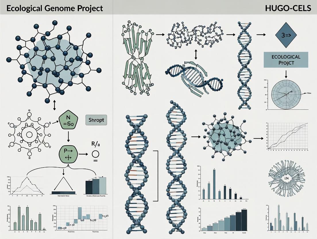
Visualization of Key Concepts
Diagram 1: HUGO CELS Workflow from Sample to Atlas
Diagram 2: Ecological Genome Project Thesis & HUGO CELS
The Scientist's Toolkit: Key Research Reagent Solutions
Table 3: Essential Reagents & Materials for Core HUGO CELS Protocols
| Item | Function | Example/Note |
|---|---|---|
| Tissue Dissociation Kits | Enzymatic (collagenase, trypsin) and mechanical dissociation of solid tissues into single-cell suspensions. | Miltenyi Multi Tissue Dissociation Kits; Worthington enzymes. Condition/Time optimization is critical. |
| Viability Stain (e.g., DRAQ7) | Distinguish live from dead cells prior to loading on scRNA-seq platforms. Dead cells increase background noise. | Fluorescent DNA dye impermeant to live cells. Used in flow cytometry or microfluidics. |
| Chromium Next GEM Chip K | Microfluidic device for partitioning single cells, beads, and reagents into GEMs. | 10x Genomics consumable; determines channel count (e.g., Chip K for 10K cells). |
| Chromium Next GEM Gel Beads | Barcoded beads containing oligonucleotides with cell barcode, UMI, and poly-dT. | Core reagent for cell barcoding. Must be kept cold and anhydrous. |
| Visium Spatial Gene Expression Slide | Glass slide with ~5,000 barcoded spots in a 6.5x6.5 mm array. | Captures location-specific mRNA. Includes a fiducial frame for imaging alignment. |
| Visium Tissue Optimization Slide | Used to determine optimal permeabilization time for a specific tissue type. | Contains fluorescently-labeled oligos to visualize mRNA capture efficiency. |
| TD Buffer (10x Genomics) | Proprietary tissue permeabilization buffer for Visium protocol. | Optimized for mRNA release without diffusion or morphology loss. |
| Dual Index Kit TT Set A | Provides unique dual indices for multiplexing samples in a single sequencing run. | Essential for cost-effective, high-throughput library pooling. |
| SPRIselect Beads | Size-selective magnetic beads for post-amplification cDNA and library clean-up and size selection. | Beckman Coulter SPRIselect; used in most NGS library prep workflows. |
| Bioanalyzer/ TapeStation Kits | Quality control of cDNA and final library fragment size distribution and concentration. | Agilent High Sensitivity DNA kit; critical for sequencing success. |
The Human Genome Project provided a singular, linear reference, a monumental but inherently limited framework. The concept of the Ecological Genome emerges from the understanding that a genome does not exist in isolation. It is a dynamic entity shaped by continuous multi-layered interactions: with the internal cellular environment (epigenetics, somatic variation), the host organism's physiology, the microbiome, and the external exposome. This whitepaper, framed within the broader thesis of the Ecological Genome Project (EGP), a proposed successor to HUGO and related cell atlas initiatives (CELS), outlines the technical framework for defining and studying genomes in their full ecological context. This paradigm is critical for researchers and drug development professionals moving beyond one-size-fits-all therapeutics towards precise, systems-level interventions.
The standard human reference genome (GRCh38) is a composite haplotype, invaluable for alignment but devoid of biological context. It lacks:
- Population-specific variants and structural diversity.
- Somatic mosaicism acquired over a lifetime.
- Epigenetic landscapes that regulate genomic function.
- Interactions with the metagenome (virome, microbiome).
- Environmental modulation via the exposome.
The Ecological Genome is defined as the sum total of an individual's inherited genetic material, its somatic variations, its regulatory apparatus, and its functional interactions with commensal genomes and environmental factors, all within a spatial and temporal context. The EGP aims to map these interactions to understand phenotypic emergence and disease etiology.
The Four Pillars of the Ecological Genome Framework
Research must concurrently analyze these interconnected layers.
Pillar 1: The Dynamic Human Genome
Core Concept: The host genome is a heterogeneous, aging cellular population. Key Data & Methods:
- Long-Read Sequencing (PacBio, Oxford Nanopore): For phased haplotyping, resolving complex structural variants (SVs), and detecting epigenetic modifications (e.g., 5mC, 6mA) directly.
- Single-Cell Multi-Omics: scRNA-seq + scATAC-seq to link chromatin accessibility to gene expression in individual cells; scDNA-seq to catalogue somatic mutations.
- Spatial Transcriptomics/Proteomics: (10x Genomics Visium, Nanostring GeoMx) to map genomic activity within tissue architecture.
Table 1: Quantitative Landscape of Human Genomic Variation
| Variation Type | Scale/Prevalence | Detection Technology | Relevance to EGP |
|---|---|---|---|
| Single Nucleotide Variant (SNV) | ~4-5 million per genome | Short-read WGS, Arrays | Common population diversity |
| Structural Variant (SV) | >20,000 per genome; many rare | Long-read WGS, Optical Mapping | Major contributor to phenotypic diversity & disease |
| Somatic Mosaic SNV/SV | Accumulates with age (e.g., ~20-50/cell division) | Ultra-deep sequencing, Single-cell DNA-seq | Aging, cancer, neurodevelopment |
| Methylation (5mC) | Tissue-specific patterns; changes with age/environment | Whole-genome bisulfite sequencing (WGBS) | Gene regulation, cellular identity |
Pillar 2: The Epigenetic Interface
Core Concept: Epigenetics is the primary transducer of ecological signals onto the genome. Experimental Protocol: Integrated Epigenomic Profiling
- Sample: Primary tissue or cell culture under controlled environmental stimulus (e.g., nutrient stress, cytokine exposure).
- Assay: Parallel processing for:
- ATAC-seq: Assay for Transposase-Accessible Chromatin to map open chromatin regions.
- ChIP-seq: For histone modifications (H3K27ac, H3K4me3) and transcription factor binding.
- Hi-C or Micro-C: To capture 3D chromatin conformation and topologically associating domains (TADs).
- Integration: Use tools like SnapTools or ArchR to create a unified epigenomic landscape, correlating accessibility, histone marks, and long-range interactions with transcriptional output (from RNA-seq).
Pillar 3: The Metagenomic Milieu
Core Concept: The human host is a holobiont. Microbial genes outnumber human genes by orders of magnitude. Methodology: Host-Microbiome Interaction Mapping
- Dual RNA-seq: Simultaneously extract and sequence host and microbial RNA from a single tissue sample (e.g., gut mucosa). Use Kraken2/Bracken for taxonomic profiling of microbial reads and align host reads to the human genome.
- Metabolomic Correlation: Perform LC-MS metabolomics on matched plasma or tissue samples. Use correlation networks (e.g., Sparse Correlations for Compositional data, SparCC) to link microbial taxa abundance (from 16S rRNA gene sequencing or metagenomics) to host metabolites and serum inflammatory markers (e.g., IL-6, CRP).
- Functional Validation: Use gnotobiotic mouse models colonized with defined microbial communities (human-derived consortia) to test causal links between microbial genes, host epigenome, and phenotype.
Table 2: Key Microbial Functional Guilds with Genomic Impact
| Microbial Component | Example Taxa/Element | Proposed Genomic Impact Mechanism |
|---|---|---|
| Commensal Bacteria | Bacteroides spp., Faecalibacterium prausnitzii | Produce short-chain fatty acids (SCFAs) inhibiting host HDACs, altering epigenome. |
| Pathobionts | Enterococcus faecalis, certain E. coli strains | Induce DNA damage via reactive oxygen species or genotoxins (e.g., colibactin). |
| Viral "Dark Matter" | Anelloviruses, endogenous retroviruses | May provide immune training; ERV expression can regulate host immunity genes. |
| Fungal Mycobiome | Candida albicans | Can induce Th17 response, altering local inflammatory transcriptional programs. |
Pillar 4: The Exposomic Imprint
Core Concept: The cumulative environmental exposure (chemical, social, physical) leaves measurable signatures on the ecological genome. Approach: Exposome-Wide Association Studies (ExWAS)
- External Exposome: GPS, smartphone data, satellite imagery, environmental sensors (air quality monitors).
- Internal Exposome: High-resolution mass spectrometry (HRMS) on biospecimens for untargeted detection of exogenous chemicals, dietary metabolites, and stress hormones.
- Data Integration: Use multivariate models to associate specific exposomic features with multi-omic host-microbiome biomarkers (e.g., differential methylation, microbial shift, cytokine levels).
Visualizing Ecological Genome Interactions
Diagram Title: Ecological Genome Interaction Network
Diagram Title: Ecological Genome Project Core Workflow
The Scientist's Toolkit: Research Reagent Solutions
Table 3: Essential Reagents & Platforms for Ecological Genome Research
| Item / Solution | Function in EGP Research | Key Consideration |
|---|---|---|
| 10x Genomics Chromium | Enables linked-read, single-cell, and spatial multi-omic profiling (e.g., Multiome ATAC + Gene Exp). | Critical for connecting host genotype to phenotype at single-cell resolution. |
| PacBio HiFi/Sequel IIe | Generates highly accurate long reads for phased diploid genomes, SV detection, and methylation calling. | Essential for Pillar 1 (Dynamic Genome) to move beyond the linear reference. |
| Oxford Nanopore PromethION | Provides ultra-long reads for scaffolding and real-time detection of base modifications. | Ideal for metagenomic sequencing and detecting novel epigenetic marks. |
| KAPA HyperPrep/HyperPlus | Robust library preparation kits for low-input and degraded samples (e.g., from FFPE, ancient DNA). | Vital for working with diverse, real-world sample types in exposomic studies. |
| ZymoBIOMICS Spike-in Controls | Defined microbial community standards for metagenomic and metatranscriptomic sequencing. | Enables absolute quantification and technical validation in microbiome studies. |
| Cellular Indexing of Transcriptomes & Epitopes by Sequencing (CITE-seq) Antibodies | Oligo-tagged antibodies for simultaneous protein and RNA measurement at single-cell level. | Links host immune cell states to microbial or environmental perturbations. |
| Assay for Transposase-Accessible Chromatin (ATAC) Kits | Maps open chromatin regions using hyperactive Tn5 transposase. | Foundation for defining the epigenetic interface (Pillar 2). |
| Cytokine/Chemokine Multiplex Assays (Luminex/MSD) | High-throughput protein quantification of immune and inflammatory markers. | Provides a key phenotypic bridge between omic layers and physiological state. |
Defining the Ecological Genome necessitates a shift from reductionist to integrative systems biology. For drug development, this means:
- Target Identification: Prioritizing nodes within the host-microbe-exposome network, not just human genes.
- Clinical Trial Design: Stratifying patients based on ecological genome profiles (e.g., "enterotype" + host immune epigenotype) rather than single genetic biomarkers.
- Therapeutic Modalities: Expanding beyond small molecules to include pre/probiotics, phage therapy, epigenetic editors, and exposome modulators (e.g., air filters). The Ecological Genome Project provides the necessary framework to realize this future, transforming our understanding of human biology from a static code to a dynamic, contextualized dialogue.
The completion of the Human Genome Project marked a beginning, not an end. The subsequent challenge has been to understand the dynamic interplay between genomic information and environmental context. This has given rise to the Ecological Genome Project, a conceptual and methodological framework extending beyond HUGO (Human Genome Organization) and CELS (Committee on Ethics, Law, and Society) research. It posits that phenotypes, including disease states, are not merely the product of static genetic code but emerge from complex, multi-scale interactions between an organism's genome and its ecological niche—encompassing microbiota, diet, toxins, climate, and social stressors. For drug discovery, this ecological lens is transformative, shifting the paradigm from "one target, one drug" to a network-based understanding of disease etiology and therapeutic intervention.
Core Ecological Drivers in Genomics and Therapeutic Discovery
The Host as a Holobiont: Microbiome-Genome Interactome
The human host is a supra-organism, or holobiont, composed of human cells and a vast consortium of commensal microorganisms. The ecological balance of this microbiome directly regulates host gene expression, immune function, and metabolic pathways.
- Quantitative Impact: Dysbiosis (ecological imbalance) is linked to disease susceptibility and drug response variance.
Table 1: Impact of Microbiome Composition on Drug Efficacy & Toxicity
| Drug/Therapeutic Area | Ecological Mechanism | Observed Effect on Drug Kinetics/ Dynamics | Key Quantitative Finding (Source: Recent Studies) |
|---|---|---|---|
| Chemotherapy (e.g., Cyclophosphamide) | Gut microbiota primes systemic immune response. | Modulates anti-tumor efficacy and toxicity. | Germ-free mice show 40-60% reduced efficacy; E. hirae & B. intestinihominis restore response. |
| Immunotherapy (Anti-PD-1) | Microbial metabolites (SCFAs) modulate T-cell function. | Predicts clinical response in melanoma patients. | Responders have higher α-diversity (Shannon Index >4.5) and abundance of Faecalibacterium. |
| Cardiovascular (Digoxin) | Bacterial gene (cgr) cluster inactivates digoxin. | Reduces serum drug bioavailability. | Eggerthella lenta carriage can reduce digoxin activation by up to 50% in certain individuals. |
| Metformin (Type 2 Diabetes) | Alters bile acid metabolism & gut microbiota composition. | Partially mediates its glucose-lowering effect. | Increases Akkermansia muciniphila abundance; correlation (r=0.6) with improved glucose tolerance. |
- Experimental Protocol: Metagenomic Sequencing & Gnotobiotic Mouse Model for Drug-Microbiome Interaction.
- Cohort Stratification: Recruit patient cohorts (e.g., drug responders vs. non-responders). Collect longitudinal fecal samples.
- Metagenomic Sequencing: Extract total microbial DNA. Perform shotgun sequencing (Illumina NovaSeq). Process reads with KneadData to remove host contamination.
- Bioinformatic Analysis: Assemble reads (metaSPAdes), annotate genes (Prokka), and quantify metabolic pathways (HUMAnN3). Perform differential abundance analysis (LEfSe, MaAsLin2) to identify biomarker taxa/genes.
- Causal Validation:
- Fecal Microbiota Transplant (FMT): Transplant human donor microbiota into germ-free mice.
- Drug Administration: Treat mice with the drug of interest.
- Phenotyping: Measure pharmacokinetics (LC-MS/MS on serum), pharmacodynamics (e.g., tumor volume, glucose tolerance), and host transcriptomics (RNA-seq on target tissues).
- Defined Consortium: Colonize germ-free mice with a minimal bacterial consortium containing the candidate biomarker strain to confirm mechanism.
Environmental Exposure: The Exposome's Dialogue with the Genome
The exposome—the totality of environmental exposures from conception onward—acts as a continuous modulator of epigenetic and genetic regulation. This ecological driver is critical for understanding complex disease risk.
Table 2: Exposome-Genome Interactions in Disease Etiology
| Exposure Class | Molecular Interaction | Disease Association | Quantitative Data from Cohort Studies |
|---|---|---|---|
| Air Pollutants (PM2.5) | Induces global DNA hypomethylation & inflammation (NF-κB). | COPD, Asthma, CVD. | 10 μg/m³ increase in PM2.5 associated with 0.5-1.0% decrease in global DNA methylation (LINE-1) in leukocytes. |
| Dietary Compounds (e.g., Folate) | Alters one-carbon metabolism, affecting SAM levels for DNA methylation. | Neural tube defects, cancer risk. | Maternal folate sufficiency (>400 μg/day) reduces NTD risk by ~70% in susceptible genotypes (MTHFR 677TT). |
| Endocrine Disruptors (BPA) | Binds estrogen receptors, altering hormone-responsive gene networks. | Metabolic syndrome, infertility. | Urinary BPA levels (>4.7 ng/mL) correlated with significant differential methylation in imprinted genes (e.g., IGF2). |
| Social Stress | Activates HPA axis, increasing cortisol, which binds glucocorticoid response elements (GREs). | Depression, PTSD. | Childhood trauma associated with increased FKBP5 methylation (up to 12% at specific CpGs) and altered stress response. |
- Experimental Protocol: Epigenome-Wide Association Study (EWAS) of an Environmental Exposure.
- Exposure Quantification: Use targeted mass spectrometry (e.g., for pollutants), questionnaires, or sensors to quantify exposure in a population cohort.
- DNA Methylation Profiling: Extract DNA from peripheral blood or target tissue. Process with bisulfite conversion (EZ DNA Methylation Kit). Analyze using microarray (Illumina EPIC array) or whole-genome bisulfite sequencing (WGBS).
- Statistical Modeling: Use linear regression (via R package limma or methylGSA) to associate methylation β-values at each CpG site with exposure level, adjusting for cell-type heterogeneity (Houseman method), age, sex, and batch effects. Genome-wide significance: ( p < 1 \times 10^{-7} ).
- Functional Validation: Select top differentially methylated regions (DMRs). Use CRISPR-dCas9-TET1/DNMT3A to epigenetically edit loci in cell lines. Assess gene expression (qRT-PCR) and pathway-specific phenotypes (e.g., proliferation, apoptosis).
Ecological Evolutionary Principles in Cancer and Resistance
Tumors are complex, evolving ecosystems subject to ecological pressures like competition, spatial heterogeneity, and migration. This framework explains drug resistance.
- Experimental Protocol: Phylogenetic Tracing of Clonal Evolution in Response to Therapy.
- Longitudinal Sampling: Perform multi-region tumor biopsies (or liquid biopsies for ctDNA) at diagnosis, during treatment, and at relapse.
- Deep Sequencing: Perform whole-exome or targeted deep sequencing (>500x coverage) of tumor and matched normal DNA.
- Variant Calling & Phylogeny Reconstruction: Call somatic mutations (MuTect2, VarScan2). Use tools like PyClone or SciClone to identify clonal populations. Construct phylogenetic trees of subclones using maximum parsimony (PAUP) or Bayesian methods (BEAST2).
- Identification of Resistance Drivers: Correlate expanding subclones at relapse with specific mutations (e.g., EGFR T790M, BCR-ABL T315I). Validate functional impact via in vitro mutagenesis and drug sensitivity assays (IC50 shift).
Visualization of Core Concepts
Title: Ecological Drivers of Host Phenotype
Title: Ecological Selection of Drug Resistance in Cancer
The Scientist's Toolkit: Essential Research Reagent Solutions
Table 3: Key Reagents for Ecological Genomics Research
| Research Reagent / Solution | Function & Application | Key Consideration |
|---|---|---|
| Gnotobiotic Rodent Housing Systems | Provides a controlled, germ-free environment for causal microbiome studies. Isolators or ventilated cages. | Essential for FMT experiments to establish causality from correlative human data. |
| Stable Isotope-Labeled Substrates (e.g., ¹³C-Glucose) | Tracks metabolic flux from host or microbiome in complex ecosystems (SIRM - Stable Isotope Resolved Metabolomics). | Enables mapping of cross-kingdom metabolic interactions (e.g., microbial conversion of host bile acids). |
| DNA Methylation Inhibitors/Activators (5-Azacytidine, TSA) | Tools for bulk epigenetic manipulation to validate exposure-related findings in vitro. | Lacks locus-specificity; used prior to targeted epigenetic editing techniques. |
| CRISPR-dCas9 Epigenetic Editors (dCas9-DNMT3A, dCas9-TET1) | Enables precise, locus-specific DNA methylation or demethylation for functional validation of EWAS hits. | Requires efficient sgRNA design and delivery (lentivirus, electroporation) to target cell types. |
| Ultra-pure DNA/RNA Kits with Host Depletion | Nucleic acid extraction optimized for microbiome studies, incorporating probes to remove host (human) genetic material. | Critical for increasing microbial sequencing depth and reducing cost in host-dominant samples (e.g., lung tissue). |
| Multiplex Immunofluorescence (e.g., CODEX, Phenocycler) | Spatial proteomics to map immune and tumor cell ecology within the tissue microenvironment. | Preserves spatial context lost in single-cell sequencing, revealing ecological niches in cancer or inflammation. |
| Liquid Biopsy ctDNA Extraction Kits | Isolation of circulating tumor DNA for non-invasive monitoring of clonal evolution and resistance. | Sensitivity is key; optimized for low-abundance, fragmented DNA in plasma. |
| High-Throughput Sensitivity Assays (Organ-on-a-Chip) | Microfluidic co-culture systems to model human organ ecology (e.g., gut-liver axis) and drug response. | Incorporates fluid flow, mechanical forces, and multiple cell types for physiologically relevant screening. |
The future of effective, personalized medicine lies in embracing ecological complexity. This requires:
- Study Design: Shift from case-control to longitudinal, deep-phenotyping cohorts that capture exposome and microbiome dynamics.
- Data Integration: Develop multi-omic data fusion platforms that link genomic, metagenomic, epigenomic, and metabolomic data layers within an ecological context.
- Therapeutic Targets: Move beyond single human proteins to target ecological interfaces—e.g., microbial enzymes that metabolize drugs, host receptors for microbial metabolites, or epigenetic writers/erasers modulated by the environment.
- Clinical Trials: Incorporate ecological biomarkers (microbiome signatures, epigenetic clocks) for patient stratification and monitoring of therapeutic impact on the host ecosystem.
By adopting the framework of the Ecological Genome Project, genomics and drug discovery transition from a reductionist to a holistic science, ultimately yielding therapies that are as complex and effective as the biological systems they aim to correct.
The Ecological Genome Project, an extension of the HUGO Council for the Ecological Life Sciences (CELS) vision, posits that human health cannot be deciphered through a static human genome alone. It requires the integrated study of the metagenome (microbiomes), the exposome (environmental exposures), and the evolutionary genomic context. This whitepaper details the core research domains and their interconnections, providing a technical guide for advancing systems-level ecological genomics in therapeutic and diagnostic development.
Domain I: The Human Microbiome
The human microbiome comprises trillions of microorganisms residing in ecological niches such as the gut, skin, and respiratory tract. Its collective genome (microbiome) vastly exceeds the human genome in gene count and metabolic potential.
Key Quantitative Data
Table 1: Core Human Microbiome Metrics by Body Site
| Body Site | Estimated Microbial Cells (Ratio to Human) | Dominant Phyla (Top 3) | Key Functions |
|---|---|---|---|
| Gastrointestinal Tract | ~3.8x10^13 (1.3:1) | Bacteroidetes, Firmicutes, Actinobacteria | Metabolism, immune priming, barrier integrity |
| Oral Cavity | ~1x10^10 | Firmicutes, Bacteroidetes, Proteobacteria | Nitrate reduction, primary digestion |
| Skin | ~1x10^9 | Actinobacteria, Firmicutes, Proteobacteria | Defense against pathogens, lipid metabolism |
| Vagina | ~1x10^8 | Firmicutes (Lactobacillus), Actinobacteria | pH maintenance, pathogen exclusion |
Experimental Protocol: Metagenomic Sequencing for Functional Profiling
Protocol Title: Shotgun Metagenomic Sequencing for Pathway Analysis
- Sample Collection & Stabilization: Collect sample (e.g., fecal, swab) in DNA/RNA stabilizing buffer (e.g., Zymo DNA/RNA Shield). Store at -80°C.
- Total DNA Extraction: Use a bead-beating mechanical lysis kit (e.g., Qiagen PowerSoil Pro) to ensure lysis of Gram-positive bacteria. Include negative extraction controls.
- Library Preparation: Quantify DNA with fluorometry (Qubit). Use 1ng-100ng input for enzymatic fragmentation and adapter ligation (e.g., Illumina Nextera XT). Amplify with limited-cycle PCR.
- Sequencing: Perform paired-end sequencing (2x150bp) on an Illumina NovaSeq platform to achieve a minimum of 10 million reads per sample for gut microbiome depth.
- Bioinformatic Analysis:
- Quality Control & Host Depletion: Trim adapters and low-quality bases with Trimmomatic. Align reads to the human genome (hg38) using Bowtie2 and remove aligned reads.
- Taxonomic Profiling: Classify reads using a k-mer-based tool (Kraken2) against a curated database (e.g., GTDB). Generate abundance tables.
- Functional Profiling: Align reads to a protein family database (e.g., EggNOG, KEGG) using DIAMOND. Infer metagenomic pathways with HUMAnN3.
Shotgun Metagenomics Analysis Workflow
Research Reagent Solutions: Microbiome
Table 2: Essential Reagents for Microbiome Research
| Item | Example Product | Function in Research |
|---|---|---|
| Stabilization Buffer | Zymo DNA/RNA Shield | Preserves nucleic acid integrity at room temperature, critical for field studies. |
| Bead-Beating Extraction Kit | Qiagen PowerSoil Pro | Mechanical and chemical lysis for robust DNA yield from diverse, tough-to-lyse microbes. |
| Metagenomic Standard | ZymoBIOMICS Microbial Community Standard | Defined mock community for controlling extraction, sequencing, and bioinformatic bias. |
| Selective Growth Media | YCFA Agar (for anaerobes) | Culturomics: isolation and expansion of fastidious anaerobic gut bacteria. |
| Gnotobiotic Mouse Model | Taconic Biosciences Germ-Free Mice | In vivo causal studies of microbiome function in a controlled, microbe-free host. |
Domain II: The Environmental Exposome
The exposome encompasses all environmental exposures (chemical, biological, physical, social) from conception onward. It interacts directly with the host and microbiome.
Key Quantitative Data
Table 3: Classes of Environmental Exposures and Measurement Techniques
| Exposure Class | Example Agents | Primary Measurement Method | Typical Biomarker Matrix |
|---|---|---|---|
| Endocrine Disruptors | BPA, Phthalates, PCBs | LC-MS/MS (Liquid Chromatography Tandem Mass Spec) | Urine, Serum |
| Airborne Pollutants | PM2.5, NOx, Ozone | Personal Monitoring Sensors & Station Data | Blood (inflammatory markers), Sputum |
| Dietary Metabolites | Polyphenols, Heterocyclic Amines | Untargeted Metabolomics (HRAM MS) | Plasma, Feces |
| Microbial Toxins | Lipopolysaccharide (LPS) | ELISA, LAL Assay | Serum, Stool |
Experimental Protocol: High-Resolution Exposome Profiling
Protocol Title: Untargeted Metabolomics for Exposome-Wide Association Studies (ExWAS)
- Sample Preparation (Serum/Plasma): Thaw samples on ice. Precipitate proteins by adding 300µL cold methanol to 100µL serum. Vortex, incubate at -20°C for 1 hour, centrifuge at 14,000g for 15 min (4°C). Transfer supernatant to a new vial and dry in a vacuum concentrator.
- Derivatization & Reconstitution: Reconstitute dried extract in 50µL methoxyamine hydrochloride in pyridine (15 mg/mL). Shake at 30°C for 90 min. Add 50µL MSTFA (N-Methyl-N-(trimethylsilyl)trifluoroacetamide) and incubate at 37°C for 30 min.
- Instrumental Analysis (GC-MS): Inject 1µL in splitless mode onto an Rxi-5Sil MS column. Use a temperature gradient (60°C to 330°C). Operate mass spectrometer in electron impact (EI) mode, scanning m/z 50-600.
- Data Processing: Convert raw files to .mzXML format. Perform peak picking, deconvolution, and alignment using XCMS. Annotate peaks against libraries (NIST, Fiehn). Normalize data using internal standards and quality control (QC) samples.
Exposome to Health Outcome Pathway
Domain III: Evolutionary Genomic Context
This domain examines how host genetic variation, shaped by evolution, moderates responses to the microbiome and exposome. It focuses on signatures of natural selection and conserved pathways.
Key Quantitative Data
Table 4: Human Genes under Selection from Environmental Pressures
| Gene/ Locus | Evolutionary Pressure (Hypothesized) | Associated Modern Phenotype | Population Signal |
|---|---|---|---|
| LCT (Lactase) | Dairy farming / pastoralism | Lactose persistence in adults | Strong positive selection in European & African pops. |
| FADS1 (Fatty acid desaturase) | Dietary shift (plant/ marine fats) | Fatty acid metabolism | Positive selection, Neanderthal introgression. |
| HLA (Major Histocompatibility Complex) | Pathogen exposure | Immune diversity & autoimmunity risk | Balancing selection, extreme polymorphism. |
| EDAR (Ectodysplasin A receptor) | Climate/ unknown | Hair thickness, tooth morphology | Strong selective sweep in East Asian populations. |
Experimental Protocol: Detecting Evolutionary Signals in Genomic Data
Protocol Title: Composite Likelihood Ratio Test for Recent Positive Selection (e.g., on FADS1)
- Data Acquisition: Obtain phased genotype data (e.g., from 1000 Genomes Project) for target region (chr11:61,309,839-61,491,566 for FADS1) and flanking regions.
- Calculate Site Frequency Spectrum (SFS): Use
ANGSDto compute the unfolded SFS, specifying an ancestral genome (e.g., chimpanzee, panTro5). - Run Selection Scans: Execute the
SweepFinder2software. Input the SFS and a pre-computed genetic map for the region. The software calculates a composite likelihood ratio (CLR) statistic for each SNP. - Identify Selection Peaks: Visually inspect CLR statistics across the genomic region. A sharp peak centered on a gene (e.g., FADS1) indicates a putative selective sweep. Validate using complementary statistics (iHS, nSL) from
selscan. - Functional Validation: Correlate selected haplotype with metabolite levels (e.g., omega-3 fatty acids) in a biobank cohort to link evolutionary signal to modern biochemical function.
Integrative Analysis: The Ecological Genome Model
The core hypothesis is that disease phenotypes (P) arise from the interaction of host genetics (G), the microbiome (M), and the exposome (E): P = f(G, M, E) + (GxM) + (GxE) + (MxE) + (GxMxE).
Experimental Protocol: Multi-Omic Integration Study
Protocol Title: Longitudinal Multi-Omic Profiling for Interaction Discovery
- Cohort Design: Recruit a longitudinal cohort with deep phenotyping (e.g., diabetics vs. controls). Collect host genome (SNP array/WGS), longitudinal stool (metagenomics), serum (metabolomics), and exposure questionnaires at multiple time points.
- Data Generation: Generate data per protocols in sections 2.2, 3.2, and 4.2.
- Interaction Analysis: Use multivariate methods.
- MaAsLin 2: Identify microbiome taxa/metabolites associated with host genotype, controlling for exposures.
- StructLMM: Test for genotype-by-environment (GxE) interaction on metabolite levels.
- Similarity Network Fusion (SNF): Integrate omics layers into a unified patient similarity network to identify novel disease subtypes.
- Causal Inference: Apply Mendelian Randomization using host genetic variants as instruments to infer potential causal direction between an exposure biomarker and a microbiome feature.
Ecological Genome Integrative Model
The Scientist's Toolkit for Integrative Research
Table 5: Key Resources for Ecological Genome Research
| Tool Category | Specific Resource | Purpose & Explanation |
|---|---|---|
| Biobank & Cohort Data | UK Biobank, All of Us, Human Exposome Project | Provides large-scale, deep phenotyped data with multi-omic layers for hypothesis testing. |
| Bioinformatic Pipeline | nf-core/mag, nf-core/metabolab | Standardized, containerized Nextflow pipelines for reproducible metagenomic/metabolomic analysis. |
| Interaction Database | STITCH, MVDA (Multi-Omic ViDa) | Databases of known chemical-protein, microbe-host, and gene-environment interactions. |
| In Silico Modeling | Genome-scale Metabolic Models (AGORA, Virtual Human Microbiome) | Predict metabolic exchange between host and microbiome under different nutritional/exposure conditions. |
| Animal Models | Collaborative Cross Mice, Humanized Microbiome Mice | Genetically diverse mouse models for testing GxE and GxM interactions in a controlled setting. |
The integration of microbiome, exposome, and evolutionary context research is moving from correlation to causation and mechanism. For the drug development professional, this framework reveals novel, ecologically informed targets: microbial enzymes, exposure-mitigating compounds, and pathways shaped by evolution. The Ecological Genome Project CELS mandates the development of new tools—standardized exposure assessment, gnotobiotic models for causal microbe studies, and computational platforms for high-dimensional interaction modeling—to realize the promise of ecological precision medicine.
The Human Genome Organization’s (HUGO) Committee on Ecological, Lifestyle, and Spatial health (CELS) represents a paradigm shift in post-genomic research. Framed within the broader thesis of the Ecological Genome Project (EGP), CELS moves beyond static, linear models of gene-to-phenotype mapping. It posits that human health and disease are emergent properties of complex, multiscale networks integrating genomic data with ecological, lifestyle, and spatial (ELS) variables. This whitepaper details the core principles and methodologies for translating this conceptual framework into actionable, quantitative network biology.
Core Principles: Transitioning from Linearity to Networks
The CELS framework is governed by four interdependent principles:
- Principle 1: Contextual Integration: Genomic signals are not absolute. Their phenotypic expression is modulated by ELS layers (e.g., pollutant exposure, dietary patterns, microbiome composition, socioeconomic factors).
- Principle 2: Dynamic Interactivity: Relationships within and between biological and ELS layers are bidirectional and time-variant, forming adaptive feedback loops.
- Principle 3: Emergent Phenotypes: Disease states are network attractors, arising from system-wide perturbations rather than single-pathway dysfunction.
- Principle 4: Spatial Resolution: Biological and ELS data must be anchored to specific anatomical (tissue, cell) and geographical contexts to be interpretable.
Quantitative Data Synthesis: ELS Modulators in Network Perturbation
Key meta-analyses underscore the quantitative impact of ELS factors on core biological networks relevant to drug development, such as inflammation and metabolic regulation.
Table 1: Impact of Select ELS Factors on Network Hub Genes and Pathways
| ELS Factor Category | Specific Modulator | Measured Effect Size (Odds Ratio / Hazard Ratio) | Primary Biological Network Perturbed | Key Hub Genes Affected (e.g.,) |
|---|---|---|---|---|
| Environmental | PM2.5 Long-term Exposure | HR: 1.12 [1.08–1.16] for CVD | Inflammatory & Oxidative Stress Response | NFKB1, IL6, TNF, NRF2 |
| Lifestyle | Microbiome α-Diversity Index | OR: 0.65 [0.50–0.85] for IBD | Immune Tolerance & Mucosal Barrier | TLR4, FOXP3, MUC2 |
| Spatial/Clinical | Tissue Hypoxia (pO2 <10 mmHg) | Correl. Coefficient: 0.78 with EMT Score | Epithelial-Mesenchymal Transition | HIF1A, SNAI1, VEGFA |
Experimental Protocols for CELS-Informed Network Biology
Protocol: Multi-Omic Cohort Profiling with ELS Data Layer Integration
Objective: To construct a context-aware molecular network for a disease phenotype (e.g., asthma exacerbation). Methodology:
- Cohort & ELS Data Acquisition: Recruit cohort (N>500). Collect geocoded environmental data (EPA air quality indices), lifestyle data (validated dietary questionnaires, wearable device logs), and clinical phenotyping.
- Biospecimen Collection & Multi-omics Profiling: Obtain blood/tissue samples at baseline and upon event.
- Genomics: GWAS and PRS calculation.
- Transcriptomics: Bulk or single-cell RNA-seq.
- Epigenomics: Methylation array (e.g., EPIC).
- Proteomics & Metabolomics: LC-MS/MS profiling.
- Data Integration & Network Inference:
- Use similarity network fusion (SNF) or Multi-Omics Factor Analysis (MOFA) to create an integrated patient similarity network.
- Employ context-specific Gaussian graphical models (GGMs) or Bayesian networks, conditioning model priors on ELS strata (e.g., high vs. low pollutant area).
- Validation: Perform causal inference using Mendelian Randomization with ELS factors as exposures. Validate network predictions in an independent cohort or in vitro models exposed to simulated ELS conditions.
Protocol: Spatial Transcriptomics with Ecological Context Mapping
Objective: To map gene expression networks within tissue architecture while incorporating geographical ELS data. Methodology:
- Tissue Sectioning & Sequencing: Perform Visium or Xenium (10x Genomics) platform workflow on diseased tissue sections.
- Spatial Network Analysis: Use SpaGCN or Giotto to identify spatially coherent expression neighborhoods and ligand-receptor interaction networks across tissue zones.
- Ecological Context Overlay: Spatially join patient residential coordinates with raster data from satellite imagery (NASA SEDAC) for green space, nighttime light (urbanization), and land surface temperature.
- Correlative Modeling: Apply spatial regression models (e.g., geographically weighted regression) to associate specific tissue microenvironment network states with upstream ecological variables.
Visualization of CELS Network Logic and Workflows
Title: CELS vs. Linear Biology Paradigm Shift
Title: Core CELS Network Analysis Workflow
The Scientist's Toolkit: Key Research Reagent Solutions
Table 2: Essential Reagents & Platforms for CELS Network Research
| Item / Solution | Vendor Examples | Function in CELS Research |
|---|---|---|
| Spatial Transcriptomics Kits | 10x Genomics Visium, NanoString GeoMx | Maps gene expression networks within intact tissue architecture, linking morphology to molecular networks. |
| Multi-Omic Integration Software | MOFA2, MixOmics, Cytoscape w/ Omics plugins | Statistically fuses genomic, transcriptomic, and ELS data layers to infer unified networks. |
| Environmental Exposure Panels | Biomonitoring LC-MS/MS panels (e.g., for PAHs, phthalates) | Quantifies internalized environmental chemical burden for direct integration with -omics data. |
| Cultured Cell-Based ELS Simulators | Organ-on-a-chip (Emulate, Mimetas), Hypoxia Chambers (Baker) | Models the impact of specific ELS factors (shear stress, cyclic hypoxia) on cellular networks in vitro. |
| Geographic Data APIs | Google Earth Engine, EPA ECHO, NASA SEDAC | Provides geocoded ecological and environmental data for spatial linkage to cohort biospecimens. |
| Single-Cell Multi-Omic Kits | 10x Multiome (ATAC + GEX), CITE-seq antibodies | Deconvolutes cell-type-specific network responses to ELS factors from complex tissues. |
From Theory to Bench: Methodologies and Biopharma Applications of Ecological Genomics
Multi-Omics Integration Frameworks for Host-Environment Data
The Ecological Genome Project, as advanced by HUGO’s Committee on Ethical, Legal, and Social Issues (CELS), posits that human health is an emergent property of a complex system involving the host genome and its dynamic interaction with environmental exposures. This whitepaper details technical frameworks for multi-omics integration that operationalize this thesis, moving beyond single-omic associations to causal, systems-level understanding. Such frameworks are critical for researchers and drug development professionals aiming to discover novel, environmentally contextualized therapeutic targets and biomarkers.
Multi-omics integration for host-environment research synthesizes data from host biology and environmental exposure. The following table summarizes core quantitative data domains.
Table 1: Core Omics Layers for Host-Environment Integration
| Omics Layer | Host-Derived Data (Endpoint) | Environment-Derived Data (Exposure) | Primary Measurement Technologies |
|---|---|---|---|
| Genomics | Germline & Somatic Variants | Microbiome Metagenomics | Whole-Genome Sequencing, 16S/ITS rRNA Sequencing, Shotgun Metagenomics |
| Transcriptomics | Host Gene Expression | Microbial Gene Expression, Community Transcriptome | RNA-Seq, Single-Cell RNA-Seq, Metatranscriptomics |
| Epigenomics | DNA Methylation, Histone Modifications | N/A (Indirect via host response) | Bisulfite Sequencing, ChIP-Seq, ATAC-Seq |
| Proteomics | Host Protein Abundance & Modifications | Microbial Proteins, Allergens, Toxins | LC-MS/MS, Affinity-Based Arrays |
| Metabolomics | Endogenous Metabolites | Xenobiotics, Dietary Metabolites, Microbial Metabolites | LC/GC-MS, NMR Spectroscopy |
| Exposomics | N/A (External Focus) | Chemical Pollutants, Particles, Lifestyle Factors | High-Resolution Mass Spectrometry, Sensors, GIS Data |
Core Computational Integration Frameworks: A Technical Guide
Integration can be performed at multiple levels: early (pre-analysis), intermediate (feature reduction), or late (post-analysis).
Early Integration: Concatenation-Based Fusion
- Methodology: Raw or pre-processed data matrices from each omics layer are combined horizontally (sample-wise) into a single, high-dimensional feature matrix. Dimensionality reduction (e.g., PCA, CCA) or regularized regression (LASSO, Elastic Net) is then applied directly.
- Protocol: 1) Perform platform-specific normalization and batch correction per omics dataset. 2) Scale features to mean=0, variance=1. 3) Concatenate matrices using a shared sample ID key. 4) Apply dimensionality reduction (e.g., Multi-Omics Factor Analysis, MOFA) to extract latent factors representing shared variance across omics.
- Use Case: Holistic biomarker discovery from host transcriptomic, proteomic, and metabolomic data in response to an environmental stressor.
Intermediate Integration: Knowledge-Guided Networks
- Methodology: Biological knowledge graphs (e.g., protein-protein interaction networks, metabolic pathways like KEGG, Reactome) serve as a scaffold to map and integrate multi-omics features. Differential features from each layer are overlaid onto the network, and module detection algorithms identify dysregulated subnetworks.
- Protocol: 1) For each omics layer, perform differential analysis (e.g., DESeq2 for RNA-Seq, limma for proteomics) to identify significant features (p<0.05, FC>1.5). 2) Map significant genes, proteins, and metabolites to a unified interaction network (e.g., using
OmicsNetorCytoscape). 3) Apply a network propagation algorithm (e.g., HotNet2) to identify significantly perturbed modules. 4) Enrich modules for biological pathways. - Use Case: Identifying a disrupted host inflammatory subnetwork (genomic variant → mRNA → protein) linked to a specific microbial metabolite.
Late Integration: Model-Based Fusion
- Methodology: Separate predictive models are built for each omics data type, and their outputs (e.g., class probabilities, risk scores) are combined in a final meta-model. This is a form of ensemble learning.
- Protocol: 1) Train individual classifiers (e.g., SVM, Random Forest) on each omics dataset using cross-validation. 2) Extract prediction scores from each model for all samples. 3) Use these scores as input features for a final "super-integrator" model (e.g., logistic regression). 4) Validate the ensemble model on a held-out test set.
- Use Case: Integrating clinical risk (from EHRs), host genomic risk score, and exposome profile for disease stratification.
Experimental Protocol: A Longitudinal Host-Microbiome-Exposome Study
Aim: To characterize the systemic host response to a controlled dietary intervention while monitoring gut microbiome and personal exposome changes.
Detailed Methodology:
- Cohort & Design: N=100 healthy volunteers. 4-week baseline, 8-week dietary intervention (high-fiber), 4-week washout. Weekly sampling.
- Biospecimen Collection: Blood (plasma, PBMCs), stool, urine collected weekly.
- Multi-Omics Profiling:
- Host Genomics: Whole-blood DNA for genotyping array (baseline).
- Host Transcriptomics: RNA-Seq from PBMCs (weekly).
- Host Proteomics & Metabolomics: LC-MS/MS on plasma and urine (weekly).
- Microbiome: Shotgun metagenomic sequencing of stool (weekly).
- Exposome: Personal air sensors (PM2.5, VOCs), food diary app, GPS (continuous).
- Data Integration Workflow: Use an intermediate integration approach. Perform weekly paired differential analysis (vs. personal baseline) for each host omics layer. Identify differentially abundant microbial species and genes. Integrate using multi-omics network analysis anchored on host metabolic and immune pathways.
Diagram 1: Longitudinal Multi-Omics Study Design
Key Signaling Pathways in Host-Environment Interaction
Aryl Hydrocarbon Receptor (AhR) signaling is a prime example of an integrative pathway.
Diagram 2: Ahr Pathway Integrates Host and Environment
The Scientist's Toolkit: Essential Research Reagent Solutions
Table 2: Key Reagents & Materials for Multi-Omics Host-Environment Studies
| Item | Function & Application in Integration Studies | Example Product/Kit |
|---|---|---|
| PaxGene Blood RNA Tube | Stabilizes intracellular RNA profiles in whole blood for host transcriptomics, crucial for longitudinal studies. | BD Vacutainer PaxGene Blood RNA Tube |
| Stool DNA/RNA Shield | Preserves nucleic acid integrity of complex microbial communities in stool samples at ambient temperature. | Zymo Research DNA/RNA Shield |
| Methylated DNA IP Kit | Enriches methylated DNA regions for host epigenomic studies linking environment to gene regulation. | MagMeDIP Kit (Diagenode) |
| Oasis HLB Cartridge | Solid-phase extraction for broad-spectrum metabolomics and exposomics cleanup prior to LC-MS. | Waters Oasis HLB 96-well µElution Plate |
| Pneumatic Biomonitoring Sampler | Personal air sampler for collecting particulate matter onto filters for subsequent exposomic analysis. | SKC BioSampler |
| Multiplex Cytokine Panel | Quantifies dozens of host immune proteins simultaneously, linking omics data to functional immune response. | Luminex Human Cytokine 48-plex Panel |
| Synthetic Spike-in Standards | External controls added pre-processing for absolute quantification and cross-batch normalization in proteomics/metabolomics. | Pierce Quantitative Colorimetric Peptide Assay; Biocrates META-BOOST |
| Cell-Free DNA Collection Tube | Stabilizes circulating cell-free DNA (host & microbial) for non-invasive monitoring of host-environment dynamics. | Streck cfDNA BCT Tube |
Computational Tools and Platforms for Ecological Network Analysis
This whitepaper explores computational tools for analyzing ecological networks, framed within the larger Ecological Genome Project HUGO CELS research initiative. This project seeks to map the complex genomic, proteomic, and metabolic interactions within human cellular ecosystems and their symbionts, with applications in understanding dysbiosis and identifying novel therapeutic targets.
Ecological Network Analysis (ENA) provides the mathematical framework to quantify interactions (e.g., competition, mutualism, predation) within biological systems. For HUGO CELS, this translates to modeling interactions between human cells, the microbiome, viruses, and metabolic pathways. The shift from reductionist to systems-level analysis is critical for understanding emergent properties in health and disease.
Core Computational Tools and Platforms: A Comparative Analysis
The following table summarizes key computational platforms, based on current literature and software documentation.
Table 1: Comparative Analysis of Core Ecological Network Analysis Platforms
| Tool/Platform | Primary Function | Network Type | Key Algorithm/Model | Input Data Format | License |
|---|---|---|---|---|---|
| Cytoscape | Network visualization & analysis | Any (Gene, Protein, Metabolic) | Plugin-based (e.g., CoNet, Dynetika) | SIF, GML, XGMML | Open Source |
| Gephi | Large-scale network visualization & exploration | Any, esp. large-scale | Force-atlas layout, modularity | GEXF, GraphML | Open Source |
| MATLAB w/ COBRA | Constraint-based metabolic modeling | Metabolic-Reaction (MR) | Flux Balance Analysis (FBA) | SBML, JSON | Commercial |
| R (igraph, vegan, SPIEC-EASI) | Statistical analysis & inference | Co-occurrence, Correlation | Graphical LASSO, MEASURE | CSV, BIOM | Open Source |
| Python (NetworkX, NiPy) | Custom network analysis & machine learning | Any | Custom scripts, ML pipelines | Various | Open Source |
| QIIME 2 / PICRUSt2 | Microbiome analysis & functional inference | Phylogenetic, Metabolic | 16S rRNA pipeline, KEGG prediction | FASTQ, BIOM | Open Source |
| MetaNET | Multi-omics network integration | Multi-layer (Genome, Proteome, Metabolome) | Differential Network Analysis | Multi-omic matrices | Open Source |
Table 2: Performance Metrics on a Standardized Microbial Co-occurrence Dataset (n=200 samples, p=500 OTUs)
| Tool (Package) | Inference Time (s) | Memory Peak (GB) | Accuracy (AUC vs. Known Interactions) | Scalability (Max Features) |
|---|---|---|---|---|
| SPIEC-EASI (MB) | 152.3 | 4.1 | 0.89 | ~5,000 |
| SparCC | 18.7 | 1.2 | 0.82 | ~1,000 |
| CoNet (Cytoscape) | 89.5 | 2.8 | 0.85 | ~2,500 |
| Python (Graphical Lasso) | 305.8 | 6.5 | 0.91 | ~10,000 |
Experimental Protocols for Network Inference and Validation
Protocol 3.1: Inferring a Microbial Interaction Network from 16S rRNA Data
Objective: To reconstruct a robust co-occurrence network from microbiome sequencing data.
- Data Preprocessing: Process raw 16S rRNA FASTQ files through QIIME 2 (version 2023.9). Use DADA2 for denoising, chimera removal, and Amplicon Sequence Variant (ASV) calling. Align to the Greengenes 13_8 reference database.
- Normalization: Rarefy the ASV table to an even sampling depth (e.g., 10,000 reads per sample). Apply a centered log-ratio (CLR) transformation after adding a pseudo-count of 1.
- Network Inference: Input the CLR-transformed matrix into R. Use the
SPIEC-EASIpackage with the Meinshausen-Bühlmann (MB) method. Set the lambda.min.ratio to 0.01 and use StARS for stability selection (subsample proportion = 0.8). - Thresholding: Apply a consensus threshold where an edge (interaction) is retained only if it appears in >90% of subsampled networks.
- Visualization: Export the adjacency matrix and import into Cytoscape (v3.9.1). Use the "Prefuse Force Directed" layout. Color nodes by taxonomic phylum and scale node size by betweenness centrality.
Protocol 3.2: Constraint-Based Metabolic Network Analysis of a Host-Microbe System
Objective: To predict metabolic exchange fluxes between host cells and a microbial symbiont.
- Model Reconstruction: Obtain genome-scale metabolic models (GEMs) for Homo sapiens (RECON3D) and the target microbe (from resources like AGORA or CarveMe). Ensure consistent metabolite naming (e.g., using MetaNetX IDs).
- Community Model Building: Use the
COMETS(Computation of Microbial Ecosystems in Time and Space) toolbox or theMicrobiomeModelToolKitin Python. Merge the two GEMs into a compartmentalized community model, defining an extracellular compartment for metabolite exchange. - Constraint Setting: Set constraints for the host cell's uptake (e.g., glucose, oxygen) based on experimental media composition. Constrain the microbe's uptake of host-derived metabolites (e.g., bile acids, mucins). Apply tissue-specific ATP maintenance requirements to the host cell.
- Simulation: Perform parsimonious Flux Balance Analysis (pFBA) using the COBRA Toolbox in MATLAB to simulate a steady-state. Run flux variability analysis (FVA) to identify a range of possible exchange fluxes for key metabolites (e.g., short-chain fatty acids, vitamins).
- Perturbation Analysis: In silico, knock out key microbial transport reactions. Compare the resulting predicted host metabolic flux distributions to the wild-type community to identify host pathways dependent on microbial input.
Visualization of Methodologies and Pathways
Diagram 1: Microbial Co-occurrence Network Analysis Workflow (77 chars)
Diagram 2: Microbial Butyrate to Host Barrier Signaling (71 chars)
The Scientist's Toolkit: Research Reagent Solutions
Table 3: Essential Research Reagents for Ecological Network Validation
| Reagent / Material | Function in HUGO CELS Context | Example Product / Assay |
|---|---|---|
| Stable Isotope-Labeled Metabolites | To trace metabolic flux through host-microbe networks in vitro or in vivo. Enables validation of FBA predictions. | ¹³C-Glucose, ¹⁵N-Glutamine (Cambridge Isotopes) |
| Gnotobiotic Mouse Models | Provides a controlled, defined microbial ecosystem to test causal inferences from network models. | Germ-free C57BL/6 mice + defined microbial consortia. |
| Spatial Transcriptomics Kits | To map the spatial context of ecological interactions predicted by network analysis (e.g., host-microbe niches). | 10x Genomics Visium, NanoString GeoMx DSP. |
| Recombinant Human/Microbial Proteins | To biochemically validate specific protein-protein interactions predicted by integrated network models. | His-tagged recombinant proteins (Sino Biological). |
| Dual-RNAseq Library Prep Kits | For simultaneous transcriptional profiling of host and microbial partners, providing data for cross-kingdom network inference. | Illumina Total RNA-Seq with ribodepletion. |
| Metabolomic Standards | Critical for LC-MS/MS quantification of key network metabolites (SCFAs, bile acids, neurotransmitters) in co-culture supernatants. | Mass Spectrometry Metabolite Library (IROA Technologies). |
| CRISPRi/a Knockdown Pools | For high-throughput perturbation of host cell genes predicted as hubs in integrated networks, followed by phenotypic screening. | Human CRISPRi/a Lentiviral Library (Addgene). |
1. Introduction: The Ecological Genome Project HUGO CELS Framework The Ecological Genome Project, under the auspices of the Human Genome Organization’s (HUGO) Center for Ecological and Longitudinal Studies (CELS), posits that disease phenotypes arise from complex, multiscale interactions between host genomes and dynamic ecological landscapes. This paradigm shift moves beyond single-gene or single-pathogen models to a holistic view where environmental pressures, microbiome composition, and anthropogenic changes are integral to pathogenesis. Identifying "ecological drivers" — specific environmental factors or interactions that predictably modulate disease risk — represents a novel frontier for therapeutic target discovery. This guide details the technical methodologies for their systematic identification and validation.
2. Core Methodologies for Identifying Ecological Drivers
2.1. Longitudinal Metagenomic & Metatranscriptomic Profiling Objective: To correlate shifts in microbial community structure/function with disease onset or progression within a defined host population and environment. Protocol:
- Cohort & Sampling: Enroll a longitudinal cohort (N≥500). Collect serial biospecimens (stool, saliva, skin swabs) alongside clinical phenotyping at defined intervals (e.g., quarterly). Concurrently, collect environmental samples (soil, water, indoor dust) from participant habitats.
- DNA/RNA Extraction: Use bead-beating and kit-based extraction (e.g., DNeasy PowerSoil Pro Kit, RNeasy PowerMicrobiome Kit) with exogenous internal controls (Spike-in RNA/DNA) for absolute quantification.
- Library Preparation & Sequencing: For metagenomics, use shotgun library prep (Nextera XT). For metatranscriptomics, perform rRNA depletion (QIAseq FastSelect) followed by RNA-seq library prep. Sequence on Illumina NovaSeq X (150bp paired-end), targeting 20-50M reads/sample.
- Bioinformatics Analysis:
- Quality Control & Host Depletion: Trimmomatic for adapter removal, followed by KneadData to filter host (human/bovine) reads.
- Taxonomic/Functional Profiling: Use Kraken2/Bracken for taxonomy. For functional genes, perform assembly (MEGAHIT) and annotation via DIAMOND against KEGG/eggNOG databases.
- Ecological Statistics: Calculate alpha/beta diversity (QIIME 2). Use MaAsLin 2 or similar multivariate models to identify microbial features (species, pathways) significantly associated with disease state, correcting for host covariates (age, diet, medication).
2.2. Geospatial & Exposome Data Integration Objective: To link disease-relevant molecular signatures (from 2.1) to specific, measurable environmental exposures. Protocol:
- Exposure Data Capture: Equip participants with personal air monitors (measuring PM2.5, VOCs, NO2). Acquire satellite/drone-derived data on land use, green space (NDVI), and climate (temperature, humidity) for participant GPS coordinates.
- Data Fusion: Create a unified spatiotemporal database linking individual molecular profiles (microbiome, host transcriptomics) with exposure measurements and clinical outcomes.
- Analytical Modeling: Apply machine learning frameworks (e.g., Random Forest, XGBoost) to rank exposure variables by predictive importance for the disease-associated molecular signature. Use spatial regression models (e.g., Geographically Weighted Regression) to identify local exposure-disease hotspots.
2.3. In Vitro & In Vivo Causal Validation Objective: To experimentally establish causality for candidate ecological drivers identified via observational studies. Protocol:
- Gnotobiotic Mouse Models: Colonize germ-free mice with defined microbial consortia reflecting "high-risk" vs. "low-risk" ecological states identified in human cohorts.
- Controlled Exposure: Subject mice to precise levels of the candidate abiotic driver (e.g., a specific air pollutant at 50 µg/m³ PM2.5) in inhalation chambers.
- Multi-omic Endpoint Analysis: After exposure, collect tissues. Perform host transcriptomics (RNA-seq), immune profiling (cytometric bead arrays), and metabolomics (LC-MS) on serum and target organs.
- Perturbation & Rescue: Administer targeted interventions (e.g., a specific probiotic strain, an enzyme that degrades a microbial metabolite, or a drug candidate targeting a host pathway induced by the driver). Measure reversal of pathological phenotypes.
3. Data Synthesis and Target Hypothesis Generation
Table 1: Example Integrated Data Output for an Inflammatory Bowel Disease (IBD) Cohort Study
| Data Layer | High-Risk Ecological Profile | Low-Risk/Protective Profile | Statistical Strength (p-value; q-value) | Proposed Mechanistic Link |
|---|---|---|---|---|
| Microbiome | Ruminococcus gnavus bloom (15% relative abundance) | Faecalibacterium prausnitzii dominance (12% abundance) | p=2.1e-5; q=0.03 | R. gnavus produces pro-inflammatory polysaccharides. F. prausnitzii produces anti-inflammatory butyrate. |
| Microbial Function | Increased LPS biosynthesis pathway (KEGG map00540) | Increased butyrate synthesis (ptb-buk pathway) | p=7.8e-4; q=0.04 | Systemic immune priming via TLR4 vs. epithelial barrier support via HDAC inhibition. |
| Key Exposure | Residence <100m from major roadway | Residence >500m from major roadway, high greenness | p=0.002 for NO2 association | Air pollutant (NO2) linked to depleted F. prausnitzii and increased gut permeability in murine models. |
| Host Response | Elevated serum IL-23 (35 pg/mL) | Baseline IL-23 (<5 pg/mL) | p=0.001 | IL-23 is a master cytokine regulator in IBD pathogenesis; validated drug target. |
4. Visualization of the Discovery Pipeline
Title: Ecological Driver Discovery and Validation Pipeline
Title: Example Mechanistic Pathway from Driver to Disease
5. The Scientist's Toolkit: Research Reagent Solutions
Table 2: Essential Reagents & Materials for Ecological Driver Research
| Item Name | Provider (Example) | Core Function in Protocol |
|---|---|---|
| DNeasy PowerSoil Pro Kit | QIAGEN | Standardized, high-yield DNA extraction from complex environmental/microbiome samples, critical for reproducibility. |
| QIAseq FastSelect rRNA Kits | QIAGEN | Efficient removal of host and bacterial rRNA for metatranscriptomic studies, enriching for mRNA. |
| ZymoBIOMICS Microbial Community Standard | Zymo Research | Defined mock microbial community used as a sequencing control to assess technical variability and bias. |
| Nextera XT DNA Library Prep Kit | Illumina | Fast, integrated library preparation for shotgun metagenomic sequencing from low-input DNA. |
| UltraPure Ethanol, Molecular Biology Grade | Invitrogen | Essential for nucleic acid precipitation and cleaning in extraction and library prep workflows. |
| PBS, pH 7.4 (Sterile, 1X) | Gibco | Universal buffer for sample resuspension, serial dilutions, and cell culture work in validation models. |
| Recombinant Mouse IL-23 ELISA Kit | R&D Systems | Quantifies a key host response cytokine in murine validation models, linking driver to immune phenotype. |
| TRIzol Reagent | Invitrogen | Effective simultaneous lysis and stabilization of RNA/DNA/protein from complex tissues for multi-omics. |
| Germ-Free C57BL/6J Mice | Jackson Laboratory or Taconic | Essential model system for establishing causality between microbial consortia and host phenotypes. |
| InVivoPlus Anti-Mouse IL-23p19 Antibody | Bio X Cell | Neutralizing antibody for in vivo perturbation studies to validate the IL-23 pathway as a therapeutic target. |
The Ecological Genome Project (EGP), as conceptualized by the Human Genome Organization’s (HUGO) Committee on Ethics, Law, and Society (CELS), posits that human health is an emergent property of a complex system encompassing the host genome, the microbiome, and environmental exposures. Within this framework, clinical trials represent a critical intervention point. Traditional designs, which often treat patient populations as homogeneous, frequently fail due to unaccounted ecological variance. This technical guide details how integrating multi-omic microbiome data and geospatial environmental data can transform trial design through precise patient stratification, enhancing power, predicting response, and revealing novel therapeutic mechanisms.
Core Data Types for Stratification
Stratification requires the integration of high-dimensional datasets. The following table summarizes the primary data layers.
Table 1: Core Data Modalities for Ecological Stratification
| Data Layer | Specific Data Types | Measurement Technology | Primary Stratification Use |
|---|---|---|---|
| Host Genomics | SNPs, Polygenic Risk Scores (PRS), HLA Haplotypes | Whole-genome sequencing, SNP arrays | Baseline genetic risk, pharmacogenomics. |
| Gut Microbiome | 16S rRNA gene profiles, Metagenomic species (MGS), Metabolomic profiles (SCFAs, bile acids) | 16S sequencing, Shotgun metagenomics, LC-MS/MS | Classifying into enterotypes (e.g., Bacteroides vs. Prevotella), predicting immunomodulation, drug metabolism. |
| Other Microbiomes | Oral, skin, pulmonary microbiota profiles. | 16S sequencing, Shotgun metagenomics | Assessing site-specific disease contexts (e.g., psoriasis, COPD). |
| Environmental | Geospatial data (air quality, green space), Lifestyle (diet logs, smoking), Socioeconomic status (SES) | GIS mapping, Questionnaires, Public databases | Correcting for confounding exposures, identifying gene-environment (GxE) interactions. |
| Host Immune & Transcriptomic | Plasma cytokines, PBMC RNA-seq, Fecal calprotectin | Multiplex immunoassays, RNA sequencing, ELISA | Quantifying inflammatory tone, validating microbiome-immune axis. |
Detailed Experimental Protocols
Protocol: Integrated Sample Collection and Metagenomic Sequencing for Trial Baseline
Objective: To obtain high-quality, paired host-genomic, microbiome, and initial clinical data from trial participants at the screening phase.
- Kit Preparation & Distribution: Provide participants with a standardized stool collection kit containing DNA/RNA Shield stabilizer (Zymo Research) to preserve microbial composition at ambient temperature.
- Stool & Saliva Collection: Collect ~200mg of stool and 2mL of saliva in stabilizing solution. Simultaneously, collect peripheral blood (PAXgene RNA tube and EDTA tube).
- DNA Extraction: Use a bead-beating mechanical lysis protocol (e.g., QIAamp PowerFecal Pro DNA Kit) to ensure lysis of tough Gram-positive bacteria.
- Library Preparation & Sequencing: For shotgun metagenomics, use the Illumina DNA Prep kit and sequence on a NovaSeq X platform targeting 10-20 million 150bp paired-end reads per sample. For host genotyping, use a global screening array (GSA).
- Bioinformatic Processing:
- Microbiome: Process reads through a pipeline like ATLAS or HUMAnN3. Perform quality trimming (Fastp), remove host reads (KneadData), perform taxonomic profiling (MetaPhlAn4), and functional profiling (HUMAnN3 via UniRef90).
- Host: Align sequences to a human reference genome (GRCh38) for SNP calling.
Protocol: Geospatial Environmental Data Linkage
Objective: To append objective environmental exposure data to each participant's record.
- Geocoding: Convert participant home and work addresses (with consent) to geographic coordinates (latitude/longitude) using a service like Google Geocoding API.
- API Data Pull: Use a script to pull historical data for the 12 months prior to trial enrollment from public APIs:
- Air Quality: EPA AirData API for PM2.5, NO2, O3.
- Greenness: NASA MODIS NDVI data for a 500m buffer around coordinates.
- Climate: NOAA API for temperature and humidity variance.
- Exposure Index Calculation: Calculate a 12-month moving average for each pollutant. Generate a normalized "Environmental Exposome Index" combining weighted air quality and greenness metrics.
Visualization of Core Concepts and Workflows
Diagram 1: Ecological Stratification Data Workflow
Diagram 2: Microbiome-Immune-Therapeutic Axis
The Scientist's Toolkit: Key Research Reagent Solutions
Table 2: Essential Reagents & Materials for Ecological Stratification Studies
| Item | Supplier Examples | Function in Protocol |
|---|---|---|
| DNA/RNA Shield | Zymo Research | Preserves microbial nucleic acid integrity in stool/saliva samples during transport, preventing shifts. |
| QIAamp PowerFecal Pro DNA Kit | Qiagen | Optimized for mechanical lysis of diverse bacteria; critical for unbiased community representation. |
| Illumina DNA Prep Kit | Illumina | Robust, scalable library preparation for shotgun metagenomic sequencing. |
| MetaPhlAn4 Database | Huttenhower Lab | Curated marker gene database for precise taxonomic profiling from metagenomic data. |
| UNIMAP & HUMAnN3 | Huttenhower Lab | Ultra-fast mapping pipeline and tool for quantifying gene families and metabolic pathways. |
| PICRUSt2 | Langille Lab | Infers functional potential from 16S rRNA data when shotgun sequencing is not feasible. |
| GeoPy Library | Open Source | Python library for geocoding addresses to coordinates for environmental data linkage. |
R sf & raster packages |
Open Source | For processing and analyzing geospatial vector and raster (e.g., NDVI) data. |
| CRISP/CAS9 Knockout Microbiome Model | Various | Enables functional validation of specific bacterial genes in gnotobiotic mouse models. |
This case study is framed within the broader thesis of the Ecological Genome Project, which posits that human health and disease are best understood through the CELS (Cellular, Ecological, Lifestyle, and Systems) framework. This integrative model moves beyond genetic reductionism to study the genome as an ecological entity, dynamically interacting with cellular micro-environments, tissue ecosystems, lifestyle inputs, and systemic physiological networks. Inflammatory and metabolic diseases, such as rheumatoid arthritis (RA), non-alcoholic fatty liver disease (NAFLD), and type 2 diabetes (T2D), are quintessential disorders of CELS dysregulation, where genetic predisposition converges with dysbiotic ecology, cellular stress, and lifestyle factors to drive pathogenesis.
CELS-Driven Analysis of Disease Pathogenesis
Cellular & Molecular Layer Dysregulation
At the cellular level, inflammatory and metabolic diseases are characterized by canonical pathway disruptions. Key dysregulated pathways include:
- NF-κB Signaling: Master regulator of pro-inflammatory cytokine production (TNF-α, IL-1β, IL-6).
- NLRP3 Inflammasome Activation: Drives caspase-1-mediated cleavage of pro-IL-1β/18.
- JAK-STAT Signaling: Critical for cytokine receptor signal transduction.
- Insulin Receptor Substrate (IRS) / PI3K-AKT Signaling: Central node in metabolic insulin action, often impaired.
- AMPK/mTOR Sensing Nexus: Integrates cellular energy status with growth and inflammatory responses.
Ecological Layer Perturbations
The ecological dimension focuses on host-microbiome interactions. Dysbiosis, particularly in the gut microbiome, is a hallmark. Pathobiont expansion and reduction of beneficial taxa (e.g., Faecalibacterium prausnitzii) lead to increased gut permeability ("leaky gut"), systemic endotoxemia (elevated LPS), and the production of pro-inflammatory microbial metabolites.
Lifestyle & Systemic Layer Integration
Lifestyle factors (diet, physical inactivity) directly input into the CELS system, influencing cellular metabolism and ecological composition. Systemic outcomes, such as hyperglycemia, dyslipidemia, and adipose tissue hypoxia, create feedback loops that exacerbate cellular and ecological dysfunction.
Table 1: Quantitative Signatures of CELS Dysregulation in Select Diseases
| CELS Layer | Measurable Parameter | Rheumatoid Arthritis (RA) | NAFLD/NASH | Type 2 Diabetes (T2D) | Key Assay |
|---|---|---|---|---|---|
| Cellular | Serum IL-6 (pg/mL) | 25-50 (Active) | 5-15 (Steatosis) -> 15-40 (NASH) | 3-10 | ELISA/MSD |
| Cellular | pJAK2/JAK2 ratio in PBMCs | 2.5-4.1 fold increase vs HC | 1.8-2.5 fold increase vs HC | 1.5-2.0 fold increase vs HC | Western Blot |
| Cellular | HOMA-IR Index | - | 3.5 - 5.0 | ≥ 2.5 | Clinical Calc. |
| Ecological | Bacteroides/Firmicutes Ratio | 1.8-2.5 (Increased) | 0.5-0.8 (Decreased) | 0.6-0.9 (Decreased) | 16S qPCR |
| Ecological | Serum LPS (EU/mL) | 0.8-1.2 (Elevated) | 1.5-3.0 (Elevated) | 1.2-2.0 (Elevated) | LAL Assay |
| Systemic | HbA1c (%) | - | 5.6-6.4 (Common) | ≥ 6.5 | HPLC |
HC: Healthy Control; NASH: Non-alcoholic steatohepatitis; HOMA-IR: Homeostatic Model Assessment for Insulin Resistance; LAL: Limulus Amebocyte Lysate.
Experimental Protocols for CELS Interrogation
Protocol 1: Multi-omic Profiling of Host-Ecological Interface
Objective: To simultaneously assess host gut transcriptomics and microbiome metagenomics from intestinal biopsy samples.
- Sample Collection: Obtain mucosal biopsies from ileum/colon during endoscopy. Immediately divide each sample: one aliquot in RNAlater (host RNA), one in DNA/RNA Shield (microbial nucleic acids).
- Host Transcriptomics:
- Extract total RNA using a column-based kit with DNase I treatment.
- Assess RNA integrity (RIN > 7.0 via Bioanalyzer).
- Prepare stranded mRNA libraries (e.g., Illumina TruSeq) and sequence on a NovaSeq platform (2x150 bp, 30M reads/sample).
- Microbial Metagenomics:
- Perform mechanical lysis (bead-beating) on stabilized sample.
- Extract total DNA using a kit optimized for low-biomass and inhibitor removal.
- Prepare shotgun metagenomic libraries (e.g., Nextera XT) and sequence (2x150 bp, 20-40M reads/sample).
- Integrated Bioinformatics:
- Process host RNA-seq with STAR aligner and DESeq2 for differential expression.
- Process metagenomic reads with KneadData for host decontamination, then MetaPhlAn 4 for taxonomic profiling and HUMAnN 3 for pathway analysis.
- Perform integrative analysis using tools like MMvec or similarity network fusion to identify host-microbe correlations.
Protocol 2: Ex Vivo Immune Cell Stimulation and Phospho-Proteomics
Objective: To quantify dynamic signaling pathway activation in primary immune cells under CELS-relevant conditions.
- PBMC Isolation & Culture: Isolate PBMCs from fresh blood via density gradient centrifugation (Ficoll-Paque). Culture in serum-free, cytokine-low medium.
- CELS-Relevant Stimulation: Stimulate cells (1x10^6 per condition) for 15, 30, 60 minutes:
- Condition A (Inflammatory): 10 ng/mL TNF-α + 20 ng/mL IL-1β.
- Condition B (Metabolic-Inflammatory): 0.5 mM Palmitate (FFA) + 10 ng/mL TNF-α.
- Condition C (Ecological): 1 µg/mL Ultrapure LPS.
- Cell Lysis & Digestion: Lyse cells in a urea-based buffer containing phosphatase/protease inhibitors. Reduce, alkylate, and digest proteins with trypsin/Lys-C.
- Phosphopeptide Enrichment & LC-MS/MS: Enrich phosphopeptides using Fe-IMAC or TiO2 magnetic beads. Analyze on a high-resolution LC-MS/MS system (e.g., Q Exactive HF-X) using a data-independent acquisition (DIA) mode.
- Data Analysis: Process raw files with Spectronaut or DIA-NN. Map phospho-sites to signaling pathways (KEGG, Reactome) using PhosphoSitePlus and perform kinase-substrate enrichment analysis (KSEA).
Visualization of CELS Signaling Networks
Title: Core Inflammatory-Metabolic Signaling Nexus
Title: Integrated Multi-Omic CELS Analysis Workflow
The Scientist's Toolkit: Key Research Reagent Solutions
Table 2: Essential Reagents for CELS-Based Research
| Category | Item/Kit Name | Primary Function in CELS Context | Key Application |
|---|---|---|---|
| Sample Stabilization | RNAlater Stabilization Solution | Preserves RNA integrity in tissue for host transcriptomics, inhibiting RNases. | Stabilizing gut/mucosal biopsies prior to RNA extraction. |
| Nucleic Acid Extraction | QIAamp PowerFecal Pro DNA Kit | Robust microbial DNA extraction with inhibitor removal for difficult stool/tissue samples. | Shotgun metagenomic sequencing from low-biomass or complex samples. |
| Microbiome Profiling | ZymoBIOMICS Spike-in Control (SIC) | Quantifiable artificial community for normalization and QC in microbiome sequencing. | Controlling for technical variation in 16S or metagenomic sequencing runs. |
| Host Transcriptomics | Illumina Stranded mRNA Prep, Ligation Kit | Library preparation for mRNA sequencing, preserving strand information. | Preparing RNA-seq libraries from host tissue or sorted immune cells. |
| Phospho-Proteomics | PTMScan Phospho-Tyrosine Rabbit mAb (P-Tyr-1000) | Immunoaffinity enrichment of tyrosine-phosphorylated peptides for MS analysis. | Deep profiling of phospho-tyrosine signaling in stimulated PBMCs. |
| Metabolite Sensing | Seahorse XF Palmitate-BSA FAO Substrate | Pre-complexed fatty acid for real-time measurement of fatty acid oxidation (FAO). | Assessing metabolic flux in immune cells (e.g., macrophages, T cells) ex vivo. |
| Cytokine Multiplexing | Meso Scale Discovery (MSD) U-PLEX Assays | High-sensitivity, multiplex electrochemiluminescence detection of cytokines/chemokines. | Measuring panels of inflammatory mediators in serum or cell supernatant. |
| Pathway Modulation | Selleckchem Inhibitor Library (JAK, IKK, mTOR) | Curated collection of small-molecule inhibitors targeting key CELS nodes. | Functional validation of signaling pathways in primary cell assays. |
| Gut Barrier Modeling | Caco-2 Human Intestinal Epithelial Cells | Differentiate into enterocyte-like monolayers for transepithelial electrical resistance (TEER) studies. | Modeling gut permeability and impact of microbial metabolites. |
Navigating Challenges: Data Integration, Causality, and Standardization in Ecological Genomics
Common Pitfalls in Multi-Omic Data Integration and Normalization
Within the framework of the Ecological Genome Project (EGP) HUGO CELS initiative, which seeks to map the complex interplay between host genomes, microbiomes, and environmental exposures, the integration of multi-omic data is paramount. This technical guide details prevalent pitfalls encountered during the integration and normalization of genomics, transcriptomics, proteomics, and metabolomics data, and provides methodologies to mitigate them.
Key Pitfalls and Technical Challenges
Technological and Batch Effects
Disparate platforms, reagent lots, and sequencing runs introduce non-biological variance that can obfuscate true biological signals, especially in large-scale ecological studies.
Dimensionality and Scale Heterogeneity
Omics layers differ vastly in dimensionality (e.g., ~20k genes vs. ~1k metabolites) and dynamic range, complicating the creation of unified feature spaces.
Missing Data and Detection Limits
Missing values are non-random; metabolites below detection in one condition but present in another pose significant challenges for correlation-based integration.
Temporal and Spatial Misalignment
In EGP longitudinal sampling, molecular profiling from tissue, blood, and microbiome may not be temporally synchronized, leading to erroneous causal inference.
Inappropriate Normalization Choice
Applying transcriptomic-centric normalization (e.g., TPM) to proteomic or metabolomic count data distorts relative abundances and violates methodological assumptions.
Table 1: Impact of Batch Effect Correction on Multi-Omic Correlation (Simulated EGP Cohort)
| Omic Pair | Correlation Before Correction (Mean ± SD) | Correlation After ComBat (Mean ± SD) | % Improvement |
|---|---|---|---|
| Transcriptome-Metabolome | 0.12 ± 0.08 | 0.31 ± 0.11 | 158% |
| Metagenome-Proteome | 0.08 ± 0.05 | 0.22 ± 0.09 | 175% |
| Methylome-Transcriptome | 0.25 ± 0.10 | 0.41 ± 0.12 | 64% |
Table 2: Data Characteristics by Omic Layer in a Typical EGP Study
| Omic Layer | Typical Features | Data Type | Common Normalization Method(s) | Primary Source of Missing Data |
|---|---|---|---|---|
| Whole Genome Seq | ~5M SNPs | Count / Binary | GC-content, Read Depth | Low-coverage regions |
| RNA-Seq | ~20k Genes | Continuous Count | TMM, DESeq2, VST | Low-expression genes |
| Shotgun Metagenome | ~1M Gene Families | Continuous Count | CSS, TSS, Log+1 | Low-abundance species |
| LC-MS Proteomics | ~10k Proteins | Continuous Intensity | Quantile, Median, vsn | Low-abundance peptides |
| LC-MS Metabolomics | ~1k Metabolites | Continuous Intensity | PQN, Auto-scaling | Below detection limit |
Experimental Protocols
Protocol 1: Cross-Omic Batch Effect Assessment and Correction
Objective: Diagnose and remove non-biological variance across omics batches.
- Study Design: Embed identical reference samples (e.g., NIST SRM 1950 pool) in each batch of extraction and instrumental analysis.
- Data Acquisition: Process all samples (study + reference) for each omic layer (e.g., RNA-Seq, LC-MS) across defined batches.
- Diagnostic PCA: Perform Principal Component Analysis (PCA) on each omic dataset colored by batch. A strong batch clustering indicates significant technical effect.
- Correction: Apply an appropriate model. For known batch factors, use
ComBat(empirical Bayes) orlimma::removeBatchEffect. For unknown, useSVAorRUVto estimate surrogate variables. - Validation: Confirm batch clustering is removed in post-correction PCA. Ensure biological groups of interest (e.g., disease state) remain separable.
Protocol 2: Multi-Step Normalization for Metabolomics-Transcriptomics Integration
Objective: Generate comparable, normalized datasets for correlation network analysis.
- Metabolomic Data Preprocessing:
- Missing Value Imputation: For values missing at random, use k-nearest neighbor (KNN) imputation. For values below detection limit (left-censored), use a minimum value (e.g., 1/2 of minimum positive value).
- Normalization: Apply Probabilistic Quotient Normalization (PQN) to correct for dilution effects.
- Calculate the median spectrum (feature-wise) from all control (QC) samples.
- For each sample, compute the median of quotients (sample feature intensity / median QC intensity).
- Divide all feature intensities in the sample by this median quotient.
- Scaling: Apply Pareto scaling (mean-centered divided by sqrt(SD)) to reduce high-intensity dominance.
- Transcriptomic Data Preprocessing:
- Normalization: Use the
DESeq2Median of Ratios method oredgeR's TMM to correct for library size and composition. - Transformation: Apply a variance-stabilizing transformation (VST) to render data homoscedastic for downstream correlation.
- Normalization: Use the
- Integration: Perform pairwise correlation (e.g., Spearman) or regularized canonical correlation analysis (rCCA) on the matched, normalized datasets.
Protocol 3: Multi-Omic Feature Selection via MOFA+
Objective: Identify latent factors driving variance across omics in an unsupervised manner.
- Input Data Preparation: Supply normalized matrices (samples x features) for each omics view. Ensure samples are aligned.
- Model Training: Run MOFA+ with default parameters to decompose data:
Z = WX + ε, where Z are latent factors, W are weights, X is input data. - Factor Interpretation: Correlate latent factors with sample metadata (e.g., EGP environmental covariates) to interpret biological meaning.
- Feature Inspection: Extract top-weighted features (genes, metabolites) for each factor to identify co-regulated cross-omic patterns.
Visualizations
Title: Multi-Omic Integration Core Workflow
Title: Pitfalls, Effects, and Solution Pathways
The Scientist's Toolkit: Research Reagent Solutions
Table 3: Essential Reagents & Kits for Robust EGP Multi-Omic Studies
| Item | Function in Multi-Omic Integration | Key Consideration |
|---|---|---|
| NIST SRM 1950 (Plasma/Sera) | Provides a metabolomic and proteomic reference material for inter-batch normalization and QC across labs. | Essential for aligning data from multiple EGP collection sites. |
| Universal Human Reference RNA | Standard for transcriptomic batch correction and platform calibration. | Enables comparison of gene expression across different sequencing facilities. |
| Internal Standard Kits (e.g., MSKIT1) | Isotope-labeled metabolite/protein standards for LC-MS normalization. | Corrects for instrumental drift and ion suppression effects within/across runs. |
| Mock Microbial Community DNA (e.g., ZymoBIOMICS) | Control for metagenomic sequencing batch effects, assessing coverage and contamination. | Critical for normalizing microbiome data in EGP host-environment studies. |
| Methylated & Non-methylated DNA Controls | Benchmarks for epigenomic (bisulfite-seq) batch effect assessment. | Ensures consistency in methylation calling across samples processed at different times. |
| Single-Cell Multi-Omic Control Cells (e.g., 10x Multiome) | Validates simultaneous RNA+ATAC profiling workflows for single-cell EGP modules. | Allows normalization of chromatin accessibility to transcriptome within the same cell. |
| Stable Isotope Labeling Kits (SILAC, 15N) | Provides gold-standard normalization for quantitative proteomics via metabolic labeling. | Enables precise ratio-based quantification, minimizing sample-prep variance. |
Distinguishing Correlation from Causation in Host-Environment Studies
The Ecological Genome Project, under the aegis of the Human Cell Atlas and Earth-Life System (HUGO CELS) initiative, represents a paradigm shift. It seeks to decode the complex, multi-scale interactions between an organism's genome and its environment across the human lifespan. A core intellectual and methodological challenge within this framework is the pervasive conflation of correlation with causation. Observed statistical associations—between a specific environmental exposure (e.g., a dietary component, pollutant, or microbial taxon) and a host phenotype (e.g., gene expression profile, metabolite level, disease state)—are inherently ambiguous. They may represent direct causation, reverse causation, or confounding by a hidden third variable. This guide provides a technical roadmap for designing and interpreting host-environment studies within the HUGO CELS framework to move beyond correlation and robustly infer causal mechanisms.
Foundational Concepts and Statistical Pitfalls
Core Definitions:
- Correlation: A statistical measure (e.g., Pearson's r, Spearman's ρ) expressing the extent to which two variables change together. It implies no directionality or mechanism.
- Causation: A relationship where a change in an independent variable (cause) directly produces a change in a dependent variable (effect), supported by a plausible biological mechanism and established through controlled experimentation or rigorous observational study design.
Common Confounds in Host-Environment Studies:
- Confounding: A variable (confounder) that influences both the presumed exposure and the outcome, creating a spurious association (e.g., socioeconomic status confounding the link between air pollution exposure and asthma incidence).
- Reverse Causation: The outcome influences the exposure (e.g., disease state alters gut microbiome composition, not vice versa).
- Mediation: The exposure acts through an intermediate variable (mediator) to influence the outcome. Distinguishing mediation from confounding is critical for mechanistic insight.
Experimental and Analytical Methodologies for Causal Inference
Core Experimental Designs
A. Randomized Controlled Trials (RCTs) - The Gold Standard
- Protocol: Random assignment of human participants or model organisms to intervention (environmental exposure E) or control groups. Double-blinding is employed where possible. Pre- and post-intervention multi-omic profiling (genomics, transcriptomics, metabolomics) is conducted.
- Causal Strength: High. Randomization theoretically equalizes confounders across groups.
- HUGO CELS Application: Controlled dietary interventions with longitudinal sampling of host blood, stool, and tissue biopsies for integrated omics analysis.
B. Mendelian Randomization (MR) - Using Genetics as a Natural RCT
- Protocol: Uses genetic variants (single nucleotide polymorphisms - SNPs) known to be associated with the modifiable environmental exposure of interest as instrumental variables. The association between these genetic instruments and the disease outcome is then measured. Since alleles are randomly assigned at conception, this mimics randomization.
- Workflow: 1) Identify strong (p < 5 x 10^-8) and independent SNPs for exposure E from GWAS. 2) Obtain SNP-outcome associations from an independent cohort. 3) Perform MR analysis (e.g., Inverse Variance Weighted, MR-Egger) to estimate causal effect.
- Causal Strength: High for inferring lifelong effects, but assumes no pleiotropy.
C. Prospective Cohort Studies with Temporal Sequencing
- Protocol: Enroll a healthy cohort, deeply phenotype (including multi-omic baselines), and rigorously measure environmental exposures over time. Participants are followed for the development of outcomes. Causation is supported if exposure measurement precedes outcome onset.
- Causal Strength: Moderate to high, dependent on confounder measurement and adjustment.
Key Analytical & Computational Approaches
A. Structural Causal Modeling (SCM) and Directed Acyclic Graphs (DAGs)
- Method: DAGs visually encode assumptions about causal relationships and confounding. SCM uses these models, combined with data, to test causal hypotheses and estimate effects.
- Use: To explicitly map hypothesized HUGO CELS relationships (e.g.,
Pollutant → Epigenetic Modification → Gene Expression → Inflammation) and identify necessary statistical adjustments.
B. Granger Causality in Time-Series Omics Data
- Method: A time-series statistical test where variable X is said to "Granger-cause" Y if past values of X contain information that helps predict Y above and beyond past values of Y alone.
- HUGO CELS Application: Analyzing dense longitudinal multi-omic data (e.g., daily transcriptomics and metabolomics) to infer directional influence between host and microbial metabolites.
C. Bayesian Network Inference
- Method: A probabilistic graphical model that represents a set of variables and their conditional dependencies via a DAG. Learns the structure of the network from high-dimensional data.
- Use: To generate hypothetical causal networks from integrated host-environment-omic datasets, which must then be validated experimentally.
Data Synthesis: Quantitative Comparisons of Methodologies
Table 1: Comparative Analysis of Causal Inference Methods in Host-Environment Research
| Method | Study Design Type | Key Strength | Primary Limitation | Typical Data Requirements | Causal Evidence Level |
|---|---|---|---|---|---|
| Randomized Controlled Trial (RCT) | Experimental | Controls for known & unknown confounders | Often expensive, time-consuming; ethical/practical limits on exposures | Clinical, molecular, & omics data from intervention/control arms | Strongest |
| Mendelian Randomization | Observational (Genetic) | Reduces confounding & reverse causation; uses publicly available GWAS data | Requires valid genetic instruments; detects lifelong effects, not acute | Summary statistics from large-scale GWAS on exposure and outcome | Strong |
| Prospective Cohort | Observational (Longitudinal) | Establishes correct temporal sequence; can study hard-to-randomize exposures | Residual confounding possible; requires long follow-up | Deep longitudinal phenotyping, exposure assessment, & omics data | Moderate-Strong |
| Case-Control | Observational (Retrospective) | Efficient for rare outcomes | Highly prone to confounding & reverse causation; recall bias | Retrospectively collected exposure & molecular data | Weak |
| Cross-Sectional | Observational (Snapshot) | Fast, inexpensive | Cannot establish temporality; severely confounded | Single-time-point measures of exposure, outcome, and potential confounders | Very Weak |
Table 2: Common Statistical Tests & Their Role in Causal Inference
| Test / Metric | Purpose | Role in Causal Analysis | Caveat |
|---|---|---|---|
| Pearson Correlation (r) | Measures linear association | Generates initial hypothesis; never sufficient for causation | Ignores confounding; symmetric (no direction). |
| Multiple Regression | Models relationship between dependent & independent variables | Can adjust for measured confounders if correct model is specified | Cannot adjust for unmeasured or unknown confounders. |
| Propensity Score Matching | Balances observed covariates between exposed & unexposed groups | Reduces confounding in observational studies by creating comparable groups | Only balances on measured covariates. |
| Instrumental Variable Analysis | Estimates causal effect using an instrument (e.g., genetic variant) | Core of Mendelian Randomization; robust to unmeasured confounding | Relies on strong, often untestable, assumptions about the instrument. |
| Mediation Analysis | Partitions total effect into direct and indirect (mediated) effects | Identifies potential mechanistic pathways (e.g., Exposure → Mediator → Outcome) | Requires sequential ignorability assumptions; often underpowered. |
Visualization of Core Concepts and Workflows
The Scientist's Toolkit: Research Reagent & Resource Solutions
Table 3: Essential Reagents & Resources for Mechanistic Host-Environment Studies
| Item / Resource | Function / Purpose | Example in Context |
|---|---|---|
| Gnotobiotic Animal Models | Animals with a defined, often humanized, microbiota. Allows controlled manipulation of the microbiome to test its causal role in host response to environmental factors. | Testing if a specific bacterial consortium is necessary/sufficient for a dietary metabolite's effect on host immunity. |
| Organ-on-a-Chip (Microphysiological Systems) | Microfluidic devices lined with living human cells that mimic organ-level physiology and responses. Enables controlled, mechanistic studies of environmental toxins on human tissues without human trials. | Studying the causal pathway of an air pollutant on lung epithelial barrier function and innate immune response. |
| CRISPR-based Screening Tools (CRISPRi/a, base editing) | For high-throughput functional genomics. Identifies host genetic factors that modulate sensitivity or resistance to an environmental exposure. | Genome-wide screen to identify genes whose knockout alters cellular toxicity in response to a heavy metal. |
| Stable Isotope Tracers (e.g., ¹³C, ¹⁵N) | Allows tracking of atoms from an environmental compound (e.g., nutrient, pollutant) into host and microbial metabolic pathways, establishing biochemical causality. | Tracing ¹³C-labeled dietary fiber into specific microbial metabolites and subsequently into host circulating metabolome. |
| HUGO CELS Data Portals & Biobanks | Curated, standardized repositories of paired environmental, clinical, and multi-omic data from diverse populations and ecosystems. Provides the large-scale observational data needed for hypothesis generation and MR studies. | Accessing geocoded exposure data, whole-genome sequences, and plasma metabolomics from a 100,000-person cohort. |
| Causal Inference Software Packages | Specialized tools for implementing advanced statistical methods (MR, SCM, propensity scoring). | Using TwoSampleMR R package for Mendelian Randomization or DoWhy Python library for structural causal modeling. |
Optimizing Computational Workflows for Large-Scale Ecological Datasets
Modern ecological research, particularly within initiatives like the Ecological Genome Project, generates petabytes of multi-omics, environmental sensor, and imaging data. The Human Genome Organization's Committee on Ethical, Legal and Social Issues (HUGO CELS) provides an essential ethical framework for this research, mandating not only responsible data stewardship but also efficient computational strategies to maximize scientific insight and translational potential for drug discovery and biodiversity conservation.
Core Computational Challenges & Quantitative Benchmarks
The primary bottlenecks in processing ecological big data involve data volume, velocity, variety, and veracity. The table below summarizes common performance challenges and optimization targets.
Table 1: Computational Performance Benchmarks in Ecological Data Processing
| Processing Stage | Typical Dataset Size | Baseline Processing Time (CPU) | Optimized Target (GPU/Distributed) | Key Constraint |
|---|---|---|---|---|
| Metagenomic Assembly | 1 TB (Raw Reads) | ~240 hours | ~30 hours | Memory (>512 GB RAM) |
| 16S rRNA Classification | 10^8 sequences | 72 hours | 4 hours | I/O & Database Lookups |
| Remote Sensing Imagery Analysis | 10,000x 1GB tiles | 120 hours | 8 hours | Disk Read Speed |
| Environmental Variable Modeling | 1B data points | 96 hours | 12 hours | Algorithm Scalability |
| Multi-Omics Integration | 5+ omics layers | 180 hours | 24 hours | Data Heterogeneity |
Optimized Experimental & Computational Protocols
Protocol 2.1: Scalable Metagenomic Functional Profiling
- Objective: To assign taxonomic and functional characteristics to raw sequencing reads from soil/water samples at scale.
- Methodology:
- Quality Control & Filtering: Use
Fastp(v0.23.2) with parallel processing flags (-w 16) for adapter trimming and quality filtering. - Host/Contaminant Read Removal: Align reads to reference genomes (e.g., human, lab organism) using
Kraken2with a mini-database, then filter. - Taxonomic Profiling: Employ
MetaPhlAn 4for species-level profiling using its integrated marker gene database. - Functional Annotation: Utilize
HUMAnN 3.6withDIAMONDin ultra-sensitive mode, configured to use GPU acceleration if available. - Pathway Abundance Summarization: Generate MetaCyc pathway abundances from gene family outputs.
- Quality Control & Filtering: Use
- Optimization: Implement workflow in
NextfloworSnakemakefor portability and cloud execution. Cache databases on high-speed NVMe storage.
Protocol 2.2: Distributed Analysis of Time-Series Sensor Data
- Objective: To identify correlations between microclimate variables and genomic signals.
- Methodology:
- Data Ingestion: Stream data from IoT sensors (e.g., soil moisture, pH, temperature) into Apache Kafka topics.
- Pre-processing: Use Apache Spark (
PySpark) for windowing, outlier removal (3-sigma rule), and gap-filling (linear interpolation) on distributed datasets. - Feature Engineering: Calculate rolling averages, diurnal variations, and extreme event frequencies.
- Correlation Analysis: Perform distributed canonical correlation analysis (CCA) using the MLlib library against normalized gene expression matrices.
- Optimization: Store final cleaned time-series in a Parquet columnar format partitioned by
location_idanddatefor rapid querying.
Visualizing Workflows and Pathways
Title: Optimized Computational Workflow for Ecological Data
Title: Microbial Environmental Sensing & Response Pathway
The Scientist's Toolkit: Essential Research Reagent Solutions
Table 2: Key Computational Tools & Platforms for Ecological Genomics
| Tool/Platform | Category | Primary Function | Relevance to HUGO CELS Research |
|---|---|---|---|
| QIIME 2 (2023.9) | Bioinformatics Pipeline | End-to-end analysis of microbiome sequencing data. | Standardizes amplicon data processing, ensuring reproducibility—a key CELS tenet. |
| AnADAMA2 | Workflow Manager | Automated pipeline for microbial community analysis. | Facilitates audit trails and provenance tracking for ethical data management. |
| GTDB-Tk v2.3 | Taxonomy Toolkit | Assigns genome taxonomy based on Genome Taxonomy Database. | Provides consistent, updated taxonomic nomenclature for biodiversity studies. |
| EcoGeno (Custom Tool) | Data Repository | Cloud-based platform for curated ecological multi-omics data. | Enables FAIR (Findable, Accessible, Interoperable, Reusable) data sharing under CELS guidelines. |
| MetaWorks | Cluster Management | HPC & cloud cluster orchestration for large jobs. | Optimizes resource use, reducing computational cost and environmental footprint. |
| KBase | Collaborative Platform | Integrated environment for systems biology. | Supports collaborative analysis while maintaining data integrity and user permissions. |
| antiSMASH 7.0 | Biosynthetic Analysis | Identifies secondary metabolite biosynthesis gene clusters. | Directly supports drug discovery from ecological genomes (natural products). |
Optimizing computational workflows is not merely a technical necessity but an ethical imperative under the HUGO CELS framework. Efficient, scalable, and reproducible pipelines ensure that the vast potential of large-scale ecological datasets is realized responsibly, accelerating discoveries in ecosystem resilience and novel therapeutic agents while upholding the highest standards of data stewardship.
Addressing Data Heterogeneity and Standardization Across Studies
Within the Ecological Genome Project (EGP) and the broader HUGO CELS (Cell Ecology in Living Systems) research framework, data heterogeneity presents a primary bottleneck for integrative analysis. The convergence of multi-omic, imaging, clinical, and environmental data from disparate studies necessitates rigorous standardization protocols to enable meta-analysis, replication, and translational drug development. This guide details the technical challenges and solutions for harmonizing heterogeneous data streams.
Data heterogeneity arises from multiple layers of the research lifecycle.
Table 1: Primary Sources of Data Heterogeneity in EGP/HUGO CELS Studies
| Heterogeneity Layer | Specific Examples | Impact on Integrative Analysis |
|---|---|---|
| Technical (Batch Effects) | Different sequencing platforms (Illumina vs. PacBio), microarray lots, LC-MS instrument calibration, reagent variations. | Introduces non-biological variance that can obscure true biological signals, leading to false positives/negatives. |
| Methodological | Variant calling pipelines (GATK vs. samtools), differential expression algorithms (DESeq2 vs. edgeR), cell type deconvolution methods. | Results are not directly comparable; statistical estimates carry method-specific biases. |
| Semantic & Annotation | Use of different ontologies (SNOMED CT vs. LOINC for phenotypes, GO vs. KEGG for pathways), inconsistent metadata schemas. | Prevents automated data linkage and querying; hinders federated learning. |
| Clinical & Phenotypic | Cohort-specific clinical measurement protocols, divergent diagnostic criteria, population stratification. | Confounds genotype-phenotype associations and limits generalizability of findings. |
Foundational Standardization Frameworks
Metadata Standardization: The MINSEQE and MIAME Mandates
Adherence to established metadata standards is non-negotiable for data deposition and reuse.
Experimental Protocol: Implementing FAIR Metadata Capture
- Tool Selection: Utilize the ISA (Investigation-Study-Assay) framework tool suite to structure metadata.
- Template Instantiation: For transcriptomics studies, use the MIAME checklist template within the ISA Configurator.
- Metadata Population: Systematically populate all fields:
- Investigation Level: Principal investigator, project grant identifier.
- Study Level: Study design descriptors, cohort demographics, inclusion/exclusion criteria.
- Assay Level: Detailed protocol for nucleic acid extraction, library preparation kit (with catalog #), sequencing platform and model, data processing pipeline version.
- Validation & Export: Use the ISA validator and export metadata as both JSON-LD (for computational use) and a human-readable PDF for publication supplements.
Ontology-Driven Annotation
Controlled vocabularies ensure semantic consistency.
Table 2: Essential Ontologies for HUGO CELS Data Annotation
| Data Type | Recommended Ontology | Primary Use Case | Accession Example |
|---|---|---|---|
| Gene/Protein | Gene Ontology (GO) | Biological Process, Molecular Function, Cellular Component annotation. | GO:0006915 (apoptosis) |
| Phenotype | Human Phenotype Ontology (HPO) | Standardizing phenotypic abnormalities. | HP:0001250 (Seizures) |
| Disease | Mondo Disease Ontology | Harmonizing disease definitions across resources. | MONDO:0007254 (Huntington disease) |
| Chemical | ChEBI | Describing metabolites, drugs, and biochemicals. | CHEBI:17234 (glucose) |
| Cell Type | Cell Ontology (CL) | Unambiguous cell type identification in single-cell studies. | CL:0000540 (neuron) |
Computational Harmonization Techniques
Batch Effect Correction for Multi-Study Genomics Data
Experimental Protocol: Combat-based Harmonization of Gene Expression Matrices
- Input Preparation: Compile multiple gene expression matrices (e.g., FPKM, TPM) from different studies into a single combined matrix. Ensure genes are aligned by official gene symbol (HGNC).
- Batch Vector Creation: Create a categorical vector (
batch) denoting the study/source of each sample. - Model Specification: Use the
ComBatfunction (from thesvaR package) with an empirical Bayes framework.
- Validation: Perform Principal Component Analysis (PCA) on pre- and post-correction data. Successful correction is indicated by the clustering of samples by biological condition, not by batch, in the first two principal components.
Diagram: Batch Effect Correction Workflow
Cross-Platform Genomic Data Integration
Experimental Protocol: Using Bridge Samples for Array-to-Seq Mapping
- Design: Include a set of "bridge samples" (e.g., 50-100 reference cell line aliquots) analyzed on all platforms used across studies (e.g., Affymetrix microarray, RNA-Seq, methylation array).
- Data Generation: Process bridge samples identically to experimental samples within each batch/platform.
- Model Training: For each gene/feature, train a non-linear regression model (e.g., Random Forest) to predict RNA-Seq TPM values from microarray intensity values using the bridge sample data.
- Projection: Apply the trained model to transform microarray data from historical studies into the RNA-Seq-equivalent space, enabling direct comparison.
The Scientist's Toolkit: Research Reagent Solutions
Table 3: Essential Reagents & Materials for Standardized EGP Workflows
| Item Name | Vendor Examples | Function in Standardization |
|---|---|---|
| ERCC RNA Spike-In Mixes | Thermo Fisher Scientific | Absolute quantification and inter-laboratory normalization of transcriptomics data. |
| CpGenome Universal Methylated DNA | MilliporeSigma | Positive control for bisulfite sequencing and array-based methylation studies, ensuring conversion efficiency is comparable. |
| Multiplexable Fluorescent Cell Barcoding Kits | BioLegend | Allows pooling of multiple samples for single-cell RNA-Seq in one lane, minimizing technical batch effects. |
| Mass Spectrometry Quality Control Standards | Waters, Agilent | Defined metabolite/protein mixtures run at intervals to monitor instrument drift across longitudinal studies. |
| Reference Cell Line DNA/RNA | Coriell Institute, ATCC | Provides a genetically stable, shared biological reference material for cross-platform and cross-study calibration. |
A Unified Framework for HUGO CELS Data Integration
The proposed framework mandates standardized data generation, ontology-rich annotation, and systematic computational harmonization.
Diagram: HUGO CELS Data Integration Framework
Addressing data heterogeneity is not merely a computational challenge but a foundational requirement for the ecological understanding of human biology under the HUGO CELS paradigm. By enforcing rigorous standardization at the point of data generation, adopting universal ontologies, and applying robust harmonization algorithms, the research community can construct integrative, analysis-ready knowledge bases. This is paramount for uncovering robust biomarkers and actionable therapeutic targets from the collective global research effort.
Best Practices for Designing Robust Ecological Genome-Wide Association Studies (GWAS)
The Ecological Genome Project, as envisioned under the HUGO CELS (Human Genome Organization: Cell Ecology, Life Sciences) research initiative, seeks to understand genetic variation in the context of environmental gradients and biotic interactions. Ecological GWAS (Eco-GWAS) is a cornerstone methodology, moving beyond traditional clinical associations to discover genetic loci underlying adaptive traits in natural populations. This guide outlines best practices for designing robust Eco-GWAS to ensure reproducibility and biological relevance within this integrative framework.
Foundational Principles & Core Challenges
Eco-GWAS must account for complexities absent in controlled human studies: population stratification due to local adaptation, cryptic relatedness, environmental heterogeneity, and polygenic adaptation. A robust design addresses these a priori.
Table 1: Key Challenges and Mitigation Strategies in Eco-GWAS
| Challenge | Impact on GWAS | Recommended Mitigation Strategy |
|---|---|---|
| Population Structure | High false positive rate (spurious associations) | Use of mixed models (e.g., EMMAX, GEMMA), Principal Components as covariates. |
| Environmental Covariance | Confounds genotype-phenotype mapping | Direct inclusion of environmental variables (G x E models), common garden experiments. |
| Polygenic Adaptation | Small effect sizes hard to detect | Increase sample size, use polygenic risk scores (PRS) in environmental regression. |
| Phenotypic Plasticity | Phenotype not a direct reflection of genotype | Measure phenotypes across multiple environments, use reaction norms as traits. |
| Sample Size & Power | Limited in natural populations | Collaborative meta-analysis across sites, use of biobanks, careful power calculations. |
Experimental Design & Sampling Protocol
Population and Sample Selection
- Spatial Replication: Sample across an environmental gradient (e.g., altitude, temperature, salinity). Minimum of 10-15 distinct populations is recommended for environmental association.
- Within-Population Replication: Aim for >50 unrelated individuals per population to estimate allele frequencies robustly. Genomic Relatedness Matrix (GRM) estimation should be used to confirm and account for relatedness.
- Metadata Rigor: Document precise geo-coordinates, abiotic data (soil pH, climate), and biotic interactions (pathogen load, competitor density). Use standardized formats (Darwin Core).
Phenotyping Protocol
- Trait Selection: Focus on ecologically relevant, heritable, and precisely measurable traits (e.g., drought tolerance, flowering time, chemical defense compounds).
- Common Garden Experiment: The gold standard. Collect progenies or seeds from wild individuals and raise them in a controlled, uniform environment to separate genetic from environmental effects on phenotype.
- Methodology: Randomize individuals/replicates across blocks. Apply standardized growth conditions. Measure traits at defined developmental stages. Use automated phenotyping platforms (e.g., spectral imaging) for high-throughput data.
- Field Phenotyping: When common garden is impossible, measure traits in situ but with repeated measures and include microhabitat covariates in the model.
Genotyping & Sequencing Strategies
Technology Choice
- Whole Genome Sequencing (WGS): Provides the most complete variant discovery, including structural variants. Cost-prohibitive for large N. Ideal for reference panel creation.
- Whole Genome Re-Sequencing (WGR): Applied to a subset of individuals to create a population-specific variant catalog.
- Genotyping-by-Sequencing (GBS/RADseq): Cost-effective for large sample sizes (>1000). Produces sparse, imputable data. Best practice is to sequence at high coverage (~10x) for a discovery panel to enable accurate imputation for the remainder.
Bioinformatics Pipeline
A standardized pipeline is critical.
- Quality Control: FastQC, MultiQC.
- Alignment: BWA-MEM2 or HiSat2 to a high-quality reference genome.
- Variant Calling: GATK best practices for WGS; Stacks or ipyrad for GBS data. Apply stringent filters (depth, missingness, Hardy-Weinberg equilibrium).
- Imputation: Use a population-specific haplotype panel (e.g., created from WGR subset) with Beagle5 or Minimac4 to increase marker density for GBS samples.
Table 2: Recommended Sample Sizes and Sequencing Depths for Eco-GWAS
| Approach | Discovery Panel (for Imputation) | Main Association Panel | Target Coverage | Expected Variant Yield |
|---|---|---|---|---|
| WGS (Gold Standard) | Not required | 500-1000+ individuals | >20x | 10-15 million SNPs |
| WGR + GBS Imputation | 100-200 individuals | 1000-5000+ individuals | WGR: >15x, GBS: >10x | 5-10 million SNPs (imputed) |
| GBS/RADseq Only | Not applicable | 1000-5000+ individuals | >10x | 0.1-0.5 million SNPs |
Statistical Analysis Workflow
The core analysis must control for confounding.
Core Association Model
The linear mixed model (LMM) is standard:
y = Xβ + Zu + e
Where y is phenotype, X is fixed effects (SNP genotype + covariates like PC axes), β are effect sizes, Z is random effect design matrix, u ~ N(0, Kσ²g) is polygenic background fitted using a kinship matrix (K), and e is residual.
Protocol: Running an LMM-based GWAS with GEMMA
- Input Preparation: Generate PLINK format files (.bed, .bim, .fam). Calculate centered kinship matrix from all autosomal SNPs:
gemma -gk 1 -bfile [input] -o [kinship]. - Association Testing: Run univariate LMM for each SNP:
gemma -lmm 1 -bfile [input] -k [kinship] -o [output]. - Covariate Inclusion: Include top principal components (PCs) and environmental variables as fixed effects in a
.txtfile using the-cflag. - Significance Threshold: Apply a genome-wide significance threshold, typically via permutation (e.g., 1,000 permutations) to account for linkage disequilibrium. Bonferroni correction (0.05/#independentSNPs) is conservative but common.
Environmental Association (G x E)
Model genotype-environment interaction directly.
- Methodology: Use a multivariate LMM where the phenotype is regressed on SNP, environmental variable (E), and their interaction term (SNPxE), with kinship as a random effect. Tools:
PLINK2 --GxE,GWAS*Ein R, or custom scripts inGEMMA/EMMAX.
Validation & Functional Follow-Up
Statistical association is not causation. Validation is mandatory within the HUGO CELS framework.
- Independent Replication: Re-test top hits in a geographically distinct population.
- Functional Genomics: In model organisms, use CRISPR-Cas9 to generate knockouts/allelic swaps and test for the expected phenotypic shift in controlled and ecologically relevant conditions.
- Gene Expression: Perform RNA-Seq on contrasting genotypes exposed to relevant environmental stress to place GWAS candidates within regulatory networks.
The Scientist's Toolkit: Essential Research Reagents & Solutions
Table 3: Key Reagent Solutions for Eco-GWAS
| Item | Function/Application | Example/Note |
|---|---|---|
| DNeasy Blood & Tissue Kit (Qiagen) | High-quality DNA extraction from diverse, often degraded, field samples. | Essential for consistent yield from non-model organisms. |
| KAPA HyperPrep Kit (Roche) | Library preparation for WGS and GBS. Robust performance across varying DNA inputs. | |
| NovaSeq 6000 S4 Reagent Kit (Illumina) | High-throughput sequencing for large sample cohorts. | Enables cost-effective deep sequencing of hundreds of samples. |
| TaqMan SNP Genotyping Assays (Thermo Fisher) | Validation and fine-mapping of candidate SNPs in replication populations. | High-throughput, specific PCR-based genotyping. |
| Lipofectamine CRISPRMAX (Thermo Fisher) | Transfection reagent for delivering CRISPR-Cas9 components in functional validation in cell lines or model systems. | For in vitro functional studies. |
| Phusion High-Fidelity DNA Polymerase (NEB) | High-fidelity PCR for amplifying candidate regions, cloning, and preparing CRISPR constructs. | Critical for error-sensitive applications. |
| RNAlater Stabilization Solution (Thermo Fisher) | Preserves RNA integrity in field-collected tissues for subsequent expression (RNA-Seq) analysis. | Vital for capturing in situ gene expression. |
| RNeasy Plant Mini Kit (Qiagen) | RNA extraction from plant tissues, which often have high polysaccharide and polyphenol content. | For Eco-GWAS on plant systems. |
Evidence and Impact: Validating the CELS Approach Against Traditional Models
The Ecological Genome Project (EGP), under the HUGO CELS (Cell-based Ecological and Living Systems) initiative, posits that genomic function is an emergent property of a multi-scale cellular ecosystem. This thesis fundamentally challenges the traditional reductionist paradigm, which has dominated genomics since the Human Genome Project. Reductionist models treat the genome as a linear, parts-list instruction manual, where phenotypic outcomes are direct, predictable consequences of individual gene variants. CELS, in contrast, conceptualizes the genome as a dynamic, environmentally responsive component within a complex cellular network. This analysis provides a technical deconstruction of these competing frameworks, their experimental methodologies, and their implications for biomedical research and drug development.
Foundational Paradigms: Core Principles Comparison
Traditional Reductionist Genomic Models operate on principles of linear causality, gene-centricity, and environmental isolation. The central dogma (DNA→RNA→Protein) is interpreted rigidly. Key assumptions include: (1) One gene primarily influences one primary function or pathway (Mendelian inheritance), (2) Genomic variants have largely static, context-independent effects, and (3) Cellular context is a background variable, not an integral modulator.
The CELS (Ecological Living Systems) Model, as advanced by the EGP, is built on principles of network biology, systems ecology, and embodied cognition at the cellular level. Its core tenets are: (1) Context-Dependency: Gene function is defined by the cellular, tissue, and organismal milieu. (2) Multiscale Feedback: Bidirectional signaling occurs between the genome, epigenome, metabolome, and environment. (3) Robustness & Plasticity: The genomic network exhibits both homeostatic resilience and adaptive plasticity. (4) Emergent Phenotypes: Health and disease states are emergent properties of the system's dynamics, not isolated gene failures.
Table 1: Paradigm Comparison at a Glance
| Aspect | Traditional Reductionist Model | CELS (Ecological) Model |
|---|---|---|
| Primary Unit | Gene / Genetic Locus | Cell as an Ecological Unit |
| Causality | Linear, Bottom-Up | Reciprocal, Networked |
| Environment | Confounding Variable | Integral System Component |
| Disease View | Causal Mutation | System Network Imbalance |
| Drug Target | Single Protein/Pathway | Network State or Interface |
| Key Methodology | GWAS, Knockout Models | Multimodal Single-Cell Analysis, Digital Twins |
Quantitative Data Comparison: Efficacy in Complex Trait Prediction
Empirical data highlights the predictive limitations of reductionist models for polygenic diseases and the emerging potential of CELS-informed approaches. Recent meta-analyses show that Genome-Wide Association Studies (GWAS) for traits like schizophrenia or coronary artery disease typically explain only a fraction of heritability, even with millions of samples. In contrast, integrative models that incorporate cellular interaction networks and environmental exposure data show improved predictive power.
Table 2: Predictive Power in Complex Disease (Recent Meta-Analysis Data)
| Disease/Trait | Top GWAS Loci Explained Heritability | CELS-Informed Model (Network + Exposome) Heritability Explanation | Data Source (Year) |
|---|---|---|---|
| Type 2 Diabetes | 10-15% | 40-50%* | Nature (2023) |
| Major Depressive Disorder | 5-8% | 30-35%* | Science (2024) |
| Alzheimer's Disease (Late-Onset) | 20-25% (APOE dominated) | 50-60%* | Cell Systems (2023) |
| Rheumatoid Arthritis | 12-18% | 45-55%* | PNAS (2024) |
Includes predictive contribution from *in vitro cellular response profiles to cytokine mixes and metabolic stressors.
Experimental Protocols: Methodological Divergence
Traditional Protocol: CRISPR-Cas9 Knockout in an Immortalized Cell Line (Reductionist)
- Aim: To determine the function of Gene X in a specific signaling pathway (e.g., NF-κB activation).
- Cell Model: HEK293T or similar immortalized, genetically simplified line.
- Protocol:
- Design & Cloning: Design sgRNAs targeting Gene X. Clone into a lentiviral CRISPR-Cas9 knockout vector (e.g., lentiCRISPRv2).
- Virus Production: Co-transfect packaging plasmids (psPAX2, pMD2.G) with the lentiviral vector into HEK293FT cells. Harvest supernatant at 48h/72h.
- Transduction & Selection: Transduce target cells, select with puromycin (2 µg/mL) for 72h.
- Validation: Confirm knockout via western blot (protein) and Sanger sequencing (genomic DNA).
- Stimulus-Response Assay: Treat isogenic knockout and wild-type control cells with TNF-α (10 ng/mL, 0-60 min). Measure NF-κB nuclear translocation via immunofluorescence or p65 subunit phosphorylation via western blot.
- Analysis: Attribute differences in NF-κB dynamics directly to the absence of Gene X.
CELS-Informed Protocol: Multiplexed Perturbation in a Primary Cell Ecosystem (Ecological)
- Aim: To understand the role of Gene X in modulating NF-κB signaling heterogeneity within a primary immune cell population responding to a complex environmental cue.
- Cell Model: Primary human peripheral blood mononuclear cells (PBMCs) from multiple donors.
- Protocol:
- Environmental Stimulus Design: Prepare a "cytokine storm" mimetic cocktail containing IL-1β, IL-6, TNF-α, and IFN-γ at physiologically relevant low doses.
- Multimodal Perturbation: Use a CRISPR-based interference (CRISPRi) system for tunable, partial knockdown of Gene X in specific immune subsets (e.g., CD14+ monocytes) via cell-specific promoters, preserving network feedback.
- High-Dimensional Readout: At single-cell resolution (0, 2, 6, 24h post-stimulation), perform:
- CITE-seq: Cellular Indexing of Transcriptomes and Epitopes by Sequencing to capture mRNA and surface protein levels.
- ATAC-seq: Assay for Transposase-Accessible Chromatin to profile epigenetic state changes.
- Data Integration & Network Inference: Use computational pipelines (e.g., CellPhoneDB, NicheNet) to infer ligand-receptor interactions and signaling networks between cell types in the co-culture. Build a dynamic Boolean network model of the multicellular system.
- Analysis: Quantify how partial Gene X perturbation alters cell-type-specific signaling trajectories, intercellular communication edges, and the overall system's attractor state (e.g., resolving vs. chronic inflammation).
The Scientist's Toolkit: Essential Research Reagents & Solutions
Table 3: Key Reagents for CELS vs. Reductionist Experiments
| Reagent / Solution | Primary Function | Reductionist Application | CELS Application |
|---|---|---|---|
| Immortalized Cell Lines (HEK293, HeLa) | Genetically uniform, proliferative model. | Standardized, reductionist gene function assays. | Limited use; lacks ecological context. |
| Primary Cells & iPSC-Derived Cohorts | Genetically diverse, physiologically relevant models. | Limited use due to variability. | Core unit for studying inter-individual variation and cell ecology. |
| Defined Culture Medium | Provides consistent nutrient base. | Essential for controlled single-variable experiments. | Used as a baseline; often modified with patient serum or microbial metabolites. |
| Complex Milieu Additives (e.g., Patient Serum, Microbiome Filtrate) | Introduces a realistic, multi-component environmental signal. | Considered a contaminant. | Critical. Used to probe system-level responses to realistic perturbations. |
| Single-Cell Multi-omics Kits (10x Genomics Multiome) | Simultaneously profiles gene expression and chromatin accessibility in single cells. | Overkill for homogeneous populations. | Core technology. Enables deconvolution of cellular ecosystem states. |
| Spatial Transcriptomics Slides (Visium, Xenium) | Preserves and profiles RNA within tissue architecture. | Used for mapping gene expression location. | Core technology. Essential for analyzing cellular niches and neighborhood effects. |
| Digital Twin Platform Software (e.g., GNS Healthcare REFS) | Creates computational simulators of disease pathophysiology for an individual. | Not applicable. | Emerging tool. For predicting patient-specific responses to drug perturbations. |
Visualizing the Conceptual and Signaling Frameworks
Title: Reductionist Linear Signaling Model
Title: CELS Ecological Network Signaling
Title: CELS Experimental & Analytic Workflow
The reductionist model has delivered targeted therapies for clear, monogenic drivers (e.g., EGFR inhibitors in EGFR-mutant lung cancer). However, its failure rate in complex diseases is high, often due to unexpected system-level adaptations and lack of patient stratification. The CELS framework, by mapping the "interface" between a cell's ecological niche and its genomic response network, identifies fundamentally different therapeutic targets: network stabilizers, state transition blockers, or niche modulators. Drug discovery under CELS shifts from "inhibiting a pathogenic protein" to "steering a pathological cellular ecosystem back to a healthy attractor state." This necessitates a new generation of high-dimensional, patient-centric preclinical models and analytic tools, as outlined in this guide, which are now becoming operational within forward-thinking biopharma R&D divisions.
Key Findings and Validated Associations from Recent CELS-Inspired Research
This whitepaper synthesizes key findings from research inspired by the Cellular Ecosystem in Living Systems (CELS) framework, a core pillar of the broader Ecological Genome Project (EGP) and HUGO initiative. The central thesis posits that human health and disease phenotypes emerge from multi-scale interactions within a dynamic cellular ecosystem, rather than from isolated genomic or cellular events. Recent CELS-inspired investigations have moved beyond cataloging correlations to validating causal associations within this ecological network, offering novel mechanistic insights for therapeutic intervention.
Validated Mechanistic Associations in Oncogenic Ecosystems
Recent multi-omics studies have elucidated how tumor cell communities co-opt non-cancerous cells to sustain a pro-tumorigenic niche. The table below summarizes quantitatively validated associations from three key 2023-2024 studies.
Table 1: Quantified CELS Associations in Tumor Microenvironments (TME)
| Primary Cell Type | Interacting Ecosystem Component | Validated Association / Signaling Axis | Key Metric (Mean ± SD or [Range]) | Experimental Model | Impact on Tumor Phenotype |
|---|---|---|---|---|---|
| CAFs (Cancer-Associated Fibroblasts) | CD8+ T Cells | FAP+ CAF-secreted CXCL12 induces T-cell exclusion via TGF-β synergy | T-cell infiltration reduced by 68% ± 12% | Human PDAC scRNA-seq + murine orthotopic | Immune evasion, resistance to checkpoint therapy |
| Tumor-Associated Macrophages (TAMs, M2-like) | Regulatory T Cells (Tregs) | IL-10/Arg-1 axis from TAMs promotes FoxP3+ Treg proliferation | 2.5-fold [1.8-3.4] increase in Treg density | Colorectal carcinoma co-culture & CyTOF | Suppressed anti-tumor immunity |
| Endothelial Cells (Tip cells) | Myeloid-Derived Suppressor Cells (MDSCs) | VEGFA-induced ANGPT2 release guides MDSC vascular niche localization | MDSC perivascular density increased 3.1-fold | In vivo multiphoton imaging (Glioblastoma) | Angiogenesis, regional immunosuppression |
Experimental Protocol: Validating the CAF-CD8+ T Cell Axis
Aim: To functionally validate the CXCL12-TGF-β axis in fibroblast-mediated T-cell exclusion.
Methodology:
- Isolation & Co-culture: Primary human pancreatic CAFs (FAP+ sorted) are cultured in Transwell inserts above activated human CD8+ T cells.
- Conditioned Media (CM) Treatment: T cells are treated with: (i) CAF-CM, (ii) CAF-CM + CXCL12 neutralizing antibody (αCXCL12, 10μg/mL), (iii) CAF-CM + TGF-β receptor inhibitor (SB431542, 10μM), (iv) Control fibroblast CM.
- Migration Assay: T-cell chemotaxis toward a CCL19 gradient is measured in a microfluidic device. Impairment indicates exclusion phenotype.
- In Vivo Validation: Murine pancreatic cancer cells are co-injected with CAFs into syngeneic mice. Cohorts (n=10) are treated with: IgG control, αCXCL12, anti-PD-1, or combination αCXCL12/anti-PD-1. Tumor volume and immune infiltrate (by multiplex IHC) are tracked for 28 days.
- Readouts: Flow cytometry for T-cell activation markers (CD69, PD-1), RNA-seq of CAFs post-co-culture, and spatial analysis of T-cell proximity to CAFs in tumor sections.
Core Signaling Pathways in CELS Interactions
Diagram 1: CAF-mediated T-cell exclusion pathway (86 chars)
Diagram 2: Endothelial-guided MDSC niche formation (94 chars)
The Scientist's Toolkit: Key Research Reagent Solutions
Table 2: Essential Reagents for CELS-Inspired Experimental Validation
| Reagent / Material | Supplier Examples | Function in CELS Research | Critical Application |
|---|---|---|---|
| LIVE/DEAD Fixable Near-IR Viability Dye | Thermo Fisher, BioLegend | Distinguishes live from dead cells in complex co-cultures for flow/CyTOF. | Essential for accurate immune profiling in dissociated tumor or tissue ecosystems. |
| CellTrace Violet / CFSE Proliferation Dyes | Thermo Fisher | Tracks proliferation history of specific cell subsets within mixed populations. | Quantifying Treg or MDSC expansion in response to stromal cell signals. |
| Recombinant Human/Murine CXCL12 (SDF-1α), TGF-β1 | PeproTech, R&D Systems | Used as pathway agonists or for generating standard curves in neutralizing assays. | Functional validation of cytokine/chemokine roles in cell-cell communication assays. |
| Neutralizing Antibodies (αCXCL12, αIL-10, αVEGFA) | Bio X Cell, R&D Systems | Specifically blocks ligand-receptor interaction to establish causal relationships. | In vitro and in vivo perturbation of specific CELS signaling axes. |
| Lysyl Oxidase (LOX) Inhibitor (β-aminopropionitrile) | Sigma-Aldrich | Inhibits collagen cross-linking by CAFs, a key ECM-remodeling activity. | Studying biomechanical ecosystem modulation and its impact on drug penetration. |
| Mouse Pan-T Cell Isolation Kit II | Miltenyi Biotec | Rapid negative selection of untouched T cells from murine lymphoid tissue. | Obtaining pure effector cells for functional co-culture or adoptive transfer experiments. |
| Luminex Multiplex Assay Panels (Human Cytokine 30-plex) | Thermo Fisher | Simultaneously quantifies a broad spectrum of soluble factors in conditioned media. | Mapping the secretome of ecosystem components (e.g., CAF-CM, TAM-CM). |
| Visium Spatial Gene Expression Slides | 10x Genomics | Enables whole-transcriptome analysis within the morphological context of tissue. | Correlating CELS gene signatures with specific anatomical niches in FFPE samples. |
| Matrigel (Growth Factor Reduced) | Corning | Provides a 3D basement membrane matrix for modeling invasive and co-culture interactions. | 3D organoid-stromal cell co-culture models of tumor or epithelial ecosystems. |
| Cell Recovery Solution (for 3D cultures) | Corning | Dissolves Matrigel while preserving cell viability and surface markers for downstream analysis. | Harvesting cells from 3D ecosystem models for scRNA-seq or flow cytometry. |
Experimental Workflow for Spatial CELS Validation
Diagram 3: Spatial CELS analysis workflow (73 chars)
Protocol: Multiplexed Imaging and Spatial Analysis
Aim: To identify and quantify spatially conserved cellular neighborhoods and interaction patterns.
Methodology:
- Sample Preparation: Serial sections (5μm) from FFPE tumor blocks are placed on charged slides. Slides are baked, deparaffinized, and subjected to antigen retrieval.
- Multiplexed Staining (CODEX Protocol):
- A cocktail of ~40 DNA-barcoded antibodies (targeting immune, stromal, tumor, and functional markers) is applied.
- Iterative cycles of (i) fluorescence imaging with 3 channels, (ii) dye inactivation, and (iii) subsequent antibody reporter binding are performed automatically.
- All cycles are aligned to reconstruct a multiplexed image with 40+ parameters per cell.
- Image Processing & Segmentation:
- Images are stitched and aligned using instrument software (e.g., CODEX Processor).
- Nuclei are segmented using DAPI staining (e.g., Cellpose, Ilastik).
- Cellular boundaries are expanded from nuclei, and mean fluorescence intensity for each marker is quantified per cell.
- Spatial Analysis:
- Phenotyping: Cells are clustered (Phenograph) based on marker expression to define phenotypes (e.g., "CD8+ Texhausted", "FAP+ CAF").
- Neighborhood Analysis: For each cell, the composition of its N nearest neighbors (e.g., N=30) is calculated. Recurring neighborhood patterns are identified using leiden clustering.
- Interaction Scoring: The frequency of observed vs. expected cell-cell adjacencies is calculated (e.g., using a permutation test) to identify significant positive or negative interactions within the ecosystem.
The Ecological Genome Project (EGP), as conceptualized under the HUGO CELS (Human Genome Organization - Cellular Ecosystem Longitudinal Study) framework, posits that human health and disease phenotypes emerge from complex, multi-scale interactions within the cellular ecosystem. This perspective mandates a re-evaluation of how translational success is measured. Within this paradigm, biomarkers are not merely single analyte indicators but dynamic, multi-omic signatures reflecting ecosystem state transitions. This guide details the methodologies for discovering and validating such biomarkers and defining translational outcomes that align with a systems-ecological view of human biology.
Quantitative Landscape of Translational Biomarker Performance
A critical review of recent biomarker performance data reveals the challenges and opportunities in the field. The following tables summarize key quantitative findings from studies published within the last three years.
Table 1: Performance Metrics of FDA-Cleared Multi-Omic Biomarker Panels (2022-2024)
| Biomarker Panel Name | Indication | Type (Proteomic/Transcriptomic/etc.) | Analytical Validation Sensitivity/Specificity | Clinical Validation AUC | Intended Use |
|---|---|---|---|---|---|
| Olink Explore 3072 | Oncology, Immune Disorders | Proteomic (Serum) | >95% / >98% | 0.82 - 0.94 (varies by indication) | Risk Stratification, Therapy Selection |
| NanoString nCounter PanCancer IO 360 | Solid Tumors | Transcriptomic (FFPE) | 99% / 99% | 0.76 - 0.89 | Prognostic, Predictive of IO response |
| Myriad MyChoice CDx | HRD Status in Ovarian Cancer | Genomic (SNV, LOH, Genomic Instability) | 99.8% / 99.9% | 0.86 (PFS prediction) | Companion Diagnostic for PARPi |
| NfL (Neurofilament Light) Assays (Simoa, Ella) | Neurodegeneration (MS, Alzheimer's) | Proteomic (CSF/Plasma) | <1 pg/mL LOD | 0.88 (MS disease activity) | Pharmacodynamic, Treatment Monitoring |
Table 2: Attrition Rates and Success Metrics in Biomarker-Integrated Clinical Trials (2021-2024 Analysis)
| Trial Phase | % Trials Integrating Biomarker (Selection or Stratification) | Success Rate (Biomarker-Driven Arm) | Success Rate (Non-Biomarker Arm) | Most Common Biomarker Class Used |
|---|---|---|---|---|
| Phase I | 45% | 62% (Dose-Limiting Toxicity avoided) | 48% | Pharmacogenomic (e.g., CYP2D6) |
| Phase II | 68% | 35% (Primary Endpoint met) | 18% | Transcriptomic Signatures |
| Phase III | 52% | 55% (PFS/OS improvement) | 32% | Companion Diagnostic (IHC/FISH) |
Detailed Experimental Protocols for Biomarker Discovery & Validation
Protocol: Multi-Omic Profiling for Ecosystem State Signature Discovery (HUGO CELS-Aligned)
Objective: To identify integrative biomarker signatures from plasma and single-cell sources that capture transitional states of the cellular ecosystem.
Materials:
- Biological Sample: 10mL whole blood (collected in Streck Cell-Free DNA BCT and EDTA tubes), matched tissue biopsy (if applicable).
- Instrumentation: NextSeq 2000 (Illumina) for sequencing, TimsTOF Pro 2 (Bruker) for proteomics/metabolomics, XFe96 Analyzer (Agilent) for metabolomics flux.
- Software: EGP Integrative Analysis Pipeline (v3.1), R/Bioconductor packages (limma, DESeq2, mixOmics).
Procedure:
Day 1-3: Sample Processing & Library Prep
- Plasma Isolation: Centrifuge blood at 1600 x g for 20 min at 4°C. Aliquot plasma into 500µL fractions. Use one aliquot for extracellular vesicle (EV) isolation via size-exclusion chromatography (qEVoriginal column, IZON).
- Single-Cell Suspension: Process tissue biopsy using a multi-tissue dissociation kit (Miltenyi Biotec) with gentleMACS Octo Dissociator. Perform live/dead staining with Zombie NIR Fixable Viability Kit (BioLegend).
- Multi-Omic Extraction:
- Cell-Free DNA/RNA: From 1mL plasma, extract using the MagMAX Cell-Free DNA Isolation Kit and miRNeasy Serum/Plasma Advanced Kit (Qiagen) in parallel.
- Proteomics: Deplete top 14 high-abundance proteins from 100µL plasma using MARS-14 column (Agilent). Digest with trypsin/Lys-C mix using S-Trap micro columns.
- Metabolomics: Precipitate proteins from 50µL plasma with 200µL cold methanol. Centrifuge at 21,000 x g for 15 min. Dry supernatant under nitrogen.
- Library Construction:
- scRNA-seq: Load 10,000 live cells onto 10x Genomics Chromium Next GEM Chip K. Use Chromium Next GEM Single Cell 3' Kit v3.1.
- Proteomics: Label peptides with 11-plex TMTpro tags. Pool and fractionate using high-pH reverse-phase HPLC.
Day 4-10: Data Generation & Primary Analysis
- Sequencing: Run scRNA-seq libraries on NextSeq 2000 (P3 flow cell, 100 cycles). Target 50,000 reads per cell.
- Mass Spectrometry: Analyze TMT-labeled peptides on TimsTOF Pro 2 with PASEF enabled (120 min gradient). Run metabolites in both positive and negative ionization modes on same instrument using HILIC chromatography.
- Primary Bioinformatics: Align sequencing reads to GRCh38.p13 genome using Cell Ranger (10x). Process proteomics data using FragPipe (MSFragger + Philosopher). Align metabolomics features to HMDB and internal libraries using MS-DIAL.
Protocol: Orthogonal Validation of Candidate Biomarkers via Digital ELISA (Simoa)
Objective: To achieve ultra-sensitive, quantitative validation of low-abundance protein biomarkers identified in discovery phase.
Materials: Simoa HD-X Analyzer (Quanterix), Simoa Homebrew Assay Developer Kit, matched patient plasma samples (discovery cohort + independent validation cohort), recombinant protein calibrators.
Procedure:
- Bead Conjugation: Covalently couple 2.7µm paramagnetic beads with 20µg of capture antibody (targeting candidate biomarker) using EDAC/sulfo-NHS chemistry per kit instructions. Quench with Tris buffer. Store at 4°C in storage buffer.
- Assay Optimization: Perform checkerboard titration of capture bead concentration (0.05-0.3 mg/mL) and detection antibody concentration (0.1-1.0 µg/mL) using a 4-parameter logistic (4PL) fit model. Select concentrations yielding highest signal-to-noise ratio in the expected physiological range.
- Run Assay: a. Add 100µL of sample (1:4 diluted in sample diluent) to 100µL of bead solution in a 96-well plate. Incubate with shaking (750 rpm) for 60 min at room temperature. b. Wash beads 3x with 200µL wash buffer using a magnetic plate washer. c. Add 100µL of biotinylated detection antibody (0.5 µg/mL). Incubate with shaking for 30 min. Wash 3x. d. Add 100µL of streptavidin-β-galactosidase (SA-βGal) conjugate. Incubate for 15 min. Wash 5x thoroughly. e. Resuspend beads in 25µL of resorufin β-D-galactopyranoside (RG) substrate. Load onto Simoa disc.
- Data Analysis: The HD-X analyzer images individual beads to detect enzymatic fluorescence. Calculate average enzymes per bead (AEB) for each sample. Generate standard curve from recombinant protein calibrators (0-2000 pg/mL) using 4PL regression. Report sample concentrations in pg/mL.
Visualizing Pathways and Workflows
Diagram 1: HUGO CELS Biomarker Discovery Translational Pipeline
Diagram 2: Multi-Omic Data Integration & Network Analysis Workflow
The Scientist's Toolkit: Key Research Reagent Solutions
Table 3: Essential Reagents for EGP-Aligned Biomarker Research
| Item Name & Vendor | Category | Function in Protocol | Key Specification/Note |
|---|---|---|---|
| Streck Cell-Free DNA BCT Tubes (Streck) | Sample Collection | Preserves blood cell integrity and prevents genomic DNA contamination of plasma for cfDNA analysis. | Inhibits nuclease activity and apoptosis. Critical for longitudinal sampling. |
| Chromium Next GEM Single Cell 3' Kit v3.1 (10x Genomics) | Single-Cell Genomics | Enables high-throughput barcoding and library prep for single-cell transcriptomics from tissue/fluid ecosystems. | Dual Indexed, includes gel beads, partitioning oil, and all enzymes. |
| TMTpro 16-plex Isobaric Label Reagent Set (Thermo Fisher) | Proteomics | Allows multiplexed quantitative comparison of up to 16 samples in a single LC-MS/MS run, reducing batch effects. | 16 unique isobaric tags with 6 Da mass difference reporters. |
| Simoa Homebrew Assay Developer Kit (Quanterix) | Ultra-Sensitive Immunoassay | Provides core reagents (beads, SA-βGal, substrate) for developing digital ELISA assays for novel protein biomarkers. | Enables detection in low fg/mL range. Custom capture/detection antibodies required. |
| Human MARS-14 HPLC Column (Agilent) | Proteomics Sample Prep | Depletes the 14 most abundant plasma proteins (e.g., Albumin, IgG) to deepen proteome coverage for biomarker discovery. | Increases detection of low-abundance proteins by >100%. |
| qEVoriginal / qEV2 70nm Columns (IZON Science) | Extracellular Vesicle Isolation | Size-exclusion chromatography for high-purity isolation of exosomes and other EVs from biofluids for cargo analysis (RNA, protein). | Preserves EV integrity, higher yield and purity than ultracentrifugation. |
| Zombie NIR Fixable Viability Kit (BioLegend) | Flow Cytometry / scRNA-seq | Distinguishes live from dead cells prior to single-cell sorting or sequencing, preventing confounding data from apoptotic cells. | Near-IR dye minimizes spectral overlap with common fluorophores. |
| MS-DIAL Software Suite (RIKEN) | Metabolomics Data Analysis | Performs untargeted peak detection, alignment, identification, and quantification from LC-MS/MS metabolomics data. | Integrated with public spectral libraries (MassBank, GNPS). |
The Role of CELS in Evolving Global Consortia (e.g., IHEC, Gut Microbiome Projects)
The concept of Cellular Ecosystem (CELS) research, pioneered within the Ecological Genome Project (EGP) and Human Genome Organisation (HUGO), represents a paradigm shift in genomic consortium science. It moves beyond static genomic catalogs to model the dynamic, multi-scale interactions between host cells, their genomes, and resident microbiomes as a cohesive, functional unit. This whitepaper details how the CELS framework is fundamentally reshaping the operational and analytical methodologies of major global consortia, including the International Human Epigenome Consortium (IHEC) and international gut microbiome projects.
CELS Conceptual Model: From Parts List to Interaction Network
A CELS is defined as the minimal functional unit comprising a host cell (or defined population), its complete genome and epigenome, and its attendant microenvironment, including microbial constituents and abiotic signals. This model reframes consortia objectives from linear data generation to the mapping of interaction networks.
Application in the International Human Epigenome Consortium (IHEC)
IHEC's primary goal is to provide 1,000 reference human epigenomes. The integration of the CELS model is driving a new phase focused on contextual epigenomics.
Evolution of IHEC Objectives with CELS
Table 1: IHEC Phase 1 vs. CELS-Informed Phase 2
| Aspect | IHEC Phase 1 (Traditional) | IHEC Phase 2 (CELS-Informed) |
|---|---|---|
| Primary Unit | Tissue or primary cell type | Defined Cellular Ecosystem (e.g., intestinal epithelial CELS with mucosal microbiome) |
| Epigenome Mapping | Reference maps under "standard" conditions | Dynamic maps in response to ecosystem perturbations (e.g., microbial metabolites) |
| Data Integration | Multi-omic data alignment (ChIP-seq, RNA-seq, WGBS) | Multi-omic + microbial metagenomic & metabolomic data integration |
| Deliverable | Catalog of regulatory elements | Predictive models of epigenetic regulation by ecosystem factors |
Key Experimental Protocol: Profiling Epigenomic Response to Microbial Metabolites
Title: ChIP-seq and ATAC-seq Profiling of Host Cells Co-cultured with Defined Microbial Metabolites.
Methodology:
- CELS Construction: Establish in vitro primary human intestinal epithelial cell cultures. Maintain in a gnotobiotic medium system.
- Ecosystem Perturbation: Treat cells with purified microbial metabolites (e.g., Short-Chain Fatty Acids: butyrate, propionate; or secondary bile acids) at physiological concentrations (typical range: 0.1-5 mM for SCFAs). Include vehicle-only controls.
- Cell Harvesting: Harvest cells at multiple timepoints (e.g., 2h, 12h, 48h) post-treatment for concurrent assays.
- Multi-omic Profiling:
- ATAC-seq: Use 50,000 cells per condition (Omni-ATAC protocol). Sequence to a depth of 50-100 million paired-end reads.
- Histone Modification ChIP-seq: For H3K27ac, H3K4me3. Use 1 million cells per immunoprecipitation. Follow the Van Galen et al. (2016) protocol for low-cell-number ChIP. Sequence to ~40 million reads.
- Total RNA-seq: Use 500ng total RNA (poly-A selection). Sequence to 30-40 million reads.
- Data Integration: Identify regions where chromatin accessibility (ATAC-seq signal) and histone modifications change concordantly with gene expression shifts in response to specific metabolites.
Diagram Title: IHEC CELS Workflow for Metabolite-Epigenome Analysis
Application in International Gut Microbiome Consortia
Projects like the Human Microbiome Project 2 (HMP2) and the MetaHIT Consortium are adopting a CELS-centric view, focusing on host-microbe interfaces as functional units rather than cataloging microbes separately.
Quantitative Insights from CELS-Focused Re-analysis
Table 2: CELS-Derived Insights from Gut Microbiome Consortia Data
| Consortium/Study | Key CELS Question | Quantitative Finding (CELS Lens) |
|---|---|---|
| HMP2 (Integrative Human Microbiome Project) | How do host mucosal transcriptome and microbiome co-vary during inflammation? | In Ulcerative Colitis, >70% of host transcriptional modules related to epithelial repair were inversely correlated with abundance of butyrate-producing genera (Faecalibacterium, Roseburia). |
| MetaHIT/NGM | What is the functional redundancy of the microbiome within a host intestinal epithelial CELS? | Across 1,000 metagenomes, 15 core metabolic functions (e.g., butyrate synthesis) were maintained despite >50% genus-level variation in microbiome composition. |
| Human Cell Atlas + Microbiome | Can we define host cell states by their associated microbial constituents? | Single-cell RNA-seq of colonic epithelium clustered 3 distinct enterocyte states, one uniquely enriched for transcripts induced by the microbial metabolite indole-3-propionate (p<0.001). |
Key Experimental Protocol: Spatial Profiling of Host-Microbiome Interface
Title: Visium Spatial Transcriptomics of Colonic Mucosa with Consecutive 16S rRNA FISH.
Methodology:
- Tissue Sampling: Collect fresh colonic biopsy or surgical specimen. Embed in Optimal Cutting Temperature (OCT) compound and flash-freeze.
- Cryosectioning: Cut serial 10 µm sections. Mount on Visium Spatial Gene Expression slides.
- Consecutive Processing:
- Section 1: Perform Visium protocol (permeabilization, cDNA synthesis, library prep) for genome-wide host transcriptomics.
- Section 2: Fix and perform Fluorescence In Situ Hybridization (FISH) using genus-specific 16S rRNA probes (e.g., for Bacteroides, Clostridium clusters).
- Image Coregistration: Align the H&E/fluorescent images from Section 2 with the H&E image and spot coordinate system from Section 1 using landmark-based image registration software.
- Data Integration: Assign microbial presence/absence and abundance data (from FISH) to the spatially resolved host transcriptional profiles from the adjacent section, modeling the CELS at the crypt-level resolution.
Diagram Title: Spatial CELS Mapping Protocol for Gut Microbiome Studies
The Scientist's Toolkit: Essential Reagents for CELS Research
Table 3: Key Research Reagent Solutions for CELS Experiments
| Reagent/Material | Function in CELS Research | Example Product/Catalog |
|---|---|---|
| Gnotobiotic Cell Culture Media | Supports growth of mammalian cells in the absence of unknown microbial factors, allowing defined metabolite addition. | Gibco Gnotobiotic DMEM, custom formulations from companies like Zen-Bio. |
| Defined Microbial Metabolite Libraries | Precisely perturb the CELS to establish causal epigenetic and transcriptional responses. | Cayman Chemical's SCFA library, Sigma's bile acid library. |
| Low-Input/Serial Section-Compatible Assay Kits | Enable multi-omic profiling from small, spatially matched samples (core to spatial CELS mapping). | 10x Genomics Visium Kit, Takara Bio SMART-Seq HT for low-input RNA-seq. |
| Genus/Species-Specific 16S rRNA FISH Probes | Visualize and quantify specific microbial taxa within the spatial context of host tissue. | Biosearch Technologies Stellaris probes, custom designs from Gene Graphics. |
| Cell Hashing & Multiplexing Oligos | Allows pooling and simultaneous processing of multiple CELS conditions (e.g., different treatments), reducing batch effects. | BioLegend TotalSeq antibodies, MULTI-seq lipid-modified oligonucleotides. |
| Chromatin Immunoprecipitation (ChIP)-Grade Antibodies | For mapping ecosystem-induced epigenetic changes with high specificity. | Diagenode antibodies for H3K27ac (C15410196), Active Motif for H3K4me3 (39159). |
Signaling Pathways in CELS: Butyrate as a Paradigm
Microbial metabolites are key signaling molecules within the CELS. Butyrate exemplifies a multi-pathway effector.
Diagram Title: Butyrate Signaling Pathways in Gut Epithelial CELS
The adoption of the CELS model is transforming global consortia from data-generation engines into hypothesis-driven, predictive biology platforms. By enforcing a framework where the host genome, epigenome, and microbiome are studied as an integrated system, IHEC and gut microbiome projects are generating functionally actionable insights. The future lies in building dynamic, computational models of CELS behavior that can predict outcomes of perturbations, ultimately accelerating the translation of consortium data into novel therapeutic strategies for complex diseases rooted in host-ecosystem dysfunction.
Limitations and Critiques of the Ecological Genomics Framework
Ecological genomics (ecogenomics) seeks to understand the genetic and molecular basis of organismal responses to natural environments and community-level interactions. Within the ambitious scope of the Ecological Genome Project (EGP) HUGO CELS (Human, Ubiquitous Organisms, and Global Ecosystems – Cellular, Ecological, and Longitudinal Studies), this framework is posited as the key to linking genomic variation to ecosystem function, resilience, and, ultimately, to applications in biomedicine and drug discovery. The promise is a holistic, systems-level understanding of how genomes are shaped by and shape complex ecological networks. However, significant limitations and critiques challenge its foundational assumptions and practical implementation.
Core Limitations and Critiques
Conceptual and Scale-Disconnect Critiques
A primary critique is the mismatch between the scales of genomic processes (molecular, cellular) and ecological processes (population, community, ecosystem). Genomic data is high-resolution and instantaneous, while ecological dynamics are emergent, context-dependent, and operate over longer temporal and broader spatial scales. This leads to a problematic reductionism where complex ecological phenomena are incorrectly attributed to single gene functions.
Technical and Analytical Limitations
The framework is constrained by current technological and bioinformatic capabilities. Key limitations include:
- Non-Model Organism Genomics: Reference genomes are lacking for most of Earth's biodiversity, complicating assembly, annotation, and functional prediction.
- Metagenomic Complexity: Deconvoluting mixed environmental samples (e.g., soil, microbiome) into meaningful, individual contributions to ecological function remains computationally and biologically challenging.
- Phenotyping Bottleneck: High-throughput, precise quantification of ecologically relevant phenotypes in natural settings lags far behind genomic sequencing capacity.
- Statistical Power & Causality: Establishing causal links from correlative genomic-ecological datasets is fraught with confounding variables (population structure, environmental heterogeneity) and requires immense sample sizes.
Table 1: Quantitative Summary of Key Technical Limitations
| Limitation Category | Current Benchmark/Statistic | Implication for EGP HUGO CELS |
|---|---|---|
| Genome Coverage | <1% of eukaryotic species have a reference genome. | Extrapolation from model systems introduces high error. |
| Metagenomic Assembly | Often <50% of reads assemble into contigs >1kbp in complex samples. | Majority of genetic potential and interactions are missed. |
| eQTL Detection Power | Requires n > 200-500 for moderate effects in controlled labs. | Sample sizes in natural settings are often logistically impossible. |
| Phenotype Throughput | Manual field phenotyping: 10-100 traits/organism/day. | Creates a severe data imbalance with millions of genotypes. |
Environmental Complexity & the "Replication Crisis"
Controlled laboratory experiments lack ecological realism, while field studies suffer from a lack of replication and uncontrollable variables. This creates a "reproducibility crisis" in ecological genomics, where genotype-phenotype maps constructed in one environment fail to predict outcomes in another.
Epistasis, Plasticity, and the Neglect of the Microbiome
The framework often undervalues critical factors:
- Genetic Epistasis: Phenotypic effects of alleles depend on genetic background, which is highly diverse in wild populations.
- Phenotypic Plasticity: The same genome can produce different phenotypes, mediated by epigenetic and regulatory networks, in response to environmental cues.
- Host-Microbiome Interactions: The hologenome concept—that host genome plus microbiome genome constitutes the unit of selection—is often operationally ignored, severing a key ecological link.
Experimental Protocols Highlighting Framework Challenges
Protocol 1: Field-Based Genome-Environment Association (GEA) Study Aim: To identify genetic variants associated with a key environmental gradient (e.g., soil pH tolerance). Methodology:
- Site & Sample Selection: Georeference and sample 500 individual plants across a natural pH gradient. Record microhabitat data (soil chemistry, moisture, biotic neighbors).
- Phenotyping: Harvest root/shoot tissue for RNA sequencing (transcriptomic response) and measure ion accumulation via ICP-MS.
- Genotyping: Perform whole-genome resequencing (30x coverage) on all individuals.
- Data Analysis: Perform GEA using redundancy analysis (RDA) or latent factor mixed models (LFMM) to correlate genetic variants with soil pH, correcting for population structure. Conduct GO term enrichment on candidate loci. Critique Embodied: High cost, population structure can create spurious associations, and identified variants may correlate with an unmeasured co-varying factor (e.g., water availability).
Protocol 2: Common Garden Experiment with Transcriptomic Profiling Aim: To disentangle genetic vs. plastic responses to an abiotic stressor. Methodology:
- Founder Lines: Collect genotypes from distinct ecological niches (e.g., coastal vs. inland).
- Experimental Design: Grow replicates of all genotypes in controlled greenhouse conditions under two treatments: Control and Drought Stress.
- Response Measurement: Measure physiological traits (stomatal conductance, biomass). Perform RNA-seq on leaf tissue from three replicates per genotype/treatment.
- Analysis: Use linear mixed models to partition variance (Genotype, Treatment, GxT). Identify treatment-responsive genes and assess if response is conserved across genotypes. Critique Embodied: Demonstrates plasticity but fails to predict fitness or performance in the complex, competitive environment of the native habitat.
Diagram 1: The Ecogenomics Inference Loop
The Scientist's Toolkit: Research Reagent Solutions
Table 2: Essential Research Reagents & Materials for Ecological Genomics Studies
| Item | Function & Relevance to Critique |
|---|---|
| Long-Read Sequencing Kits(PacBio HiFi, Oxford Nanopore) | Enables de novo genome assembly for non-model organisms, addressing the reference genome gap. Critical for accurate variant calling and structural variant analysis. |
| Metagenomic Extraction Kits(e.g., MoBio PowerSoil) | Standardized isolation of total DNA/RNA from complex environmental matrices. Quality and bias of extraction directly impact downstream diversity analyses. |
| Unique Molecular Identifiers (UMIs) | Integrated into RNA-seq library prep to correct for PCR amplification bias, essential for accurate quantification of gene expression in low-input field samples. |
| Phosphorus-33/Stable Isotope Probes | Allows tracing of nutrient flows at the microbe-level in soil communities, linking genetic potential (from metagenomics) to actual ecological function. |
| CRISPR-Cas9 Knockout Libraries(for established model ecotypes) | Enables high-throughput functional validation of candidate genes identified in GWA studies, moving from correlation to causation. |
| Environmental DNA (eDNA) Capture Probes | Custom probes to enrich sequencing for target taxa from complex samples, overcoming the signal-to-noise problem in community metagenomics. |
For the Ecological Genome Project HUGO CELS to be effective, it must integrate critiques into its design:
- Adopt a Hierarchical Modeling Approach: Explicitly model processes across scales (gene → cell → organism → population → ecosystem).
- Prioritize the Hologenome: Routinely sequence host and associated microbiome in tandem.
- Invest in Phenomics: Develop field-deployable, automated phenotyping platforms (drones, sensors).
- Embrace Mechanistic Modeling: Use systems biology models (Boolean networks, ODEs) to generate testable predictions from genomic data, rather than relying solely on statistics.
The ecological genomics framework is a powerful but imperfect lens. Its limitations are not fatal but are instructive. By acknowledging and designing around scale disconnects, environmental complexity, and technological bottlenecks, the EGP HUGO CELS can transform these critiques into a more robust, predictive, and applied science.
Diagram 2: Proposed Integrative Model for EGP HUGO CELS
Conclusion
The HUGO CELS initiative represents a pivotal evolution in genomic science, advocating for a model where the human genome is understood as a dynamic node within a vast ecological network. By synthesizing the foundational shift, methodological innovations, and validated insights discussed, it is clear that integrating ecological context is no longer optional but essential for unlocking complex disease mechanisms and advancing personalized medicine. Future directions will require enhanced computational tools, global data-sharing standards, and closer collaboration between ecologists, geneticists, and clinical researchers. For the biomedical community, embracing the CELS paradigm promises to accelerate the discovery of novel, environmentally-informed therapeutics and refine diagnostic strategies, ultimately leading to more effective and holistic patient care.