Essential Genes for Infection: A Complete Guide to TnSeq in Bacterial Pathogenesis Research
This article provides a comprehensive guide for researchers and drug development professionals on using Transposon Sequencing (TnSeq) to identify bacterial genes essential for infection.
Essential Genes for Infection: A Complete Guide to TnSeq in Bacterial Pathogenesis Research
Abstract
This article provides a comprehensive guide for researchers and drug development professionals on using Transposon Sequencing (TnSeq) to identify bacterial genes essential for infection. It covers the foundational principles of TnSeq, including transposon mutagenesis and high-throughput sequencing. We detail current methodological workflows for in vitro and in vivo infection models, from library preparation and host infection to data analysis. The guide addresses common troubleshooting and optimization strategies for library complexity, host model selection, and statistical thresholds. Finally, we explore validation techniques and compare TnSeq to alternative methods like CRISPRi and TraDIS. The synthesis offers actionable insights for applying TnSeq to discover novel antimicrobial targets and understand infection biology.
What is TnSeq? Core Principles for Mapping Bacterial Fitness During Infection
Within the context of a thesis on bacterial pathogenesis, TnSeq (Transposon Sequencing) has emerged as a foundational tool for identifying genes essential for bacterial growth and survival in vivo. By coupling high-density transposon mutagenesis with next-generation sequencing, researchers can systematically assess the contribution of nearly every non-essential gene in a bacterial genome to fitness under selective conditions, such as during host infection. This application note details the protocols and analytical frameworks for applying TnSeq to map bacterial genes essential for infection, directly informing antimicrobial target discovery.
TnSeq Core Principles and Quantitative Outputs
TnSeq generates quantitative fitness data for each insertion mutant in a complex pool. The key metric is the relative abundance of insertions in a given gene before and after a selection, like passage through an animal model.
Table 1: Core TnSeq Data Outputs and Interpretations
| Metric | Calculation | Interpretation in Infection Context | Typical Threshold |
|---|---|---|---|
| Read Count (TA site) | Raw sequencing reads aligned to a specific TA dinucleotide site. | Baseline measure of mutant abundance in the input pool. | N/A |
| Insertion Index | (Number of TA sites with insertions) / (Total TA sites in gene). | Saturation of mutagenesis; <20% may indicate essentiality. | <20% suggests essential gene. |
| Fitness Score (ω) | log₂(Output Count/Input Count) normalized by total library size. | Negative score indicates mutant depleted during infection (fitness defect). | ω < -2 with p < 0.05. |
| q-value (FDR) | Adjusted p-value from statistical testing of fitness scores. | Confidence in fitness defect; lower q-value = higher confidence. | q < 0.05 is significant. |
Table 2: Example TnSeq Results for Staphylococcus aureus in a Murine Infection Model
| Locus Tag | Gene Name | Function | Input Reads | Output Reads | Fitness Score (ω) | q-value | Interpretation |
|---|---|---|---|---|---|---|---|
| SAOUHSC_00001 | fabH | Fatty acid biosynthesis | 15,245 | 312 | -5.87 | 1.2E-15 | Essential in vivo |
| SAOUHSC_00567 | hlgA | Gamma-hemolysin | 8,112 | 7,890 | -0.04 | 0.78 | Non-essential |
| SAOUHSC_01234 | purA | Purine biosynthesis | 9,876 | 450 | -4.45 | 3.5E-12 | Essential in vivo |
| SAOUHSC_03030 | Unknown | Membrane protein | 7,650 | 21,045 | +1.46 | 0.02 | Advantage during infection |
Detailed Protocols
Protocol 1: Construction of a High-Density Transposon Mutant Library
Objective: Create a comprehensive library of transposon insertions in the bacterial genome of interest.
- Transformation/Conjugation: Introduce a mariner-based transposon (e.g., himar1) carried on a suicide plasmid into the target bacterium via electroporation or conjugation. Use a hyperactive transposase for high efficiency.
- Selection and Pooling: Plate transformations on solid media containing appropriate antibiotics to select for transposon insertions. Scrape and pool all colonies (~200,000-500,000 CFU) to ensure ~10-20x coverage of all possible TA sites.
- Library Expansion: Grow the pooled library in liquid culture to mid-log phase. Harvest genomic DNA (gDNA) from ~10^10 cells using a phenol-chloroform or column-based method. Assess gDNA quality via spectrophotometry and agarose gel.
- Storage: Create multiple cryostocks of the library at -80°C in media with 25% glycerol.
Protocol 2:In VivoSelection and Sample Preparation for Sequencing
Objective: Subject the mutant library to selective pressure (e.g., host infection) and prepare DNA for sequencing.
- Infection: Thaw the library, grow to mid-log phase. Infect an animal model (e.g., mouse, IV or IP injection) with a high inoculum (~10^7 CFU) to maintain library complexity. Include an "input" control sample harvested directly from the culture pre-infection.
- Harvesting: After a defined period (e.g., 24-72 hours), euthanize animals and homogenize target organs (spleen, liver). Plate homogenate dilutions to determine bacterial burden.
- gDNA Extraction: Pool bacterial colonies from the output plates or directly process organ homogenates with pathogen-selective lysis methods to extract bacterial gDNA.
- Fragmentation and Adapter Ligation: Fragment 1-2 µg of gDNA (input and output) via sonication or enzymatic digestion. Repair ends and ligate to double-stranded sequencing adapters using a commercial library prep kit.
Protocol 3: Transposon Junction Amplification & Sequencing (TraDIS)
Objective: Specifically amplify and sequence the transposon-genome junctions.
- PCR Amplification: Perform a primary PCR using one primer binding the transposon end and another binding the ligated adapter. Use a high-fidelity polymerase and limit cycles (~15-18) to prevent bias.
- Indexing PCR: Add sample-specific index barcodes and full Illumina adapter sequences in a second, limited-cycle PCR.
- Purification and Pooling: Clean PCR products using size-selection beads (e.g., SPRIselect) to remove primer dimers. Quantify by fluorometry, pool equimolar amounts of indexed libraries.
- Sequencing: Sequence on an Illumina platform (MiSeq, NextSeq, or HiSeq) using a single-end 75-150 bp run. Aim for 50-100 reads per expected insertion site for robust quantification.
Protocol 4: Bioinformatic Analysis Pipeline
Objective: Process raw sequencing reads to generate fitness scores for each gene.
- Pre-processing: Use
FastQCfor quality control. Trim adapter sequences and low-quality bases withTrimmomatic. - Alignment: Map reads to the reference genome using
Bowtie2orBWA, allowing only one alignment (--no-mixed, --no-discordant). The transposon sequence must be trimmed prior to or specified during alignment. - Counting: Use a tool like
TSASor a custom Python script to count reads aligning exactly one base downstream of each TA dinucleotide site in the genome. - Fitness Analysis: Input normalized read counts into a specialized TnSeq analysis tool:
- ESSENTIALS: Identifies essential genes under the input condition.
- ARTIST: Uses a hidden Markov model (HMM) to identify conditionally essential genes by comparing input vs. output counts.
- Transit: Performs resampling-based statistical testing (ZINB, Gumbel) to calculate fitness scores and q-values.
The Scientist's Toolkit: Research Reagent Solutions
Table 3: Essential Materials for TnSeq in Infection Research
| Item | Function | Example Product/Catalog |
|---|---|---|
| Hyperactive mariner Transposase | Catalyzes high-efficiency, random integration at TA sites. | pKRMit-1 Plasmid (Addgene #126974) |
| Suicide Delivery Vector | Plasmid that replicates only in donor strain, delivers transposon. | pSC189 (for E. coli conjugation) |
| Magnetic Beads for gDNA Cleanup | Size-selection and purification of sequencing libraries. | Beckman Coulter SPRIselect |
| High-Fidelity PCR Master Mix | Reduces amplification errors during library prep. | NEB Q5 High-Fidelity 2X Master Mix |
| Dual-Index Barcode Adapters | Allows multiplexing of multiple samples in one sequencing run. | Illumina IDT for Illumina UD Indexes |
| Pathogen DNA Isolation Kit | Extracts bacterial gDNA from complex host tissue. | Qiagen DNeasy Blood & Tissue Kit |
| TnSeq Analysis Software | Essential for statistical analysis of fitness. | ARTIST Pipeline (http://artist.unt.edu) |
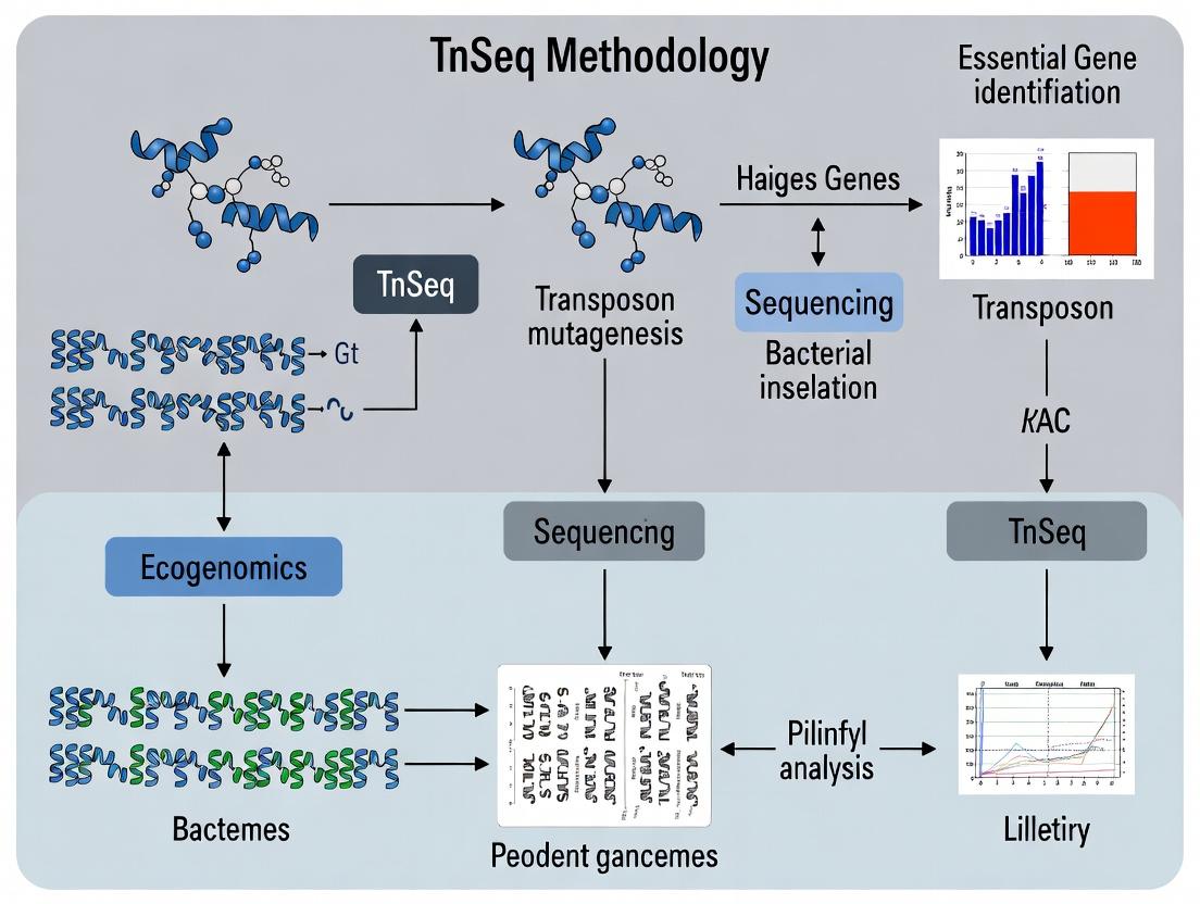
Visualization of TnSeq Workflow and Analysis
Diagram Title: TnSeq Workflow from Library to Data
Diagram Title: Identifying Essential Genes from TnSeq Data
Application Notes
This application note details the experimental and computational framework for testing the core hypothesis in bacterial pathogenesis: that genes essential for in vivo fitness, as identified by TnSeq, are high-value targets for therapeutic intervention. Within a thesis on TnSeq for infection research, this work provides the critical link between genomic-scale disruption libraries and quantitative, host-relevant phenotypic data.
Core Principles and Quantitative Foundations
The central hypothesis posits that a significant fitness defect of a mutant in vivo, relative to its growth in vitro, indicates the gene's specific role in infection. The fitness defect is quantified using the relative fitness metric (w) and the log2 fold-change (LFC) in mutant abundance.
Table 1: Key Quantitative Metrics for Fitness Analysis
| Metric | Formula / Description | Interpretation | Typical Threshold for Essentiality In Vivo |
|---|---|---|---|
| Read Count | Raw sequencing reads mapped to a TA site. | Measures mutant abundance. | N/A |
| Total Reads per Gene | Σ (Reads for all TA sites within a gene). | Represents gene-level abundance. | N/A |
| Fitness (w) | w = ln(Nfinal/Ninitial)mutant / ln(Nfinal/Ninitial)population | Normalized growth rate relative to population. | w < ~0.5 indicates severe defect |
| Log2 Fold Change (LFC) | LFC = log2( (Countoutput + pseudocount) / (Countinput + pseudocount) ) | Change in abundance from input to output pool. | LFC < -2 to -3 suggests essentiality |
| q-value / FDR | Adjusted p-value controlling for false discoveries. | Statistical confidence in hit. | < 0.05 or < 0.01 |
Table 2: Classification of Gene Essentiality from TnSeq Data
| Classification | In Vitro Fitness | In Vivo Fitness | Implication for Infection |
|---|---|---|---|
| Generally Essential | Defective | Defective | Required for basic cellular processes. Poor drug target. |
| Conditionally Essential (for In Vivo) | Normal | Defective | High-Value Target: Specifically required during infection. |
| Non-Essential / Advantageous | Normal | Normal or Increased | Not required; may contribute to virulence regulation. |
| Auxiliary | Slight Defect | Severe Defect | Important in both conditions, but critical under host stress. |
Integrated Experimental-Data Analysis Workflow
Testing the hypothesis requires a closed-loop workflow from library preparation through in vivo challenge and bioinformatic analysis.
Workflow for In Vivo Fitness Analysis
Signaling Pathways ImpactingIn VivoFitness
Conditionally essential genes often cluster in pathways critical for surviving host defenses. Two primary pathways are frequently identified.
Host Stressors and Bacterial Response Pathways
Protocols
Protocol 1: Preparation of High-Complexity Transposon Library forIn VivoPassage
Objective: Generate a saturating Mariner Himar1 transposon mutant library in the target bacterial pathogen (e.g., Salmonella enterica serovar Typhimurium).
Materials: See "Research Reagent Solutions" below. Procedure:
- Electrocompetent Cell Preparation: Grow target strain to mid-log phase (OD600 ~0.5-0.6) in appropriate broth. Wash cells 3x in ice-cold 10% glycerol. Concentrate 100-fold.
- Electroporation: Mix 50 µL competent cells with 100-200 ng of purified Himar1 transposome complex. Electroporate at 1.8 kV, 200 Ω, 25 µF. Immediately add 1 mL SOC, recover at 37°C for 1 hour.
- Library Expansion: Plate recovery culture on selective agar plates (e.g., Kanamycin) at a density of ~50,000 CFU per large (150 mm) plate. Incubate until colonies are distinct.
- Harvesting Input Pool: Scrape all colonies into 10 mL of PBS + 20% glycerol per plate. Pool suspensions, homogenize thoroughly, aliquot, and freeze at -80°C as the Input Pool Master Stock. Determine titer by serial dilution.
- Genomic DNA Extraction: Thaw an aliquot of the input pool. Isolate gDNA from ≥10^10 cells using a phenol-chloroform or commercial kit method optimized for high yield and high molecular weight DNA.
Protocol 2:In VivoSelection in a Murine Model of Systemic Infection
Objective: Subject the mutant library to a selective bottleneck within a live host to deplete mutants with fitness defects.
Materials: 6-8 week old, sex-matched mice (e.g., C57BL/6); library aliquots; appropriate animal biosafety level (ABSL) facilities. Procedure:
- Preparation of Inoculum: Thaw an aliquot of the Input Pool. Grow in 50 mL of selective broth to mid-log phase to ensure all mutants are represented. Wash 2x in PBS. Resuspend to the desired concentration (e.g., 10^8 CFU/mL in PBS).
- Infection: For the In Vivo condition, inject mice intraperitoneally (IP) with 100 µL of inoculum (e.g., 10^7 CFU). For the paired In Vitro control, inoculate 10 mL of broth in a flask with an equal number of bacteria from the same washed inoculum.
- Passage and Recovery: In Vitro: Grow for the same number of generations as expected in vivo (typically ~15-20). In Vivo: At a pre-determined endpoint (e.g., 48 hours), euthanize mice, aseptically remove the target organ (e.g., spleen, liver). Homogenize the organ in PBS.
- Output Pool Harvest: Plate a dilution of the in vitro culture and the organ homogenate onto selective agar plates to obtain ~500,000 colonies per condition. Incubate and harvest all colonies into glycerol stock as in Protocol 1, Step 4. These are the Output Pools.
Protocol 3: TnSeq Library Preparation and Sequencing (TraDIS-based)
Objective: Generate sequencing libraries from the Input and Output Pool gDNA to map transposon insertion sites.
Procedure:
- Fragmentation and Size Selection: Shear 5 µg of gDNA (from Input, In Vitro Output, In Vivo Output) to an average size of 300-500 bp (e.g., using a Covaris sonicator). Size-select fragments >200 bp using SPRI beads.
- End Repair and A-tailing: Perform end-repair and dA-tailing reactions using a standard enzyme mix (e.g., NEBNext Ultra II). Clean up with SPRI beads.
- Adapter Ligation: Ligate double-stranded Y-shaped sequencing adapters containing unique barcode sequences for each pool (Input, Vitro, Vivo). Use a high-efficiency DNA ligase.
- Transposon-Specific PCR: Perform PCR using:
- Forward Primer: Complementary to the Illumina adapter.
- Reverse Primer: Complementary to the end of the Mariner transposon, containing a 6-bp random sequence to mitigate amplification bias.
- Cycle: 12-15 cycles to minimize jackpot effects.
- Sequencing: Pool barcoded libraries. Sequence on an Illumina platform (e.g., NextSeq 2000) using a 75 bp single-end run, with the read starting from the transposon end into the genomic DNA.
Protocol 4: Bioinformatic Analysis for Conditionally Essential Genes
Objective: Process sequencing data to calculate fitness defects and identify genes specifically essential in vivo.
Software: bioinformatics tools like TRANSIT, ESSENTIALS, or a custom pipeline using Bowtie2, DESeq2/edgeR. Procedure:
- Mapping and Counting: Trim reads to remove transposon sequence. Map reads to the reference genome using Bowtie2 (--very-sensitive-local). Count reads mapping to each TA site using a script (e.g.,
count_Tn_reads.py). - Normalization and Filtering: Normalize counts by total library size (e.g., counts per million). Filter out TA sites with <10 reads in the Input pool.
- Fitness Calculation: For each gene, calculate the log2 fold-change (LFC) in abundance from Input to each Output pool using a statistical model (e.g., in TRANSIT: resampling or hidden Markov model). Account for differences in total population expansion.
- Statistical Testing: Compare the in vivo LFC to the in vitro LFC using a condition-aware method (e.g., the interaction term in a generalized linear model). This identifies genes where the fitness defect is significantly greater in vivo than in vitro.
- Hit Calling: Define conditionally essential genes as those with: i) in vitro LFC not significant (q > 0.1), ii) in vivo LFC < -2, iii) interaction term q-value < 0.05. Perform enrichment analysis (GO, KEGG) on the hit list.
The Scientist's Toolkit: Research Reagent Solutions
Table 3: Essential Materials for TnSeq-Based In Vivo Fitness Studies
| Item | Function in Experiment | Example / Specification |
|---|---|---|
| Mariner Himar1 Transposome | Enzyme-DNA complex for random genomic insertion. Provides selective marker (e.g., KanR). | Purified Himar1 transposase pre-complexed with donor DNA. |
| Electrocompetent Cells | High-efficiency bacterial cells for transposon delivery via electroporation. | Prepared in-house from target pathogen strain in 10% glycerol. |
| Selective Growth Media | Maintains selective pressure for transposon-containing mutants during library expansion and passage. | LB Agar + Kanamycin (50 µg/mL). |
| Animal Infection Model | Provides the in vivo selective environment. Must be relevant to human disease. | C57BL/6 mouse model of systemic salmonellosis. |
| High-Yield gDNA Extraction Kit | Isolates pure, high-molecular-weight genomic DNA from complex bacterial pools for sequencing. | Qiagen Genomic-tip 100/G or phenol-chloroform-isoamyl alcohol. |
| Illumina-Compatible Adapters with Barcodes | Allows multiplexing of Input, In Vitro, and In Vivo libraries in a single sequencing run. | IDT for Illumina UD Indexes. |
| Transposon-Specific PCR Primers | Amplifies only fragments containing the transposon-genome junction, enriching the library. | Rev: 5'-[Phos]NNNNNNCTGTCTCTTATACACATCT[Transposon Seq]-3'. |
| Bioinformatics Pipeline | Maps reads, counts insertions, calculates fitness, and performs statistical comparisons. | TRANSIT Software, Bowtie2, R/DESeq2. |
| Next-Generation Sequencer | Generates millions of reads to map insertions at high saturation and depth. | Illumina NextSeq 2000 (P3 flow cell, 100 cycles). |
Application Notes
This document details the application of TnSeq (Transposon Sequencing) for identifying bacterial genes essential for in vivo infection, a critical step in anti-infective drug target discovery. The core methodology leverages the high-efficiency Mariner/Himar1 transposon system to generate saturated mutant libraries. These libraries are then subjected to selection under infection-relevant conditions (e.g., animal models), and the fitness of each mutant is quantified via high-throughput sequencing of transposon junction sites. Essentiality metrics are calculated to statistically distinguish genes required for survival in vivo from dispensable ones.
Table 1: Common Essentiality Metrics in TnSeq Analysis
| Metric | Formula/Description | Interpretation | Typical Threshold (Essential) |
|---|---|---|---|
| Read Count Fold-Change (Log₂FC) | Log₂(Output Counts / Input Counts) | Negative values indicate depletion under selection. | ≤ -2 to -3 |
| Tn-seq Essentiality Index (TEI) | 1 - (Observed Insertions / Possible Insertions) | Ranges from 0 (non-essential) to 1 (essential). | ≥ 0.8 |
| Resampling-based Essentiality (Rbᵉ) | Probability of observed insertion density by chance, assessed via Monte Carlo resampling. | Low p-value indicates significant lack of insertions. | p < 0.05 |
| Transit | Gaussian mixture model to classify genes into essential, non-essential, or growth-defect states. | Provides a probabilistic assignment. | Probability(essential) > 0.9 |
| Hidden Markov Model (HMM) | Models the observed insertion pattern across the genome to call genomic regions of essentiality. | Identifies both whole genes and small essential domains. | State assignment = "Essential" |
Table 2: Typical Mariner/Himar1 TnSeq Library Parameters
| Parameter | Typical Range/Value | Notes |
|---|---|---|
| Average Insertion Density | 1 insertion per 100-500 bp | Aim for near-saturation for robust statistics. |
| Library Complexity | 10⁵ - 10⁶ unique mutants | Ensures coverage of non-essential genome. |
| Himar1 Recognition Site | TA dinucleotide | Target site duplication; occurs ~1/16 bp in AT-rich genomes. |
| Mapping Efficiency | > 80% of reads | Crucial for accurate essentiality calling. |
Detailed Protocols
Protocol 1: Generation of a BarcodedHimar1Transposon Mutant Library
Objective: Create a saturating, uniquely barcoded transposon mutant library in the target bacterial pathogen.
Materials: See "Scientist's Toolkit" below.
Procedure:
- In Vitro Transposition Reaction:
- Assemble a 50 µL reaction containing: 200 ng of target genomic DNA, 100 ng of pKMW3 or similar Himar1 transposon donor plasmid, 1x reaction buffer, and 1 µL of purified Himar1 C9 transposase.
- Incubate at 30°C for 4 hours, then heat-inactivate at 75°C for 10 min.
Transformation and Pooling:
- Electroporate the entire in vitro transposition mix into electrocompetent cells of your target bacterium.
- Immediately add 1 mL of recovery broth, incubate with shaking for 2-3 hours.
- Plate transformations onto selective agar plates (e.g., containing kanamycin) at a density to yield ~200-300 colonies per plate.
- Scrape all colonies from plates into a single suspension using 1x PBS + 20% glycerol. This is your Master Library.
Library Amplification and DNA Preparation:
- Dilute the Master Library and grow to mid-exponential phase in selective liquid medium to maintain all mutants.
- Extract genomic DNA from a 50 mL culture using a bacterial genomic DNA isolation kit. This DNA serves as the Input Pool for sequencing and selection experiments.
Protocol 2:In VivoSelection and Sequencing Library Preparation
Objective: Subject the mutant library to an animal model of infection and prepare sequencing libraries to quantify mutant fitness.
Procedure:
- Infection and Harvest:
- Infect cohorts of animals (e.g., mice) with ~10⁷ CFU of the mutant library from the Master Library via the relevant route (IV, IP, intranasal).
- After a defined period (e.g., 48-72 hours), euthanize animals and harvest the target organ(s).
- Homogenize organs, plate serial dilutions to determine total bacterial burden, and resuspend the remainder for genomic DNA extraction. This represents the Output Pool.
Tn Junction Amplification (PCR1 - Add Adapters):
- Set up 100 µL PCR reactions on Input and Output gDNA (100 ng each) using a biotinylated primer specific to the transposon end and a primer targeting a MmeI site adapter.
- Cycle: 95°C 3 min; [95°C 30s, 60°C 30s, 72°C 1 min] x 25 cycles; 72°C 5 min.
MmeI Digestion and Purification:
- Bind PCR products to streptavidin magnetic beads. Wash.
- On-bead, digest with MmeI (cuts 20/18 bp downstream of its recognition site) to release a 38-40 bp fragment containing the transposon-genome junction and the inline barcode.
- Elute the digested fragments.
Library Completion (PCR2 - Add Sequencing Handles):
- Perform a second PCR to add Illumina flow cell adapters and sample-specific indices using the eluted MmeI fragments as template.
- Purify the final library using double-sided size selection beads (e.g., SPRIselect).
Sequencing:
- Quantify libraries by qPCR. Sequence on an Illumina MiSeq or HiSeq platform using a 50-75 bp single-read run to capture the junction fragment.
Protocol 3: Bioinformatic Analysis and Essentiality Calling
Objective: Process sequencing data to calculate essentiality metrics for every gene.
Procedure:
- Demultiplexing and Preprocessing:
- Use
tn-seqpipelines (FASTX-Toolkit,Cutadapt) to demultiplex by sample index and trim transposon/primer sequences.
- Use
Mapping and Counting:
- Map reads to the reference genome using
Bowtie2orBWA, allowing no mismatches in the genomic portion. - Count the number of unique insertions and total reads per TA site in each condition using custom scripts (e.g.,
Tnpipeline).
- Map reads to the reference genome using
Essentiality Calculation:
- Normalize read counts by total reads per sample (e.g., counts per million).
- For each gene, calculate the log₂ fold-change (Output/Input) of insertion density or read count.
- Run the
TRANSITsoftware (or equivalent) using the resampling or HMM method to assign statistical significance (p-values) and essentiality calls. - Classify genes: Essential (significantly depleted), Growth-Defect (partially depleted), Non-essential (unchanged), or Advantageous (enriched).
Diagrams
TnSeq Workflow for Infection Studies
Himar1 Transposon Structure & Integration
The Scientist's Toolkit: Key Research Reagent Solutions
| Item | Function/Description | Example/Supplier |
|---|---|---|
| pKMW3 or pSAM_Bc Plasmid | Donor vector containing a Himar1 transposon with a selectable marker, MmeI site, and a barcode region for downstream sequencing. | Addgene #TODO; Lab-constructed. |
| Himar1 C9 Purified Transposase | Engineered hyperactive mutant of the Mariner transposase that excises and integrates the transposon in vitro at TA sites. | Purified in-house from E. coli expression; commercial enzyme suppliers. |
| MmeI Restriction Endonuclease | Type IIS enzyme that cuts 20/18 bp away from its recognition site, used to generate uniform fragments for sequencing library prep. | New England Biolabs (NEB). |
| Streptavidin Magnetic Beads | Used to capture biotinylated PCR products during the library prep protocol, enabling clean on-bead enzymatic steps. | Dynabeads (Thermo Fisher), Sera-Mag beads. |
| Phusion High-Fidelity DNA Polymerase | Used for high-fidelity amplification of transposon-genome junctions during library construction to minimize PCR errors. | Thermo Fisher, NEB. |
| Next-Generation Sequencer | Platform for high-throughput sequencing of the barcoded insertion libraries. | Illumina MiSeq/NextSeq (short-read). |
| TRANSIT Software | A standard open-source software package for the statistical analysis of TnSeq data, including resampling and HMM methods. | Available at sourceforge.net/projects/transit-tnseq/. |
| SPRIselect Beads | Paramagnetic beads for precise size selection and purification of DNA fragments during NGS library preparation. | Beckman Coulter. |
Application Notes: Evolution of Essential Gene Mapping in Infection Biology
The systematic identification of bacterial genes essential for survival and growth during infection has been a cornerstone of pathogenesis research and antibacterial drug target discovery. The field has evolved from low-throughput, in vivo-centric methods to genome-saturating, quantitative approaches.
Signature-Tagged Mutagenesis (STM) was a pioneering in vivo technique developed in the 1990s. It enabled the parallel screening of pools of uniquely "tagged" mutants in an animal model of infection. Mutants absent from output pools were deemed attenuated. While revolutionary, STM had limitations: it was semi-quantitative, low-resolution, and labor-intensive.
The transition to TnSeq (Transposon Sequencing) represented a paradigm shift, coupling high-density transposon mutagenesis with next-generation sequencing. This allowed for the quantitative assessment of the fitness contribution of nearly every non-essential gene in the genome under a given condition in vitro or in vivo.
Modern High-Resolution TnSeq leverages improved transposon designs (e.g., Himar1 mariner), optimized library construction protocols, sophisticated bioinformatics pipelines (e.g., TRANSIT, Bio-Tradis), and the application of conditionally essential gene analysis in complex host environments. The integration of INSeq (Insertion Sequencing) and TraDIS (Transposon Directed Insertion-site Sequencing) methodologies has standardized the field. Current applications extend to genetic interaction mapping (TnSeq of double mutants), resistance gene discovery, and profiling gene essentiality across hundreds of in vitro conditions.
Table 1: Quantitative Comparison of STM vs. Modern TnSeq
| Feature | Signature-Tagged Mutagenesis (STM) | Modern High-Resolution TnSeq |
|---|---|---|
| Throughput | ~96 mutants/pool | >100,000 mutants/library |
| Quantitation | Semi-quantitative (present/absent) | Highly quantitative (read counts per insertion) |
| Resolution | Gene-level (if insertion mapped) | Near single-nucleotide (insertion site) |
| Key Metric | Attenuation | Fitness Index/Essentiality q-value |
| Primary Screen | In vivo infection model | In vitro and/or In vivo |
| Data Output | List of attenuated mutants | Genome-wide fitness landscape |
Detailed Protocols
Protocol 2.1: Construction of a High-Complexity Mariner Transposon Library
Objective: Generate a saturated mutant library for Staphylococcus aureus with >100,000 unique insertions. Materials: See "Scientist's Toolkit" below. Procedure:
- Electrocompetent Cell Preparation: Grow S. aureus RN4220 to mid-log phase (OD600 ~0.5). Wash cells 3x in ice-cold 0.5M sucrose.
- Electroporation: Mix 50 µL cells with 100-500 ng of purified pMarA* or similar mariner transposon plasmid. Electroporate at 2.5 kV, 100 Ω, 25 µF.
- Recovery & Selection: Recover cells in 1 mL SOC + 0.5M sucrose for 1.5h at 37°C. Plate entire recovery on 150mm agar plates containing chloramphenicol (10 µg/mL). Incubate 48h at 37°C.
- Library Harvesting: Scrape all colonies into 10 mL of PBS + 20% glycerol. Mix thoroughly, aliquot, and store at -80°C. Determine library titer (CFU/mL).
- Complexity Validation: Isolate genomic DNA from a pool of ~200,000 CFU. Perform sequencing library prep using a MmeI-based protocol (see below). Sequence to a depth of ~50-100 reads per expected insertion. Analyze with TRANSIT software to confirm uniform genome coverage.
Protocol 2.2:In VivoTnSeq Screen in a Murine Infection Model
Objective: Identify conditionally essential genes required for S. aureus systemic infection. Procedure:
- Input Pool Preparation: Thaw library aliquot and grow in 50 mL TSB + Cm to mid-log phase. Wash 2x in PBS. Resuspend to ~10^9 CFU/mL.
- Animal Infection: Infect 6-8 week old BALB/c mice (n=5) intravenously with 100 µL of cell suspension (~10^8 CFU). Maintain control in vitro culture in parallel.
- Output Pool Recovery: At 48h post-infection, euthanize mice. Harvest spleens and livers, homogenize, and plate homogenate serial dilutions on selective agar to recover bacterial cells. Incubate 24h.
- Genomic DNA Extraction: Pool all colonies from each mouse organ and the in vitro control. Extract gDNA using a bacterial genomic DNA kit.
- Sequencing Library Prep (MmeI Method): a. Fragment gDNA by sonication to ~500 bp. b. End-repair, A-tail, and ligate to a double-stranded adapter. c. Digest with MmeI (cuts 20 bp downstream of the transposon end). d. Purify the ~120 bp fragment containing the transposon junction. e. Amplify with primers adding Illumina indices. Size-select and purify the final library.
- Sequencing & Analysis: Sequence on Illumina MiSeq (2x150bp). Map reads to the reference genome. Calculate normalized read counts per TA site for input ( in vitro ) and output ( in vivo ) pools. Analyze using the TRANSIT resampling or HMM method to identify genes with statistically significant fitness defects in vivo.
Diagrams
Title: Evolution from STM to Modern TnSeq
Title: Standard TnSeq Experimental Workflow
The Scientist's Toolkit
Table 2: Essential Research Reagents & Materials for TnSeq
| Item | Function/Description | Example/Note |
|---|---|---|
| Mariner Transposon Plasmid | Delivery vector containing the Himar1 transposase and a selective marker flanked by inverted repeats. | pMarA*, pKMS1; provides chloramphenicol or kanamycin resistance. |
| Electrocompetent Cells | Genetically tractable strain for library construction, often lacking restriction systems. | S. aureus RN4220, E. coli BW29427. |
| Selection Antibiotics | To select for transposon insertion and maintain library diversity. | Chloramphenicol (10 µg/mL), Kanamycin (50 µg/mL). |
| MmeI Type IIS Restriction Enzyme | Cuts at a fixed distance from its recognition site, enabling precise junction fragment capture. | Critical for efficient sequencing library prep. |
| Illumina-Compatible Adapters & Primers | For amplifying transposon-genome junctions for sequencing. | Must include indices for multiplexing. |
| Genomic DNA Extraction Kit | For high-yield, pure gDNA from bacterial pools. | Qiagen DNeasy, Promega Wizard. |
| Bioinformatics Software | For mapping reads, counting insertions, and calculating essentiality. | TRANSIT, Bio-Tradis, ESSENTIALS. |
| Animal Model | For in vivo essentiality screens. | Typically murine (e.g., BALB/c for systemic infection). |
Application Notes
This Application Note details the use of Transposon Sequencing (TnSeq) to identify conditionally essential bacterial genes required for host colonization and survival. The methodology is contextualized within a broader thesis on functional genomics for infection research, aiming to pinpoint novel, host-specific drug targets.
Core Principle: TnSeq combines high-density transposon mutagenesis with next-generation sequencing to quantitatively assess the contribution of each gene to fitness under a specific condition (e.g., in vivo infection) compared to a reference condition (e.g., in vitro growth).
Key Quantitative Outcomes: Recent studies (2023-2024) consistently demonstrate that 10-25% of a bacterial genome comprises conditionally essential genes during infection. The following table summarizes data from representative pathogens:
Table 1: Quantitative Output of TnSeq in Infection Models (Recent Data)
| Pathogen | Infection Model | Total Genes Screened | Conditionally Essential Genes (In Vivo) | % of Genome | Primary Functional Categories Enriched | Reference (Type) |
|---|---|---|---|---|---|---|
| Salmonella Typhimurium | Murine colitis model | 4,489 | ~550 | 12.3% | Nutrient acquisition (C, N, Mg), anaerobic metabolism, host defense evasion | PMID: 38113047 |
| Klebsiella pneumoniae | Murine pneumonia model | 5,432 | ~1,210 | 22.3% | Capsule biosynthesis, purine/pyrimidine synthesis, cell envelope integrity | PMID: 38262935 |
| Acinetobacter baumannii | Murine septicemia model | 3,950 | ~400 | 10.1% | Iron acquisition, lipid metabolism, stress response regulators | PMID: 38055214 |
| Pseudomonas aeruginosa | Ex vivo human sputum | 5,570 | ~680 | 12.2% | Biofilm formation, quorum sensing, proteolytic enzyme secretion | PMID: 38345622 |
Data Interpretation: Genes are classified using a statistical framework (often a negative binomial model) to calculate a Fitness Defect (FD) score. A gene with an FD ≤ -2.0 (log² scale) and a false-discovery rate (FDR) < 5% is typically deemed conditionally essential. The resulting gene set reveals metabolic pathways and virulence factors uniquely required within the host niche.
Experimental Protocols
Protocol 1: High-Complexity Transposon Library Construction & Preparation
Objective: Create a saturated mariner-based Himar1 transposon mutant library in the target bacterial strain.
Materials:
- Target bacterial strain (e.g., K. pneumoniae ATCC 43816).
- Himar1 transposase expression plasmid (e.g., pSC189, temperature-sensitive origin).
- Transposon donor plasmid containing Himar1 inverted repeats flanking a selectable marker (e.g., kanamycin resistance) and a unique molecular barcode (UMB) for each insertion.
- Mueller-Hinton Broth (MHB) and agar plates with appropriate antibiotics.
Procedure:
- Electroporation: Introduce the transposase plasmid into the target strain via electroporation. Recover at permissive temperature (30°C).
- Transposition: Transform the transposon donor plasmid into the strain carrying the transposase plasmid. Plate on selective agar at the restrictive temperature (37°C) to select for transposon integration and loss of the transposase plasmid.
- Library Expansion: Pool all colonies (≥ 200,000 CFU) and grow in liquid culture under selection. Isolate genomic DNA using a kit optimized for high-molecular-weight DNA (e.g., Qiagen Genomic-tip).
- Complexity Verification: Perform Illumina sequencing on a barcoded fragment of the library to confirm insertion density. Aim for a library where > 90% of non-essential genes have at least 15-20 insertions.
Protocol 2:In VivoSelection and Sample Processing for TnSeq
Objective: Subject the mutant library to selective pressure in an infection model and recover bacterial genomes for sequencing.
Materials:
- Prepared transposon library.
- Animal infection model (e.g., 8-week-old C57BL/6 mice, n=5 per group).
- Homogenizer (e.g., GentleMACS).
- Lysis buffer (20 mg/mL lysozyme, 1% SDS).
- Phenol:chloroform:isoamyl alcohol (25:24:1).
- Magnetic beads for DNA clean-up (e.g., SPRIselect beads).
Procedure:
- Input (T0) Sample: Harvest 10⁹ CFU from the in vitro library pre-inoculation. Pellet cells and freeze at -80°C.
- In Vivo Passage: Infect mice via the relevant route (e.g., intranasal for pneumonia) with ~10⁷ CFU of the library. After 48-72 hours, euthanize and harvest the target organ (e.g., lungs, liver).
- Output (T1) Sample: Homogenize the organ. Plate homogenate dilutions to determine bacterial burden. Resuspend the remaining homogenate in lysis buffer and incubate at 37°C for 1 hour.
- gDNA Isolation: Perform phenol-chloroform extraction on homogenized tissue lysate. Precipitate gDNA with isopropanol. Treat with RNase A. Purify using magnetic beads. Quantify by Qubit.
Protocol 3: TnSeq Library Preparation & Sequencing
Objective: Amplify and barcode transposon-genome junctions for multiplexed Illumina sequencing.
Materials:
- Fragmentation enzyme (e.g., Covaris shearing or enzymatic fragmentase).
- End-repair, A-tailing, and ligation module (e.g., NEBNext Ultra II).
- Custom Y-adapter containing Illumina sequencing primer sites and sample index.
- Primers specific to the transposon ends.
- PCR purification kit and size-selection beads.
Procedure:
- Fragmentation & Adapter Ligation: Shear 1 µg gDNA to ~300 bp. Perform end-repair, A-tailing, and ligation of the Y-adapter.
- Transposon-Specific PCR: Perform a primary PCR (12-15 cycles) using a primer complementary to the transposon end and a primer complementary to the adapter. This enriches for fragments containing the junction.
- Indexing PCR: Perform a secondary PCR (8-10 cycles) using Illumina index primers to add unique dual indices for each sample (T0 and T1).
- Purification & QC: Clean PCR products with size-selection beads (0.7x ratio) to remove primer dimers. Validate library size (~400 bp) on a Bioanalyzer. Pool libraries and sequence on an Illumina MiSeq or HiSeq (2x150 bp), targeting 20-50 million reads per sample.
Visualizations
Title: TnSeq Workflow for Conditional Essentiality
Title: Host Signal Activates Essential Genes
The Scientist's Toolkit
Table 2: Essential Research Reagents and Solutions
| Item | Function in TnSeq Experiment | Example/Supplier |
|---|---|---|
| Himar1 Transposon System | Source of mariner transposase and engineered transposon for random, stable insertion mutagenesis. | pSC189/pSAM_Bt plasmids; Dharmacon. |
| Magnetic Size Selection Beads | Critical for clean PCR product purification and accurate size selection post-library amplification. | SPRIselect (Beckman Coulter), AMPure XP. |
| High-Fidelity PCR Master Mix | Amplifies transposon junctions with minimal bias and error for accurate insertion counting. | NEBNext Q5, KAPA HiFi. |
| Dual-Indexed Illumina Adapters | Enables multiplexing of multiple T0 and T1 samples in a single sequencing run. | IDT for Illumina UD Indexes. |
| Tissue Homogenization Kit | Efficiently lyses host tissue to recover bacterial cells for downstream gDNA isolation. | GentleMACS (Miltenyi), Precellys tubes. |
| gDNA Clean-Up Kit | Removes host DNA contamination and PCR inhibitors from in vivo samples. | QIAamp DNA Microbiome Kit (Qiagen). |
| Bioanalyzer/Pico Chip | Provides precise quality control of final TnSeq library fragment size distribution. | Agilent 2100 Bioanalyzer. |
| TnSeq Analysis Pipeline | Software for mapping reads, counting insertions, and calculating fitness defects. | TRANSIT, ARTIST, Bio-Tradis. |
TnSeq Workflow: Step-by-Step Protocol from Library Construction to In Vivo Analysis
Application Notes
In the broader context of a thesis on TnSeq for mapping bacterial genes essential for host infection, the construction of a high-quality, saturated mutant library is the foundational step. This library enables genome-wide, quantitative assessment of gene fitness under selective conditions, such as during in vitro or in vivo infection models. A saturated library, where transposon insertions are distributed across all non-essential genomic regions, allows for the statistical identification of genes essential for growth in vitro and those conditionally essential for infection. This approach directly informs drug discovery by pinpointing vulnerable, pathogen-specific pathways.
Key Quantitative Considerations for Library Design
The following table summarizes critical parameters for achieving library saturation and the associated statistical confidence.
Table 1: Key Parameters for Saturated Library Construction and Analysis
| Parameter | Typical Target Value | Rationale & Calculation |
|---|---|---|
| Insertion Density | 1 insertion every 20-50 bp (on average) | Ensures multiple insertions per gene for robust statistical analysis. |
| Library Size (Mutant Count) | 100,000 - 500,000 unique mutants | For a 5 Mb genome with 50% essential genes, ~150,000 unique insertions provide ~95% probability of hitting a given 300 bp non-essential region. |
| Saturation Threshold | >99% of TA sites (or other insertion motif) occupied | Assessed by sequencing a naive library; high saturation reduces "jackpot" effects and sampling noise. |
| Read Depth per Condition | >200-500 reads per insertion site | Provides statistical power to detect significant fitness defects (e.g., using a negative binomial model). |
| Essential Gene Cutoff (for in vitro growth) | Fitness defect ≤ -2 to -3 (log2 fold-change) & q-value < 0.05 | Identifies genes where insertions are severely depleted in the output pool compared to the input library. |
Experimental Protocols
Protocol 1:In VitroTransposon Delivery and Mutant Library Construction
Objective: To generate a complex, random transposon insertion library in the target bacterial pathogen. Materials: See "Research Reagent Solutions" below. Method:
- Transposome Complex Assembly: Combine purified hyperactive Himar1 C9 transposase with a custom-designed mariner-based transposon DNA fragment (containing a selectable marker, e.g., kanamycin resistance, and outward-facing primers for sequencing) at a molar ratio of 1:1 in a buffer containing 25% (v/v) glycerol. Incubate at 30°C for 1 hour.
- Electroporation: Thaw electrocompetent cells of the target bacterial strain (e.g., Pseudomonas aeruginosa PAO1) on ice. Mix 50 µL of cells with 1-2 µL of transposome complex. Electroporate using standard parameters for the organism (e.g., 2.5 kV, 200 Ω, 25 µF for E. coli; optimize for others). Immediately add 1 mL of rich recovery medium (e.g., SOC).
- Outgrowth and Selection: Recover cells with shaking at 37°C for 1-3 hours to allow expression of the antibiotic resistance marker. Plate the entire culture volume across 10-20 large (150 mm) agar plates containing the appropriate selective antibiotic. Incubate until colonies are visible (typically 12-48 hours).
- Library Harvesting: Scrape all colonies from plates using 2-3 mL of liquid medium + 20% glycerol per plate. Pool into a single sterile tube. Homogenize thoroughly by vortexing and/or pipetting. Measure the OD600 and aliquot into cryovials. Flash-freeze in a dry-ice/ethanol bath and store at -80°C. This is the Master Library Stock.
- Titration: Serially dilute the recovery culture from Step 3 and plate on selective agar to determine the total library diversity (CFU/mL x total recovery volume).
Protocol 2: Library Quality Control and Genomic DNA Preparation for TnSeq
Objective: To extract high-quality, pooled genomic DNA (gDNA) from the mutant library for sequencing library preparation. Method:
- Library Expansion: Thaw a Master Library Stock aliquot and dilute into fresh, selective medium at a low OD600 (~0.005) to maintain all mutants. Grow to mid-exponential phase (OD600 ~0.5-0.8). This culture serves as the "input pool" for subsequent experiments.
- Genomic DNA Extraction: Harvest cells from 5-10 mL of culture (≥ 5 x 10^9 cells) by centrifugation. Use a magnetic bead-based gDNA extraction kit (e.g., NucleoBond HPT) designed for high molecular weight DNA. Follow manufacturer's protocol, including RNase A treatment. Elute DNA in 10 mM Tris-HCl, pH 8.5.
- DNA Quantification and Quality Assessment: Measure DNA concentration using a fluorometric assay (e.g., Qubit dsDNA BR Assay). Assess integrity by pulsed-field or standard agarose gel electrophoresis. The DNA should appear as a high molecular weight smear > 20 kb. Store at -20°C.
- Fragmentation and Size Selection (Alternative Shear-by-Sequencing): For protocols requiring fragmentation, shear 1-2 µg of gDNA to an average size of 300-500 bp using a focused-ultrasonicator (e.g., Covaris). Size-select using solid-phase reversible immobilization (SPRI) beads.
Diagrams
Title: Transposon Mutant Library Construction Workflow
Title: TnSeq Analysis for Essential Gene Discovery
The Scientist's Toolkit
Table 2: Key Research Reagent Solutions for Transposon Library Construction
| Item | Function in Protocol | Example & Notes |
|---|---|---|
| Hyperactive Transposase | Catalyzes random genomic integration of the transposon. | Himar1 C9 mutant: High efficiency for broad GC-content range in bacteria. |
| Synthetic Transposon Donor DNA | Provides transposon ends for transposase binding and contains selectable marker/sequencing adapters. | pKMW3-derived fragment: Contains kanR, MmeI site for sequencing, outward primers. |
| Electrocompetent Cells | High-efficiency bacterial cells for DNA uptake via electroporation. | Prepared in-house for target strain; critical for achieving high diversity. |
| Selection Antibiotic | Selects for mutants with successful chromosomal transposon integration. | Kanamycin (50-100 µg/mL) or other strain-appropriate antibiotic. |
| Magnetic Bead gDNA Kit | High-yield, high-purity genomic DNA extraction from pooled bacterial cells. | NucleoBond HPT Kit (Macherey-Nagel) or MagAttract HMW DNA Kit (Qiagen). |
| TnSeq Sequencing Primers | Amplify transposon-genome junctions for Illumina sequencing. | Custom primers containing Illumina adapters, indices, and transposon-specific sequence. |
| Analysis Software | Map sequencing reads, count insertions, and calculate fitness statistics. | TRANSIT, ESSENTIALS, or ARTIST pipelines. |
Following TnSeq-based identification of putative essential bacterial genes for in vitro growth, Stage 2 validates their role in the infection context. This stage employs three complementary biological models to map host-pathogen interactions: animal models (gold standard for systemic physiology), organoids (3D human-relevant tissue), and cell-based assays (high-throughput screening). The selection dictates the mechanistic depth and translational relevance of findings for therapeutic development.
Comparative Model Analysis
The choice of model balances physiological relevance, throughput, cost, and ethical considerations.
Table 1: Quantitative Comparison of Infection Models
| Parameter | Murine (Animal) Models | Human Organoids | Immortalized Cell Lines (2D) |
|---|---|---|---|
| Physiological Relevance | High (whole organism, immune system) | High (human, 3D tissue structure) | Low (monolayer, often cancerous origin) |
| Throughput | Low (weeks/months, n<50 typical) | Medium (weeks, n=10-100) | High (days, n>1000) |
| Cost per Experiment | High ($500-$5000+) | Medium ($200-$2000) | Low ($10-$500) |
| Genetic Manipulability | Medium (host transgenic models) | High (CRISPR on host cells) | Very High (easy transfection/knockdown) |
| Key Readouts | Survival, bacterial burden (CFU/organ), histopathology | Bacterial invasion, host cell damage, cytokine secretion | Adhesion, invasion, intracellular survival, cytotoxicity |
| Primary Application | Validation of virulence in vivo, pharmacokinetics/pharmacodynamics | Human-specific pathogenesis mechanisms | High-throughput mutant screening, initial mechanism |
Detailed Protocols
Protocol 3.1: Murine Acute Pneumonia Model forPseudomonas aeruginosa
Application: Validating genes essential for lung infection identified by TnSeq. Objective: To compare bacterial burden and host survival between wild-type and TnSeq-identified mutant strains.
Materials:
- 6-8 week old, sex-matched C57BL/6 mice.
- P. aeruginosa wild-type and mutant strains (grown to mid-log phase in LB).
- PBS for washing.
- Isoflurane anesthesia system.
- Intranasal instillation setup.
- Sterile surgical tools for organ harvest.
- Homogenizer.
- LB agar plates for colony-forming unit (CFU) enumeration.
Procedure:
- Bacterial Preparation: Grow strains to OD600 = 0.8. Wash twice in PBS, resuspend to 2 x 10^8 CFU/mL (confirmed by plating serial dilutions).
- Animal Infection: Anesthetize mouse with isoflurane. Instill 50 µL of bacterial suspension (10^7 CFU) slowly into the nostrils. Hold mouse upright for 30 seconds post-instillation.
- Monitoring: Monitor mice at least twice daily for signs of morbidity (weight loss >20%, lethargy, ruffled fur). Euthanize moribund mice for survival curve analysis.
- Bacterial Burden (24h): At 24h post-infection, euthanize cohort (n=5/group). Aseptically harvest lungs and spleen. Homogenize organs in 1 mL PBS.
- CFU Enumeration: Perform 10-fold serial dilutions of homogenates in PBS. Plate 100 µL of each dilution on LB agar. Incubate plates at 37°C overnight. Count colonies and calculate CFU per organ.
- Statistical Analysis: Compare mutant vs. wild-type CFU using Mann-Whitney U test. Survival analysis via Log-rank test.
Protocol 3.2: Human Intestinal Organoid Infection withSalmonella enterica
Application: Assessing human epithelial-specific invasion and damage by bacterial mutants. Objective: To quantify invasion efficiency and epithelial integrity disruption of TnSeq-derived mutants.
Materials:
- Established human intestinal organoids (derived from stem cells).
- Matrigel.
- Intestinal organoid growth medium (with growth factors).
- Organoid dissociation reagent (e.g., TrypLE).
- 24-well tissue culture plate.
- S. enterica strains (wild-type and mutant, grown to late-log phase).
- Gentamicin (100 mg/mL stock).
- CellTiter-Glo 3D for viability assay.
- Lysis buffer (1% Triton X-100) for bacterial recovery.
Procedure:
- Organoid Preparation: Dissociate mature organoids into single cells/small clusters using TrypLE. Seed 10,000 cells in 20 µL Matrigel droplets per well of a 24-well plate. Overlay with growth medium. Culture for 3-4 days to form mature, lumen-containing organoids.
- Bacterial Infection: Grow bacteria to OD600 = 1.0, wash, and resuspend in antibiotic-free organoid medium. Remove growth medium from organoids and add 500 µL of bacterial suspension (MOI ~10:1). Centrifuge plate at 300 x g for 5 min to facilitate contact.
- Invasion Assay (2h): Infect for 2h at 37°C. Wash organoids 3x with PBS. Add medium containing 100 µg/mL gentamicin to kill extracellular bacteria. Incubate for 1h.
- Intracellular Bacterial Recovery: Wash organoids 3x with PBS. Lyse organoids with 500 µL of 1% Triton X-100 for 10 min. Vortex vigorously. Serially dilute lysate in PBS and plate on LB agar to enumerate intracellular CFU.
- Epithelial Damage Assay (24h): For a separate set of infected organoids, after 24h, aspirate medium. Add 200 µL CellTiter-Glo 3D reagent. Lyse organoids by shaking for 5 min. Measure luminescence (relative to uninfected controls) as a proxy for viability/tissue damage.
- Analysis: Normalize mutant intracellular CFU and luminescence to wild-type values (set at 100%). Compare using Student's t-test.
Protocol 3.3: High-Throughput Intracellular Survival Assay in Macrophages
Application: Rapid screening of TnSeq hits for defects in immune evasion. Objective: To measure survival of bacterial mutants within immortalized macrophages over 24 hours.
Materials:
- RAW 264.7 murine macrophages.
- 96-well tissue culture-treated plates.
- Cell culture medium (DMEM + 10% FBS).
- Bacterial strains.
- Gentamicin.
- PBS.
- Lysis buffer (0.1% Deoxycholate in PBS).
- LB agar plates or an automated colony counter.
Procedure:
- Cell Seeding: Seed RAW 264.7 cells at 5 x 10^4 cells/well in 100 µL medium. Incubate overnight at 37°C, 5% CO2.
- Infection: Grow bacteria to mid-log phase. Wash and resuspend in DMEM. Add 10 µL of bacterial suspension (MOI ~5:1) to each well (total 110 µL). Centrifuge plate at 500 x g for 5 min. Incubate for 30 min at 37°C.
- Gentamicin Protection: Wash wells 2x with PBS. Add 200 µL of medium containing 50 µg/mL gentamicin. Incubate for 1h to kill extracellular bacteria.
- Timepoint Harvest:
- T = 2h: Wash 3x with PBS. Lyse cells with 100 µL of 0.1% deoxycholate for 10 min. Vortex. Perform serial dilutions and plate for CFU (initial internalized count).
- T = 24h: After the 1h gentamicin treatment, replace medium with fresh medium containing 10 µg/mL gentamicin (maintenance dose). At 24h post-infection, lyse cells and plate as above.
- Calculation: Calculate intracellular survival ratio as (CFU at 24h / CFU at 2h) * 100%. A mutant with a survival ratio significantly lower than wild-type indicates a defect in intracellular persistence.
Visualizations
Title: Infection Model Selection and Output Workflow
Title: Organoid Infection and Assay Protocol Flow
The Scientist's Toolkit
Table 2: Key Research Reagent Solutions for Infection Models
| Reagent/Material | Primary Function | Example in Protocol |
|---|---|---|
| Matrigel (or equivalent ECM) | Provides a 3D extracellular matrix scaffold to support organoid growth and polarization. | Protocol 3.2: Base for intestinal organoid culture. |
| Gentamicin (or other non-cell-penetrant antibiotic) | Selective killing of extracellular bacteria while sparing intracellular populations for invasion/survival assays. | Protocols 3.2 & 3.3: "Gentamicin protection" assay. |
| CellTiter-Glo 3D / 2D | Luminescent assay quantifying ATP, proportional to metabolically active cells; used for viability/cytotoxicity. | Protocol 3.2: Measuring organoid epithelial damage. |
| Triton X-100 / Deoxycholate | Mild detergents used to lyse eukaryotic host cells without completely inactivating recovered bacteria for plating. | Protocols 3.2 & 3.3: Lysing organoids/macrophages. |
| Isoflurane System | Volatile inhalant anesthetic for safe and reversible sedation of rodents during infection procedures. | Protocol 3.1: Mouse anesthesia for intranasal infection. |
| Defined Organoid Growth Medium | Contains essential growth factors (Wnt, R-spondin, Noggin) to maintain stemness and drive intestinal crypt differentiation. | Protocol 3.2: Culturing human intestinal organoids. |
Application Notes
Within the broader context of a TnSeq-based thesis for mapping bacterial genes essential for infection, this stage is the critical translational link between in vivo infection models and high-throughput sequencing. Successful execution ensures that the relative abundance of each bacterial transposon mutant, as established within the complex environment of host tissues, is accurately preserved and converted into a sequencing-ready library. The primary challenge lies in maximizing bacterial DNA yield and purity while minimizing contamination from host genomic DNA, which can severely impact library complexity and sequencing depth. Recent methodologies emphasize the use of differential lysis and enzymatic digestion steps to selectively degrade mammalian cells and DNA, coupled with optimized bacterial DNA extraction protocols designed for low-biomass samples. The quality and quantity of DNA output at this stage directly determine the sensitivity and statistical power of subsequent essential gene identification.
Detailed Protocol: Harvesting & Differential Lysis
Objective: To recover bacteria from infected host tissue, lyse host cells, and digest host genomic DNA with minimal impact on bacterial integrity.
Materials:
- Infected tissue samples (e.g., spleen, liver, lung) harvested at defined time points post-infection.
- Sterile 1X Phosphate-Buffered Saline (PBS), ice-cold.
- Homogenizer (e.g., gentleMACS Dissociator or manual Dounce homogenizer).
- Proteinase K (20 mg/mL).
- DNase I (RNase-free).
- Lysozyme solution (10-50 mg/mL in TE buffer).
- Nuclease-free water.
- Centrifuges (refrigerated microcentrifuge and low-speed centrifuge).
Procedure:
- Tissue Homogenization: Place harvested tissue (e.g., ~100 mg) in a tube containing 1 mL of ice-cold PBS. Homogenize thoroughly using a mechanical homogenizer on a pre-cooled setting until no visible tissue fragments remain.
- Differential Centrifugation: Centrifuge the homogenate at 500 x g for 10 minutes at 4°C to pellet host cell debris and nuclei. Carefully transfer the supernatant, containing bacteria and some host components, to a new microcentrifuge tube.
- Host Cell Lysis: Add Proteinase K to the supernatant to a final concentration of 0.5 mg/mL. Incubate at 56°C for 30 minutes to degrade host proteins.
- Host DNA Digestion: Add 10-20 units of DNase I directly to the lysate. Incubate at 37°C for 30 minutes to degrade accessible host genomic DNA. This step is crucial for reducing host DNA contamination.
- Bacterial Pellet Recovery: Centrifuge the treated supernatant at 16,000 x g for 5 minutes at 4°C to pellet the bacterial cells. Discard the supernatant.
- Bacterial Cell Wall Weakening: Resuspend the bacterial pellet in 200 µL of TE buffer containing Lysozyme (1 mg/mL final concentration). Incubate at 37°C for 30 minutes.
Detailed Protocol: Bacterial Genomic DNA Extraction & Shearing
Objective: To isolate high-purity, high-molecular-weight bacterial gDNA and fragment it to an appropriate size for NGS library construction.
Materials:
- Commercial bacterial DNA extraction kit (e.g., DNeasy Blood & Tissue Kit, QIAGEN).
- RNase A.
- Absolute ethanol (96-100%).
- Magnetic bead-based DNA clean-up system (e.g., AMPure XP beads).
- Covaris ultrasonicator or focused-ultrasonicator (e.g., M220 Focused-ultrasonicator, Covaris).
- TapeStation or Bioanalyzer (Agilent).
Procedure:
- DNA Extraction: Proceed from the lysozyme-treated bacterial suspension using a column-based bacterial DNA extraction kit according to the manufacturer's instructions, including the recommended RNase A treatment step. Elute DNA in a low-EDTA TE buffer or nuclease-free water (e.g., 50 µL).
- DNA Quantification & Quality Control: Quantify DNA using a fluorometric assay (e.g., Qubit dsDNA HS Assay). Assess purity via Nanodrop (A260/280 ~1.8) and integrity by agarose gel electrophoresis or fragment analyzer.
- DNA Shearing: Fragment 500 ng - 1 µg of purified gDNA to a target size of 300-500 bp using a focused-ultrasonicator. Typical Covaris M220 settings: Peak Incident Power: 50W, Duty Factor: 20%, Cycles per Burst: 200, Treatment Time: 45 seconds.
- Size Selection: Purify and select the sheared DNA fragments using magnetic beads. Perform a double-sided size selection (e.g., 0.5X bead-to-sample ratio to remove large fragments, followed by a 0.8X ratio to the supernatant to recover the target size range) to ensure a tight fragment distribution. Elute in 20-30 µL of buffer.
- Final QC: Confirm fragment size distribution using a TapeStation (Agilent High Sensitivity D1000 tape).
Data Presentation
Table 1: Typical DNA Yield and Quality Metrics from Murine Spleen Infected with Salmonella Typhimurium Tn Library
| Sample (n=5 mice) | Tissue Weight (mg) | Total DNA Yield (ng) | Bacterial DNA Purity (A260/280) | Host DNA Contamination (% by qPCR) | Post-Shearing Size (bp) |
|---|---|---|---|---|---|
| Mouse 1 | 120 | 850 | 1.82 | 4.2 | 385 |
| Mouse 2 | 115 | 790 | 1.79 | 5.1 | 410 |
| Mouse 3 | 135 | 910 | 1.85 | 3.8 | 395 |
| Mouse 4 | 110 | 735 | 1.80 | 6.0 | 400 |
| Mouse 5 | 125 | 880 | 1.83 | 4.5 | 390 |
| Mean (±SD) | 121 ± 9 | 833 ± 68 | 1.82 ± 0.02 | 4.7 ± 0.9 | 396 ± 10 |
Table 2: Critical Steps and Optimization Parameters for Host DNA Depletion
| Step | Reagent/Instrument | Key Parameter | Optimal Value/Range | Function & Rationale |
|---|---|---|---|---|
| Host Cell Lysis | Proteinase K | Concentration | 0.5 - 1.0 mg/mL | Degrades host structural proteins and nucleases without damaging bacterial cell walls. |
| Host DNA Digestion | DNase I | Incubation Time | 30 - 45 min at 37°C | Selectively degrades exposed host DNA post-lysis. Mg2+ cofactor is essential. |
| Bacterial Recovery | Centrifugation | Speed (x g) | 16,000 - 20,000 x g | Pellets bacterial cells while leaving smaller host nucleic acid fragments in supernatant. |
| Bacterial Lysis | Lysozyme | Concentration | 1 - 2 mg/mL | Weakens Gram-negative/positive cell walls prior to kit-based lysis, increasing yield. |
| Final Clean-up | AMPure XP Beads | Bead:Sample Ratio | 0.8X - 1.0X | Removes enzymes, salts, and very short fragments to prepare DNA for library prep. |
Visualizations
The Scientist's Toolkit: Research Reagent Solutions
| Item/Category | Specific Example(s) | Function in Context |
|---|---|---|
| Tissue Homogenizer | gentleMACS Dissociator (Miltenyi), Dounce Homogenizer | Provides rapid, reproducible mechanical disruption of host tissue to release bacterial cells into suspension. |
| Host Depletion Enzyme | DNase I (RNase-free) | Critical for degrading host genomic DNA exposed after proteinase K treatment, drastically reducing contamination. |
| Bacterial Lysis Enzyme | Lysozyme from chicken egg white | Weakens the bacterial cell wall, increasing the efficiency of subsequent chemical/proteolytic lysis steps. |
| gDNA Extraction Kit | DNeasy Blood & Tissue Kit (QIAGEN) | Silica-membrane based purification optimized for bacterial DNA, removing contaminants and enzyme inhibitors. |
| DNA Shearing Instrument | Covaris M220 Focused-ultrasonicator | Provides consistent, reproducible acoustic shearing of gDNA to the ideal fragment size for NGS library prep. |
| Size Selection Beads | AMPure XP Beads (Beckman Coulter) | Magnetic bead-based purification for precise selection of DNA fragments by size and removal of unwanted byproducts. |
| DNA QC Instrument | Agilent 4200 TapeStation | Provides accurate sizing and quantification of sheared DNA fragments prior to library construction. |
In the context of TnSeq for mapping bacterial genes essential for infection, the amplification of transposon-genome junctions is the critical step that converts a pooled mutant library into a sequencing-ready sample. This stage selectively enriches the short DNA fragments containing the transposon end and the adjacent genomic sequence, which serve as unique markers for each insertion event. The efficiency and fidelity of this amplification directly determine the sensitivity and accuracy of essential gene identification in complex host infection models.
Core Principles and Considerations
Objective: To generate sufficient quantities of the transposon junction region from a complex genomic DNA pool for high-throughput sequencing, while minimizing amplification bias.
Key Challenge: The genomic DNA is sheared into fragments, of which only a small subset contains the transposon end. The amplification must be highly specific to these junctions to ensure the sequencing data accurately reflects insertion abundance.
Common Strategies:
- Adapter Ligation-Based PCR: A biotinylated adapter is ligated to sheared DNA, fragments containing the transposon are captured using streptavidin beads, and PCR is performed with one primer specific to the transposon end and one specific to the adapter.
- Transposon-Specific PCR: PCR is performed directly on sheared DNA using one primer binding within the transposon and one binding at a known distance within the engineered transposon sequence or using a semi-degenerate primer for the genomic side.
Quantitative Comparison of Amplification Approaches
Table 1: Comparison of Key Amplification Methods for Transposon Junction Enrichment
| Method | Principle | Advantages | Disadvantages | Typical Yield | Best Suited For |
|---|---|---|---|---|---|
| Single Primer PCR | Uses a single primer that binds the transposon end; relies on self-hairpin formation of sheared ends. | Simple, fewer steps. | Lower specificity, high background, prone to amplification bias. | Variable, often lower | Low-complexity libraries, pilot studies. |
| Adapter Ligation & Capture PCR (Classical TraDIS) | Biotinylated adapter ligation, streptavidin capture of transposon-containing fragments, then PCR. | High specificity, low background, excellent for complex pools. | More steps, requires careful adapter cleanup. | High, consistent | Large-scale TraDIS/HITS, in vivo infection studies. |
| Two-Step Nested/Semi-Nested PCR | Two consecutive PCRs with primer sets that bind progressively closer to the junction. | Increases specificity and yield from low-input samples. | Higher risk of contamination, more hands-on time. | High | HITS, samples with low mutant abundance. |
| Tagmentation-Based (Nextera) | Use of Tn5 transposase to fragment and simultaneously add sequencing adapters. | Fast, integrated fragmentation and adapter addition. | Optimization required to avoid fragment size bias, proprietary enzyme. | High | High-throughput workflows, rapid library prep. |
Table 2: Common PCR Components and Optimizations
| Component | Standard Concentration | Purpose & Optimization Notes |
|---|---|---|
| Polymerase | 1.25 U/50 µL rxn | Use high-fidelity, hot-start polymerase to minimize errors and primer-dimer. |
| dNTPs | 200 µM each | Quality is critical for efficient amplification. |
| MgCl₂ | 1.5 - 2.0 mM | Optimize to enhance specificity and yield. |
| Transposon-Specific Primer | 0.2 - 0.5 µM | Must be specific to the constant end of the transposon. HPLC purification recommended. |
| Adapter/Genomic Primer | 0.2 - 0.5 µM | For adapter-based methods, this primer binds the ligated adapter sequence. |
| Template gDNA | 100 pg - 100 ng | Input depends on library complexity; too much can increase background. |
| PCR Cycles | 18 - 25 cycles | Minimize cycles to reduce bias and chimera formation; determine cycle number empirically. |
Detailed Protocols
Protocol 4.1: Standard Adapter Ligation and Capture PCR (for TraDIS/HITS)
This protocol follows genomic DNA shearing and cleanup.
I. Materials & Reagents
- Purified, sheared genomic DNA (200-500 bp fragments).
- T4 DNA Ligase and Buffer (with 10 mM ATP).
- Biotinylated Double-Stranded Adapter (e.g., 5'-[BIOTIN]ACACTCTTTCCCTACACGACGCTCTTCCGATCT-3').
- Streptavidin-coated magnetic beads (e.g., MyOne Streptavidin C1).
- Binding & Wash Buffer (5 mM Tris-HCl pH 7.5, 0.5 mM EDTA, 1 M NaCl).
- High-Fidelity PCR Master Mix.
- Transposon-specific primer (TnSP).
- Adapter-specific primer (ASP).
- Nuclease-free water.
- Magnetic rack.
II. Procedure
- Adapter Ligation:
- Set up a ligation reaction:
- Sheared gDNA: 50-100 ng
- Biotinylated Adapter (15 µM): 2.5 µL
- T4 DNA Ligase Buffer (10X): 5 µL
- T4 DNA Ligase: 2.5 µL
- Nuclease-free water to 50 µL.
- Incubate at 20°C for 2 hours or overnight at 16°C.
- Set up a ligation reaction:
Clean-up and Bead Capture:
- Purify the ligated product using a PCR clean-up kit, eluting in 50 µL nuclease-free water.
- Wash 50 µL of streptavidin beads twice with 200 µL Binding & Wash Buffer.
- Resuspend beads in 100 µL of Binding & Wash Buffer.
- Add the entire purified ligation product to the beads. Mix gently and incubate at room temperature for 30 minutes with occasional mixing.
- Place on magnetic rack. Once clear, discard supernatant.
- Wash beads twice with 200 µL Binding & Wash Buffer, then twice with 200 µL nuclease-free water. Keep beads on the magnet during wash changes.
On-Bead PCR Amplification:
- Prepare a PCR mix on ice:
- High-Fidelity PCR Master Mix (2X): 25 µL
- TnSP (10 µM): 2.5 µL
- ASP (10 µM): 2.5 µL
- Nuclease-free water: 15 µL
- Total: 45 µL
- Resuspend the washed beads in the 45 µL PCR mix. Transfer to a PCR tube.
- Run the following PCR program:
- 98°C for 2 min (initial denaturation/bead release).
- 18-22 cycles of: 98°C for 20 sec, 60°C for 30 sec, 72°C for 45 sec.
- 72°C for 5 min.
- Hold at 4°C.
- Prepare a PCR mix on ice:
Product Recovery:
- Place the PCR tube on a magnetic rack for 2 minutes.
- Carefully transfer the supernatant (amplified library) to a new tube.
- Purify the library using a PCR clean-up kit or size-selection beads. Quantify by Qubit and analyze fragment size by Bioanalyzer/TapeStation.
Protocol 4.2: Two-Step Nested PCR for Enhanced Specificity
I. Materials & Reagents
- Purified, sheared genomic DNA.
- Two sets of primers:
- Outer Set: TnSPOuter, GenomicOuter (or Adapter_Outer).
- Inner Set (Nested): TnSPInner, AdapterInner. Inner primers must bind inside the amplicon generated by the outer primers.
- Two separate High-Fidelity PCR Master Mixes.
- PCR clean-up kit.
II. Procedure
- First PCR (Outer):
- Set up a 50 µL reaction with outer primers (0.2 µM each) and 10-50 ng sheared gDNA.
- Run 15-18 cycles of amplification (98°C/20s, 60°C/30s, 72°C/45s).
- Purify the product with a PCR clean-up kit, eluting in 30 µL.
- Second PCR (Nested):
- Use 1-5 µL of the purified first PCR product as template in a 50 µL reaction with the inner primers (0.2 µM each).
- Run 12-15 cycles of amplification with the same cycling conditions.
- Purify the final library as in Protocol 4.1.
Visualization of Workflows
Title: TraDIS Junction Amplification Workflow
Title: Nested PCR Library Amplification Steps
The Scientist's Toolkit: Key Research Reagent Solutions
Table 3: Essential Materials for Transposon Junction Amplification
| Item / Reagent | Function & Importance in the Protocol | Example Product(s) |
|---|---|---|
| High-Fidelity PCR Enzyme | Amplifies junction fragments with minimal errors, crucial for accurate sequence mapping. Hot-start prevents non-specific amplification. | Q5 High-Fidelity DNA Polymerase (NEB), KAPA HiFi HotStart ReadyMix. |
| Biotinylated Adapter Oligos | Provides a universal sequence for capture and subsequent amplification of all transposon-containing fragments, enabling multiplexing. | IDT Duplex oligos with 5' Biotin modification. |
| Streptavidin Magnetic Beads | Selectively captures biotin-tagged adapter-ligated DNA fragments, enabling stringent washing to remove background genomic DNA. | Thermo Fisher MyOne Streptavidin C1 beads, Dynabeads MyOne Streptavidin T1. |
| Size-Selective Beads | Cleans up PCR reactions and performs precise size selection (e.g., 200-500 bp) to ensure uniform library fragment length for sequencing. | Beckman Coulter SPRIselect beads, KAPA Pure Beads. |
| DNA Clean-Up Kits | For intermediate purification steps (post-ligation, post-PCR) to remove enzymes, salts, and primers. | Qiagen MinElute PCR Purification Kit, Zymo DNA Clean & Concentrator. |
| Fluorometric DNA Quantitation Kit | Accurately measures double-stranded DNA library concentration prior to sequencing pooling. Critical for ensuring balanced representation. | Thermo Fisher Qubit dsDNA HS Assay, Invitrogen Picogreen. |
| Library Quality Control Analyzer | Assesses library fragment size distribution and detects adapter dimer or other contaminants before costly sequencing runs. | Agilent Bioanalyzer (HS DNA chip), Agilent TapeStation (D1000/HS ScreenTape). |
Application Notes
Following the generation of TnSeq data from bacterial pools extracted from an in vivo infection model, bioinformatic analysis is critical to identify conditionally essential genes. This stage translates raw sequencing counts into statistically robust lists of genes required for survival and fitness in the host environment. Three primary, specialized pipelines are employed, each with distinct methodological strengths. This analysis is the cornerstone of target prioritization in therapeutic development.
Pipeline Comparison & Quantitative Outputs
| Pipeline | Core Algorithm | Key Output | Optimal Use Case in Infection Research | Typical Run Time* (for 10^6 reads) | Primary Statistical Metric |
|---|---|---|---|---|---|
| TRANSIT | Re-sampling, HMM, Gumbel | Gene essentiality calls (p-value), log2 fold-change (condition vs. input) | Analysis of single in vivo condition vs. pooled in vitro input; detection of essential regions. | ~30 minutes | Permutation p-value, q-value (FDR) |
| Bio-Tradis | Tradis (Traditional Tn-seq) | Insertion index, fold-change, essentiality score | Rapid, standardized analysis of simple condition comparisons (e.g., host vs. culture medium). | ~15 minutes | Essentiality Score (ES) |
| ESSENTIALS | Poisson Model, Bayesian | Normalized read counts, growth rate estimate (φ), probability of essentiality (Péss) | Complex time-series or multi-condition infection studies; quantitative fitness estimates. | ~45 minutes | Posterior Probability of Essentiality (Péss) |
*Run times are approximate for a standard bacterial genome (~4 Mb) on a high-performance workstation.
Detailed Experimental Protocols
Protocol 1: Analysis with TRANSIT forIn VivoEssentiality
Objective: To identify genes essential for bacterial survival in a mouse lung infection model compared to a rich in vitro starting pool.
Materials (Research Reagent Solutions):
| Item | Function |
|---|---|
| FASTQ Files | Raw sequencing reads from the TnSeq library pre-infection (in vitro input) and post-recovery from infected lungs (in vivo output). |
| Reference Genome (FASTA & GFF3) | The complete genomic sequence and annotation file for the bacterial strain used, essential for mapping insertions. |
| TRANSIT Software (v4.0.2+) | The integrated analysis pipeline that performs normalization, statistical testing, and visualization. |
| Python 3.10+ Environment | Required runtime for TRANSIT. |
| Bowtie2 or SMALT | Read alignment tools packaged within TRANSIT for mapping sequences to the genome. |
Procedure:
- Data Preparation: Ensure your paired-end FASTQ files are demultiplexed and labeled clearly (e.g.,
Input_Rep1_R1.fq,Lung_Rep3_R2.fq). - File Conversion: Convert your GFF3 annotation file to a TRANSIT-compatible ProtTable format using the transit command:
transit convert gff_to_prot_table [GFF_PATH] [PROTTABLE_PATH]. - Alignment & Counting: Run the
transit tn5command to align reads and count insertions at each TA site for all samples. Example: - Essentiality Analysis: Perform the conditionally essential gene analysis using the Resampling method. Example: This will generate a tab-separated file with gene names, log2 fold-change, p-values, and q-values (corrected for multiple hypothesis testing).
- Visualization: Use TRANSIT's GUI to generate histograms of insertion counts and genome-track plots to visualize essential regions.
Protocol 2: Time-Course Analysis with ESSENTIALS
Objective: To model bacterial fitness and identify essential genes across multiple time points during a systemic infection.
Materials (Research Reagent Solutions):
| Item | Function |
|---|---|
| WIG Files | Pre-processed files of insertion site counts per genomic position for each time-point sample. |
| Genome Annotation (NCBI .ptt or GFF) | Gene coordinate information. |
| ESSENTIALS R Package | Implements the Bayesian model for fitness inference. |
| R Environment (v4.1+) | Statistical computing platform required to run ESSENTIALS. |
Procedure:
- Input Data Generation: Prepare WIG files from your alignment files (BAM) using a script like
bam2wig.py. Organize WIG files by time point (e.g., T0, T24, T48). - Load Package in R: Install and load the ESSENTIALS package:
library(ESSENTIALS). - Run Fitness Estimation: Use the
fitEssentialsfunction to calculate the growth rate parameter (φ) and probability of essentiality for each gene across the time series. - Extract Results: The
results$gene_essdataframe contains the key outputs:P_ess(probability of essentiality),phi(fitness estimate), and credible intervals. - Thresholding: Apply a conservative threshold (e.g.,
P_ess > 0.95) to generate a high-confidence list of conditionally essential genes for further validation.
Visualization: TnSeq Analysis Workflow & Pathway Logic
TnSeq Bioinformatics Pipeline Flow
Pipeline Selection Logic
Transposon sequencing (TnSeq) is a powerful functional genomics technique for identifying bacterial genes essential for growth in vitro and for survival and proliferation within host environments. Within the broader thesis on TnSeq for mapping infection-related genes, this application note presents case studies on three major pathogens: Salmonella enterica serovar Typhimurium, Mycobacterium tuberculosis, and uropathogenic Escherichia coli (UPEC). The protocols and data herein provide a roadmap for applying TnSeq to uncover novel therapeutic targets.
TnSeq Experimental Protocol: Core Workflow
This protocol details the generation and analysis of a saturated transposon mutant library.
Materials & Key Reagents:
- Transposon Vector: A mariner-based Himar1 transposon is preferred for its near-random insertion bias.
- Delivery Mechanism: Electrocompetent target bacteria and electroporation for vector delivery.
- Selection Antibiotics: For the transposon marker (e.g., kanamycin) and counter-selection against the delivery vector.
- Growth Media: Rich media (e.g., LB) for input library expansion; defined or in vivo media for condition-specific selection.
- DNA Extraction Kit: For high-quality genomic DNA from pooled mutant libraries.
- Sequencing Primers: Primers complementary to the transposon ends for amplifying insertion junctions.
- High-Throughput Sequencer: Illumina platforms are standard for generating millions of sequencing reads.
- Bioinformatics Pipeline: ESSENTIALS, TRANSIT, or ARTIST for sequence alignment and statistical analysis of insertion densities.
Procedure:
- Library Construction: Electroporate the transposome complex into the target bacterium. Plate on selective agar to generate ~10^5-10^6 independent mutants. Pool all colonies to create the master "Input Library."
- Conditional Selection (Passage): Dilute the input library and grow it under the condition of interest (e.g., in minimal media, under antibiotic stress, or during host infection in an animal model). Harvest genomic DNA from the "Output Pool" after 10-20 generations.
- Library Preparation for Sequencing: Fragment genomic DNA and perform a digestion-ligation or PCR-based method (e.g., Nextera tagmentation) to enrich for transposon-genome junctions. Add sequencing adapters and barcodes.
- Sequencing & Data Analysis: Sequence pooled libraries on an Illumina MiSeq or HiSeq. Map reads to the reference genome. Calculate the insertion index (reads per TA site) for each gene in Input and Output pools.
- Fitness Calculation: Use a statistical model (e.g, a hidden Markov model in the TRANSIT software) to compare insertion densities. Calculate a fitness defect score for each gene. Genes with statistically significant depletion in the output pool are deemed essential for the tested condition.
Case Study 1: Salmonella Typhimurium in the Murine Model
Application: Identification of genes required for systemic infection in BALB/c mice.
Protocol Highlights:
- Input Library: S. Typhimurium Himar1 Tn library (~100,000 mutants).
- Infection Model: Competitive pool infection via intraperitoneal injection. Spleens harvested 2-3 days post-infection.
- Control: In vitro cultured input library.
Key Quantitative Findings:
Table 1: S. Typhimurium Genes Essential for Systemic Infection
| Gene Category | Example Genes | Fitness Defect Score (Range) | Confirmed Role |
|---|---|---|---|
| Salmonella Pathogenicity Island 2 (SPI-2) | ssaV, sseD | -4.5 to -6.2 | Intracellular survival & replication |
| Purine Biosynthesis | purD, purH | -3.8 to -5.1 | De novo nucleotide synthesis in host |
| Lipopolysaccharide Core Biosynthesis | rfaG, rfaP | -3.2 to -4.5 | Serum resistance & membrane integrity |
| Metal Ion Acquisition | mntH, sitABCD | -2.5 to -3.8 | Manganese & iron scavenging |
Title: Salmonella TnSeq In Vivo Workflow
Case Study 2: Mycobacterium tuberculosis under Hypoxia
Application: Mapping genes essential for non-replicating persistence, mimicking the granuloma environment.
Protocol Highlights:
- Strain & Library: M. tuberculosis Himar1 Tn library in H37Rv strain.
- Condition: Wayne model of hypoxia. Library shifted to anaerobic conditions for ~3 weeks.
- Control: Aerobically grown library.
Key Quantitative Findings:
Table 2: M. tuberculosis Genes Essential for Hypoxic Survival
| Functional Pathway | Essential Genes | Read Depletion (Log2 Fold-Change) | Hypothesized Function in Dormancy |
|---|---|---|---|
| Respiratory Shift | cydC, cydD | -5.2 | Cytochrome bd oxidase assembly (low-O2 respiration) |
| Redox Homeostasis | ahpC, trxB2 | -4.1 | Defense against reactive nitrogen intermediates |
| DosR Regulon | rv3133c, tgs1 | -3.5 to -4.8 | Transition to dormancy & lipid metabolism |
| Cell Wall Maintenance | iniA, iniB | -3.0 | Stress-induced cell wall thickening |
Title: M. tuberculosis DosR Hypoxia Response Pathway
Case Study 3: Uropathogenic E. coli (UPEC) in Urine
Application: Identifying fitness factors for growth in human urine, a key step in cystitis.
Protocol Highlights:
- Library: UPEC strain CFT073 Himar1 Tn library.
- Condition: Filter-sterilized, pooled human urine as sole growth medium. Passaged for ~15 generations.
- Control: Library grown in rich LB medium.
Key Quantitative Findings:
Table 3: UPEC Genes Essential for Growth in Human Urine
| Nutrient Category | Essential Genes | Fitness Defect (ω) | Nutrient Scavenged |
|---|---|---|---|
| Peptides/Amino Acids | oppA, dppA | -2.8 | Oligopeptides, Dipeptides |
| Iron | fyuA, irp2 | -2.5 | Yersiniabactin siderophore system |
| Zinc | znuA, znuC | -2.1 | High-affinity zinc uptake |
| Osmoprotectants | proP, proVWX | -1.8 | Glycine betaine, Proline |
The Scientist's Toolkit: Key Reagent Solutions
Table 4: Essential Research Reagents for TnSeq in Infection Studies
| Reagent/Material | Function/Application | Example Product/Catalog |
|---|---|---|
| Mariner Himar1 Transposon | High-efficiency, near-random insertion for library generation. | pKMW3 or pSAM_Bt vectors. |
| Electrocompetent Cells Preparation Kit | High-efficiency transformation for library construction. | Lucigen E. coli or Mycobacteria kits. |
| Nextera XT DNA Library Prep Kit | Efficient tagmentation-based preparation of Tn-seq libraries. | Illumina FC-131-1096. |
| Mag-Bind Total Pure NGS Beads | For PCR cleanup and library size selection. | Omega Bio-tek M1378. |
| TRANSIT Software Package | Statistical analysis of TnSeq data for fitness calculations. | Open source (http://transit.readthedocs.io). |
| Murine Macrophage Cell Line (RAW 264.7) | For in vitro intracellular survival assays (Salmonella, UPEC). | ATCC TIB-71. |
| Wayne Hypoxia Culture Apparatus | For inducing non-replicating persistence in M. tuberculosis. | Custom or specialized glassware setup. |
| Pooled, Filtered Human Urine | Physiologically relevant medium for UPEC fitness studies. | Collected per IRB protocol, 0.22µm filtered. |
Solving Common TnSeq Challenges: Optimizing Library Complexity, Bias, and Statistical Power
In TnSeq for mapping bacterial genes essential for infection, a foundational challenge is the construction of a highly saturated mutant library. Inadequate library saturation—where not every possible genomic insertion site is represented—leads to statistical noise and false negatives in essential gene identification. Compounding this, bottleneck effects during animal infection, where only a subset of the input library establishes infection, drastically reduce library complexity, exacerbating sampling error and obscuring true fitness phenotypes. This application note details protocols to quantify, mitigate, and analyze these critical issues.
Quantifying Library Saturation and Bottlenecks
Table 1: Key Metrics for Library Quality Assessment
| Metric | Calculation | Target Value | Interpretation |
|---|---|---|---|
| Saturation | (Unique Insertion Sites / Theoretical TA Sites) x 100 | >50-70% | Higher is better; <50% indicates poor coverage. |
| Reads per Insertion | Total Reads / Unique Insertion Sites | >20-50 | Ensures statistical power for fitness calls. |
| Bottleneck Severity (Ne) | Estimated from loss of unique insertions pre- vs. post-infection | As high as possible; often 10^3-10^5 | Low Ne (<10^3) leads to high stochastic noise. |
| Essential Gene Concordance | % overlap with known essential genes (e.g., from DEG) | >80-90% | Validates library and assay performance. |
Table 2: Impact of Bottleneck Size on Detection Power
| Estimated Bottleneck (Ne) | Detectable Fitness Defect (min. | s | ) | Risk of False Essential Calls |
|---|---|---|---|---|
| 100 | >0.5 | Very High | ||
| 1,000 | ~0.2 | High | ||
| 10,000 | ~0.06 | Moderate | ||
| 100,000 | ~0.02 | Low |
Protocols
Protocol 1: Assessing Pre-Infection Library Saturation
Objective: Quantify the complexity and uniformity of your input transposon mutant library.
- Culture & Harvest: Grow the pooled Tn library in rich medium to mid-log phase. Harvest genomic DNA from ≥10^9 CFU using a kit optimized for Gram-negative/positive bacteria.
- Library Preparation for Sequencing: Follow a standardized TnSeq protocol (e.g., TraDIS, INSeq). Key steps:
- Fragment DNA (sonication or enzymatic).
- Use a biotinylated primer specific to the transposon end to enrich for junction fragments via streptavidin beads.
- Ligate sequencing adaptors, amplify with 12-16 PCR cycles.
- Purify amplicons and quantify by qPCR. Sequence on an Illumina platform to achieve a minimum of 50 million reads.
- Bioinformatic Analysis:
- Map reads to the reference genome using a tool like
Bowtie2orBWA. - Count unique insertions at each TA site using
Bio-TradisorTransit. - Calculate saturation: (Unique TA sites with insertions / Total genomic TA sites) x 100.
- Map reads to the reference genome using a tool like
Protocol 2: Quantifying the In Vivo Bottleneck
Objective: Measure the effective population size (Ne) that establishes infection.
- Animal Infection: Use a relevant murine infection model (e.g., intravenous for systemic, intranasal for pulmonary).
- Prepare the library as for Protocol 1. Determine input CFU by plating.
- Infect cohorts of mice (n≥5) with a standardized inoculum (e.g., 10^7 CFU in 100 µL).
- Harvest & Recovery: At a defined timepoint (e.g., 24-48h), euthanize animals, aseptically harvest target organs (spleen, liver), and homogenize.
- Output Library Preparation: Plate homogenate dilutions to determine output CFU. Pool remaining homogenate, plate on selective media to recover bacterial output, and harvest genomic DNA from the pooled colonies.
- Sequencing & Calculation: Prepare sequencing libraries as in Protocol 1. Calculate Ne using a genetic drift model: Ne ≈ -N0 * ln(1 - (Uout/Uin)), where N0 is output CFU, Uin and Uout are unique insertion counts pre- and post-infection.
Protocol 3: Statistical Correction for Bottleneck Effects
Objective: Apply a resampling-based analysis to distinguish true fitness defects from stochastic loss.
- Generate Resampled Datasets: Using a script (e.g., in R or Python), simulate the bottleneck process.
- For in silico resampling, randomly draw Ne mutants from the input pool (with probabilities weighted by input read counts) without replacement. Repeat this process 1000-10,000 times to generate a null distribution of output counts for each gene.
- Calculate p-values: For each gene, determine the proportion of resampled datasets where the gene's insertion count is less than or equal to the experimentally observed output count. This yields a p-value for gene essentiality.
- Correct for Multiple Testing: Apply a False Discovery Rate (FDR) correction (e.g., Benjamini-Hochberg) to the p-values. Genes with an FDR < 0.05 and a fold-change below a threshold (e.g., >10x depletion) are considered high-confidence essentials.
Diagrams
Diagram 1: TnSeq bottleneck effect and analysis
Diagram 2: Protocol: Bottleneck quantification
The Scientist's Toolkit
Table 3: Essential Research Reagent Solutions
| Item | Function | Example/Notes |
|---|---|---|
| Mariner-based Transposon | Creates random, stable insertions at TA dinucleotide sites. | pSAM_Ec: Contains hyperactive Himar1 C9 transposase; allows for antibiotic selection. |
| High-Efficiency Electrocompetent Cells | For library transformation to achieve maximum diversity. | E. coli EC100D pir-116; supports R6Kγ origin replication. |
| Magnetic Streptavidin Beads | Enriches transposon-genome junction fragments for sequencing. | Dynabeads MyOne Streptavidin C1; for binding biotinylated primers. |
| NEBNext Ultra II FS DNA Library Prep Kit | Fragments and prepares high-yield sequencing libraries. | Used for non-enrichment based TnSeq methods. |
| Transit or Bio-Tradis Software | Maps sequencing reads, counts insertions, and calculates fitness indices. | Transit: Includes resampling (TTR) module for bottleneck correction. |
| In Vivo Animal Model | Provides the host environment for the infection bottleneck. | C57BL/6 mice; specific pathogen-free, age and sex-matched. |
| Tissue Homogenizer | Efficiently lyses organ tissue to recover bacterial cells. | GentleMACS Octo Dissociator with C tubes. |
| Selective Growth Agar | Maintains selection for transposon marker during library expansion. | LB Agar + appropriate antibiotic (e.g., Kanamycin 50 µg/mL). |
In the context of a thesis employing Transposon Sequencing (TnSeq) to map bacterial genes essential for host infection, a significant methodological challenge is the contamination of bacterial DNA samples with host genetic material. During in vivo infections or ex vivo host-cell assays, bacterial pathogens are intimately associated with or internalized by eukaryotic host cells. Upon DNA extraction, host DNA constitutes a substantial, often overwhelming, majority of the total nucleic acids. This host-induced bias and contamination directly compromise TnSeq library quality and data integrity.
The primary impacts are:
- Reduced Sequencing Depth for Transposon Junctions: Sequencing reads are consumed by host DNA, drastically lowering the coverage of bacterial transposon insertion sites, leading to poor statistical power.
- Biased Representation of Bacterial Mutants: The physical association of certain bacterial mutants with host cells (e.g., adherent vs. non-adherent) can lead to their over- or under-representation during DNA extraction and subsequent steps.
- Increased Cost and Complexity: Additional steps are required to deplete host DNA or enrich for bacterial DNA, increasing reagent costs and protocol length.
Table 1: Typical Host DNA Contamination Levels in Infection Models
| Infection Model / Sample Type | Approximate % Host DNA (Pre-enrichment) | Impact on TnSeq Library Complexity | Key Citation (Example) |
|---|---|---|---|
| Sputum from CF P. aeruginosa infection | 70 - 95% | Severe. Requires enrichment. | (PMID: 31040279) |
| Bacterial cells from infected macrophages | 80 - 99.9% | Severe. Mandatory enrichment/depletion. | (PMID: 25870283) |
| Murine splenic homogenate | 90 - 99% | Severe. Mandatory enrichment/depletion. | (PMID: 29700299) |
| In vitro bacterial culture (control) | < 1% | Minimal. Standard protocol sufficient. | N/A |
Table 2: Comparison of Host DNA Depletion/Bacterial DNA Enrichment Methods
| Method | Principle | Approximate Bacterial DNA Yield | Host DNA Depletion Efficiency | Suitability for TnSeq |
|---|---|---|---|---|
| Propidium Monoazide (PMA) Treatment | Crosslinks free DNA (from lysed host cells); inhibits its amplification. | Moderate to High | Moderate (~1-2 log reduction) | Good for samples with many dead/damaged host cells. |
| Selective Lysis + Column Filtration | Gentle lysis of host cells, filter retention of bacteria, then bacterial lysis. | Low to Moderate | High (>99% reduction) | Good if bacterial viability/cell integrity is high. |
| Methylation-Based Enrichment (e.g., MBD2) | Binding of methylated CpG motifs (abundant in vertebrate hosts). | Moderate | Very High (>99.9% reduction) | Excellent for vertebrate hosts, requires specialized kits. |
| Oligonucleotide Hybridization Depletion | Probe-based capture and removal of host rRNA and DNA sequences. | High | Very High (>99% reduction) | Excellent but costly; best for defined host species. |
Detailed Experimental Protocols
Protocol 3.1: Selective Lysis and Filtration for Bacterial DNA Enrichment from Infected Cells
This protocol enriches intact bacteria from lysed host cell material prior to DNA extraction.
Key Reagents: HEPES-buffered saline with osmotic protectant (e.g., 300mM sucrose), gentle detergent (e.g., 0.1% Triton X-100 in HEPES-sucrose), DNase I (optional), syringe filter units (1.2µm and 0.45µm pore size), bacterial DNA extraction kit.
- Harvest Infected Cells: Terminate infection. For adherent cells, scrape into cold PBS. Pellet cells (500 x g, 10 min, 4°C).
- Host Cell Lysis: Resuspend pellet in 1ml ice-cold HEPES-sucrose buffer with 0.1% Triton X-100. Incubate on ice for 5-10 min with gentle pipetting. Visually confirm host cell lysis under microscope.
- Filtration (Size Exclusion): Pass lysate through a 1.2µm pore syringe filter to capture large host debris. Collect flow-through.
- Bacterial Capture: Pass the 1.2µm flow-through through a 0.45µm pore syringe filter. This retains most bacteria while allowing soluble host DNA to pass through.
- Wash: Wash the 0.45µm filter membrane with 2ml of HEPES-sucrose buffer.
- Bacterial Lysis and DNA Extraction: Place the filter (membrane-side down) directly into a tube containing lysis buffer from a bacterial DNA extraction kit (e.g., Qiagen DNeasy Blood & Tissue Kit with Gram-negative or Gram-positive specific pre-treatment). Proceed with kit protocol.
- DNase Treatment (Optional): Prior to bacterial lysis, consider a mild DNase I treatment (on the filter) to degrade any residual adherent host DNA, followed by enzyme inactivation.
Protocol 3.2: Methylation-Dependent Host DNA Depletion using MBD2-Fc
This protocol depletes methylated host DNA post-extraction, leaving bacterial DNA (largely unmethylated) in solution.
Key Reagents: MBD2-Fc protein or commercial kit (e.g., NEBNext Microbiome DNA Enrichment Kit), magnetic beads coupled to Protein A/G, binding/wash buffer (high salt), elution buffer (low salt or containing competitor like free biotin).
- DNA Extraction: Perform a total DNA extraction from the infection sample using a standard phenol-chloroform or column-based method.
- MBD2-Fc Binding: Incubate the extracted DNA with recombinant MBD2-Fc protein in a high-salt binding buffer (e.g., 1.5M NaCl) for 15-30 minutes at room temperature. The MBD2 domain binds methylated CpG dinucleotides.
- Capture Complex: Add magnetic beads conjugated to Protein A/G (which binds the Fc portion of the fusion protein) to the mixture. Incubate for 15 min.
- Separation: Place tube on a magnetic rack. The bead complex, now bound to methylated host DNA, will migrate to the magnet.
- Recovery of Bacterial DNA: Carefully transfer the supernatant, which contains the enriched, unmethylated bacterial DNA, to a fresh tube.
- Cleanup: Concentrate and clean the supernatant using a DNA clean-up kit (e.g., PCR purification column) to remove salts.
- Quality Control: Quantify DNA by fluorometry (e.g., Qubit) and assess host/bacterial ratio by qPCR of a conserved single-copy gene from each genome (e.g., host GAPDH vs. bacterial rpoB).
Visualizations
Title: Selective Lysis & Filtration Workflow for Host DNA Depletion
Title: Logical Impact & Solutions for Host DNA Challenge
The Scientist's Toolkit: Essential Research Reagents
Table 3: Key Reagents for Mitigating Host DNA Contamination in TnSeq
| Reagent / Material | Function in Protocol | Key Consideration for TnSeq |
|---|---|---|
| Propidium Monoazide (PMA) | Photosensitive dye that penetrates compromised membranes and crosslinks DNA upon light exposure, preventing PCR amplification. | Effective for samples with high degrees of host cell death; may not penetrate all eukaryotic nuclei equally. |
| Recombinant MBD2-Fc Protein | Binds methylated CpG motifs prevalent in vertebrate host DNA, enabling magnetic separation from unmethylated bacterial DNA. | Highly effective for mouse/human infection models; less effective for hosts with low genomic methylation. |
| Sucrose/HEPES Osmotic Buffer | Maintains isotonicity to protect bacterial cell walls during gentle detergent lysis of eukaryotic host cells. | Critical for maintaining bacterial integrity in selective lysis protocols. |
| Syringe Filters (PES membrane) | Size-based separation of intact bacteria (≥0.45µm) from host cell lysate and debris. | Pore size (0.45µm vs. 0.2µm) must be validated for the bacterial species of interest. |
| Host-Specific Depletion Probes | Biotinylated oligonucleotides that hybridize to host rRNA/DNA for streptavidin-bead removal. | Most comprehensive depletion but requires species-specific probe sets; can be expensive. |
| Dual-Quandrant qPCR Assay | Simultaneous quantification of host (e.g., GAPDH) and bacterial (e.g., rpoB) DNA to calculate enrichment/depletion efficiency. | Essential quality control step before proceeding to costly TnSeq library preparation and sequencing. |
In TnSeq studies aimed at identifying bacterial genes essential for successful infection in vivo, a primary analytical challenge is distinguishing between two classes of gene disruption phenotypes:
- Infection-Specific Defects (ISD): Mutants that are attenuated specifically within the host environment but grow normally in vitro. These genes represent virulence factors, host-adaptation machinery, or niche-specific metabolic pathways.
- General Growth Defects (GGD): Mutants with inherent growth defects under both in vitro and in vivo conditions. These represent core housekeeping genes and are typically poor therapeutic targets.
Accurate differentiation is critical for prioritizing genes that are essential for infection but not for general survival, as these are promising targets for novel antimicrobials that may exert less selective pressure for resistance.
Quantitative Data Framework
Table 1: Comparative Phenotype Classification in TnSeq Analysis
| Phenotype Category | In Vitro Fitness (Fvitro) | In Vivo Fitness (Fvivo) | Fitness Ratio (FR = Fvivo/Fvitro) | Interpretation & Target Potential |
|---|---|---|---|---|
| Infection-Specific Defect (ISD) | Near 1.0 (Neutral) | < 0.5 (Severe Defect) | << 1.0 (e.g., < 0.5) | High-priority virulence/niche-essential gene. Ideal target. |
| General Growth Defect (GGD) | < 0.5 (Severe Defect) | < 0.5 (Severe Defect) | ~ 1.0 | Core cellular process gene. Poor selective target. |
| Conditionally Attenuated | Variable (e.g., < 0.8) | < 0.5 (Severe Defect) | < 1.0 | May have combined defect; requires validation. |
| Neutral / Non-Essential | ~ 1.0 | ~ 1.0 | ~ 1.0 | Not required in vitro or in vivo. |
| Hyper-Fitness In Vivo | ~ 1.0 | > 1.5 | > 1.0 | Possible gain-of-function or colonization advantage. |
Table 2: Statistical Cutoffs and Validation Rates from Recent Studies
| Study (Organism) | Primary FR Cutoff for ISD | False Discovery Rate (FDR) | Secondary Validation Rate (e.g., in vivo competition) | Key Confounding Factor Addressed |
|---|---|---|---|---|
| S. aureus Murine Bacteremia (2023) | FR < 0.3 | 5% | 85% | Corrected for bottleneck effect via input pool normalization. |
| P. aeruginosa Pneumonia (2024) | FR < 0.4 & Fvivo < 0.2 | 10% | 78% | Used in vitro conditions mimicking host (low iron, acidic). |
| S. Typhimurium GI Infection (2023) | FR < 0.5 & p < 0.01 | 15% | 70% | Included neutrophil-mediated clearance control. |
Experimental Protocols
Protocol 1: Parallel TnSeq Library Preparation & Fitness Calculation
Objective: Generate comparable fitness values for each mutant under in vitro and in vivo conditions.
- Library Preparation: Create a saturating Mariner Himar1 or Tn5 transposon mutant library (> 105 unique insertions).
- Parallel Passaging:
- In Vitro Condition: Dilute library 1:1000 into rich medium (e.g., LB) and grow to mid-log phase (OD600 ~0.5) for 5-10 generations. Perform in biological triplicate.
- In Vivo Condition: Inoculate animal model (e.g., mouse, Galleria) with ~106 CFU from the library intraperitoneally or intranasally. After 24-48 hours, harvest bacteria from target organ (spleen, lungs).
- Genomic DNA Extraction & Sequencing: Extract gDNA from input pool, in vitro output, and in vivo output samples. Prepare sequencing libraries using a protocol specific for amplifying transposon-genome junctions (e.g., Sheared Fragment or Restriction Enzyme-based).
- Fitness Calculation:
- Map sequencing reads to the reference genome and count insertions per gene using established pipelines (e.g., TRANSIT, Bio-Tradis).
- Calculate Fitness (F) for gene i:
F_i = log2( (Count_i_output / TotalCount_output) / (Count_i_input / TotalCount_input) ) / Number_of_Generations - Fvitro and Fvivo are calculated separately from their respective outputs.
Protocol 2: Competitive Index (CI) Validation for High-Priority ISD Mutants
Objective: Confirm infection-specific attenuation of individual gene knockout mutants.
- Strain Preparation: Generate clean, marked deletions (e.g., kanamycin resistance) for 5-10 candidate ISD genes. Include a known GGD mutant (e.g., rpoB) and a neutral mutant as controls.
- Competition Assay: Mix each mutant 1:1 with a fluorescently tagged (e.g., GFP) or antibiotic-marked wild-type strain.
- In Vivo Competition: Infect animal model with the mixture (~106 total CFU). At endpoint, homogenize the target organ and plate dilutions on selective and non-selective media.
- In Vitro Competition: Similarly, inoculate the mixture into culture medium and passage for equivalent generations.
- CI Calculation:
CI = (Mutant_CFU_output / WT_CFU_output) / (Mutant_CFU_input / WT_CFU_input)- ISD Confirmatory Result: In vivo CI < 0.1; In vitro CI ≈ 1.0.
- GGD Confirmatory Result: In vivo CI < 0.1; In vitro CI < 0.1.
Visualizations
Workflow for Distinguishing ISD from GGD in TnSeq
Competitive Index Validation Protocol Flow
The Scientist's Toolkit
Table 3: Key Research Reagent Solutions
| Item | Function in Experiment | Example Product/Catalog |
|---|---|---|
| Mariner Himar1 Transposase | Catalyzes random genomic integration of transposon for library generation. | Purified Himar1 C9 variant (Thermo Fisher, EP0111). |
| TnSeq-Compatible Transposon Donor | Contains transposon with outward-facing promoters and sequencing adapters. | pSAM_Bc vector (Addgene #126469) or pKMW3 (KanR). |
| High-Fidelity Polymerase for Junction PCR | Amplifies transposon-genome junctions for sequencing with minimal bias. | Q5 Hot Start (NEB, M0493S) or KAPA HiFi. |
| Dual-Indexed Sequencing Adapters | Enables multiplexed sequencing of multiple condition samples. | Illumina TruSeq CD Indexes or IDT for Illumina UD Indexes. |
| In Vivo Animal Model | Provides the host environment for infection-specific selection. | C57BL/6 mice, Galleria mellonella larvae, or zebrafish embryo. |
| Host-Mimicking In Vitro Media | Medium designed to partially mimic host conditions (low Fe, acidic, etc.). | RPMI 1640 + 1% Casamino Acids, or TB + 100 µM Dipyridyl (low Fe). |
| Automated Colony Picker | Essential for constructing large, arrayed mutant libraries for validation. | Singer Instruments Rotor HDA or BioMatrix PIXL. |
| Bioinformatics Pipeline | Maps sequencing reads, counts insertions, and calculates fitness statistics. | TRANSIT (http://transit.readthedocs.io) or Bio-Tradis. |
Within the broader thesis on TnSeq (Transposon Sequencing) for mapping bacterial genes essential for host infection, a critical experimental variable is the construction of a high-quality, saturated mutant library. This Application Note addresses the foundational optimization strategy for determining the Optimal Multiplicity of Infection (MOI) and the necessary biological replication numbers for in vitro and in vivo TnSeq experiments. The goal is to ensure sufficient library complexity and statistical power to reliably distinguish essential genes from non-essential ones under infection-mimicking conditions, thereby identifying high-value targets for therapeutic intervention.
Table 1: Recommended MOI Ranges for Common TnSeq Delivery Methods
| Delivery Method | Target MOI (CFU per cell) | Rationale & Consequence |
|---|---|---|
| Conjugation | 0.1 - 0.3 | Prevents multiple transposon insertions per genome, ensuring library saturation without bias. Higher MOI (>1) risks multiple insertions. |
| Phage Transduction | 0.05 - 0.2 | Phage can deliver multiple transposons; low MOI is crucial for single insertions. Critical for libraries like M. tuberculosis TnSeq. |
| Electroporation | N/A (Transformant count) | Goal is >200,000 independent transformants to ensure >95% genome saturation for a 5,000-gene bacterium. |
Table 2: Statistical Power Analysis for Determining Replication Numbers
| Experimental Condition | Suggested Minimum Replicates | Key Statistical Consideration |
|---|---|---|
| In vitro Rich Medium (Control) | 4 | Provides baseline essential gene set. Higher replicates reduce noise in read count data. |
| In vitro Infection-Mimicking (e.g., low Mg²⁺, acidic pH) | 6 | Increased biological variability of stress conditions necessitates more replicates for robust detection of conditionally essential genes. |
| In vivo Animal Model (e.g., mouse infection) | 5-8 per time point | High host-to-host variability mandates increased n. Pooling input (library) replicates is common; output (recovered) should be highly replicated. |
| Pre-treatment vs. Post-treatment (Drug) | 6-8 per group | Essential for achieving sufficient power to detect subtle fitness defects induced by sub-lethal antibiotic concentrations. |
Experimental Protocols
Protocol 1: Empirical Determination of Optimal Input MOI
Objective: To establish the transposon delivery ratio that maximizes the yield of independent, single-insertion mutants.
Materials:
- Recipient bacterial strain (e.g., target pathogen for infection studies).
- Donor strain or vector containing the mariner-based transposon (e.g., himar1).
- Selective agar plates (antibiotics for transposon and counter-selection against donor).
- Sterile PBS or saline for dilutions.
Procedure:
- Co-culture Setup: Perform conjugation or infect with phage across a range of dilutions (e.g., donor:recipient ratios of 1:10, 1:1, 10:1). For electroporation, use varying amounts of transposon DNA.
- Selection: Plate appropriate dilutions of the output on selective media. Also plate on donor- and recipient-only control plates.
- Enumeration: Count CFU for recipient, donor, and transposon-containing exconjugants/transformants.
- Calculation: MOI = (Number of Donor CFU) / (Number of Recipient CFU) at time of mixing. Plot the Number of Independent Transposon Insertion Mutants against the Calculated MOI.
- Analysis: The optimal MOI is at the point just before the curve plateaus, indicating maximum library diversity before the onset of multiple insertions per genome. Typically, this is between MOI 0.1 and 0.3 for conjugation.
Protocol 2: Pilot Study for Determining Replication Number
Objective: To perform a power analysis using preliminary data to determine the number of biological replicates required for a definitive TnSeq experiment.
Materials:
- Preliminary TnSeq library (from optimal MOI).
- In vitro stress condition or a small cohort of animals for in vivo pilot.
- DNA extraction, library preparation, and sequencing reagents.
- Bioinformatics pipeline for TA site read count mapping.
Procedure:
- Pilot Experiment: Conduct the infection or stress experiment with a feasible but moderate number of replicates (e.g., n=3).
- Sequencing & Mapping: Sequence the resulting libraries to a depth of >100 reads per TA site on average. Map reads to TA sites in the genome.
- Fitness Calculation: Calculate the log₂ fold-change (output/input) for each gene using a method like the Mann-Whitney test per gene (as in the TRANSIT software).
- Power Simulation: Use resampling statistics (e.g., in R). Repeatedly subsample different numbers of replicates (e.g., n=2, 3, 4... from your pilot data) and re-analyze.
- Determine n: Identify the point where adding more replicates no longer significantly increases the number of conditionally essential genes detected at your chosen significance threshold (e.g., FDR < 0.05). This is your optimal replicate number.
Visualizations
Diagram 1: Workflow for Optimizing MOI and Replication Number.
Diagram 2: Impact of MOI on Library Saturation and Quality.
The Scientist's Toolkit: Research Reagent Solutions
Table 3: Essential Materials for MOI & Replication Optimization
| Item | Function in Optimization | Example/Supplier Note |
|---|---|---|
| Mariner-based Transposon System (e.g., himar1 C9) | Delivers random, stable insertions at TA dinucleotide sites. Essential for creating the mutant library. | Standard for high-saturation libraries in diverse bacteria. |
| Mobilizable Donor Strain (e.g., E. coli S17-1 λ pir) | For conjugation delivery. Contains the transposon on a suicide vector and transfer genes. | Allows efficient conjugation into many Gram-negative pathogens. |
| Counter-Selection Antibiotics | Selects for transposon-containing recipients while killing donor cells. Critical for accurate MOI calculation. | e.g., Kanamycin for transposon + Streptomycin for recipient vs. donor. |
| High-Fidelity DNA Polymerase & Nextera XT Kit | For accurate amplification and barcoding of transposon-genome junctions before sequencing. | Ensures minimal bias during library prep for replication comparisons. |
| Bioinformatics Software (TRANSIT, ARTIST) | Statistical analysis of read counts to determine gene essentiality and perform power/resampling analysis. | Open-source tools specifically designed for TnSeq data. |
| Automated Colony Picker & Liquid Handler | For high-throughput library replication and inoculation in multi-well plates for replicate experiments. | Enables precise and scalable handling of hundreds of replicate cultures. |
Within the broader thesis on utilizing TnSeq for mapping bacterial genes essential for in vivo infection, data analysis optimization is paramount. Raw insertion count data is confounded by variables like genomic regional bias, variation in local transposition efficiency, and differences in input library preparation. This Application Note details a robust optimization strategy combining Input Pool Normalization with stringent statistical cut-offs (False Discovery Rate, FDR <2%) to accurately distinguish conditionally essential genes from non-essential and essential genes, thereby identifying high-confidence therapeutic targets.
Table 1: Comparative Impact of Analysis Strategies on Hypothetical S. aureus In Vivo TnSeq Data
| Analysis Step | Raw TA Site Counts | After Input Pool Normalization | After FDR<2% Cut-off |
|---|---|---|---|
| Total Genes Assessed | 2,800 | 2,800 | 2,800 |
| Genes Called Essential (In Vitro) | 350 | 350 | 350 |
| Genes Called Conditionally Essential (In Vivo) | 450 | 295 | 240 |
| Putative False Positives (Estimated) | 125 | 40 | <5 |
| Key Statistical Metric | p-value < 0.05 | p-value < 0.05 | q-value < 0.02 |
Table 2: Key Reagent Solutions for TnSeq Library Prep & Analysis
| Item | Function in Experiment |
|---|---|
| Mariner-based Transposon (e.g., himar1) | Engineered for random, high-efficiency insertion at TA dinucleotide sites. |
| Hyperactive Transposase | Catalyzes in vitro transposition for library construction. |
| MmeI Type IIS Restriction Enzyme | Generates short, sequence-specific fragments adjacent to the transposon for sequencing. |
| High-Fidelity PCR Master Mix | Amplifies library fragments with minimal bias for deep sequencing. |
| Next-Generation Sequencing Kit (Illumina) | For high-throughput sequencing of pooled TnSeq libraries. |
| Barcoded Sequencing Adapters | Enable multiplexing of multiple input/output pool samples in one run. |
| Statistical Software (e.g., ARTIST, TRANSIT) | Performs essentiality analysis, normalization, and FDR calculation. |
Detailed Experimental Protocols
Protocol 3.1: Construction of Saturated Transposon Mutant Library
- In Vitro Transposition: Combine 1 µg of purified bacterial genomic DNA, 100 ng of himar1 transposon donor DNA, and 200 ng of hyperactive transposase in 1x reaction buffer. Incubate at 37°C for 2 hours.
- Transformation: Electroporate the in vitro transposition reaction into competent E. coli, then conjugate or electroporate into the target bacterial strain (e.g., S. aureus).
- Selection & Pooling: Plate transformations on solid medium containing appropriate antibiotic(s) for transposon selection. Incubate until colonies are visible. Harvest >500,000 colonies by scraping plates into saline-glycerol solution to create the Master Input Pool. Aliquot and store at -80°C.
- Genomic DNA Extraction: Thaw an aliquot of the Master Input Pool and inoculate into liquid medium. Grow to mid-log phase. Extract genomic DNA from ~10^9 cells using a phenol-chloroform method or commercial kit.
Protocol 3.2: Library Preparation for Illumina Sequencing
- Fragmentation & Size Selection: Digest 5 µg of Input Pool gDNA with MmeI (protocol as per manufacturer). Run digest on a 2% agarose gel and excise the ~150-200 bp fragment containing the transposon junction.
- Adapter Ligation: Purify gel slice. Ligate Illumina TruSeq adapters with sample-specific barcodes to the purified fragments using T4 DNA Ligase.
- PCR Enrichment: Amplify the adapter-ligated fragments using primers complementary to the adapter and transposon ends. Use 12-15 cycles of PCR with a high-fidelity polymerase.
- Pooling & Sequencing: Quantify libraries by qPCR. Pool equimolar amounts of barcoded Input and corresponding Output (e.g., in vivo recovered) libraries. Sequence on an Illumina MiSeq or HiSeq platform using a 150-cycle kit to obtain single-end reads.
Protocol 3.3: Computational Analysis with Input Pool Normalization & FDR Control
- Read Mapping & Counting: Trim reads to remove transposon sequence. Map remaining sequences to the reference genome using Bowtie2 or BWA. Count the number of unique insertions at each TA site for each sample (Input and Output pools).
- Input Pool Normalization:
- For each gene i, calculate the read count in the Output pool (Ti) and Input pool (Ci).
- Normalize counts to total reads per million (RPM) to account for sequencing depth:
T_norm = (T_i / total_T) * 10^6. - Calculate a normalized insertion index:
I_i = (T_norm + 1) / (C_norm + 1). The "+1" pseudocount prevents division by zero. - Genes with very low I_i values in the Output relative to Input are candidate conditionally essential genes.
- Statistical Testing & FDR Cut-off:
- Using a tool like TRANSIT, perform a resampling-based statistical test (e.g., Gumbel test) or a negative binomial model (e.g., in ARTIST) on the normalized counts to assign a p-value to each gene.
- Apply Benjamini-Hochberg correction to the list of p-values to estimate the False Discovery Rate (q-value).
- Apply a stringent cut-off of q-value < 0.02 (2% FDR). Genes with I_i << 1 and q < 0.02 are designated as high-confidence conditionally essential genes.
Visualization of Workflows & Analysis
Diagram 1: TnSeq Experimental & Analysis Workflow
Diagram 2: Logic of Statistical Optimization Strategy
This document presents advanced methodologies within a broader thesis on applying Transposon Sequencing (TnSeq) to map bacterial genetic determinants essential for infection. Moving beyond standard in vitro fitness profiling, this protocol details the integration of Dual-RNA Sequencing (Dual-RNA Seq) with temporal TnSeq to enable a simultaneous, high-resolution view of bacterial genetic requirements and the host transcriptional response during infection. This systems-biology approach is critical for identifying virulence mechanisms, host-pathogen interactions, and novel targets for therapeutic intervention in drug development.
Application Notes: Integrating TnSeq with Dual-RNA Seq
The concurrent application of TnSeq and Dual-RNA Seq during an infection time-course yields multidimensional data:
- TnSeq Component: Identifies bacterial genes essential for in vivo survival and growth at specific time points (e.g., adherence, invasion, immune evasion, persistence).
- Dual-RNA Seq Component: Quantifies genome-wide transcriptional changes in both the pathogen and the infected host cells at matched time points.
- Integrated Analysis: Correlations between the depletion of specific bacterial mutants (TnSeq) and the induction/repression of specific bacterial or host pathways (RNA-Seq) reveal mechanistic links and validate target importance.
Key Quantitative Outcomes from Integrated Studies
Table 1: Representative Quantitative Data from a Macrophage Infection Model (S. Typhimurium)
| Time Point Post-Infection | TnSeq: Essential Bacterial Loci (#) | Dual-RNA Seq: Upregulated Host Pathways (#) | Key Correlated Finding |
|---|---|---|---|
| 2 hours (Adhesion/Invasion) | 145 | 22 (e.g., Cytoskeleton remodeling) | Transposon mutants in SPI-1 T3SS genes are depleted; host NF-κB pathway is activated. |
| 8 hours (Intracellular replication) | 89 | 15 (e.g., Autophagy, IFN response) | Mutants in Mg²+ transporter mgtB are depleted; bacterial Mg²+ uptake genes are upregulated. |
| 24 hours (Persistence/Dissemination) | 210 | 38 (e.g., Apoptosis, Inflammasome) | Mutants in purine biosynthesis genes are severely depleted; host antimicrobial peptide genes are highly expressed. |
Detailed Experimental Protocols
Protocol 3.1: Generation of High-Complexity Saturated Mariner Transposon Library
Objective: Create a comprehensive Himar1 mariner transposon mutant library in the target bacterial pathogen. Materials: pSAM Ec suicide plasmid, target bacterial strain, appropriate selective antibiotics (Kanamycin, Chloramphenicol), conjugative E. coli strain.
- Conjugation: Mix donor (E. coli carrying pSAM) and recipient bacteria at a 1:3 ratio on a filter placed on non-selective agar. Incubate 6-8 hours.
- Library Recovery: Resuspend cells and plate on selective agar containing antibiotics to select for transposon integration and counter-select against the donor.
- Library Expansion: Scrape all colonies, pool into freezing media, and aliquot. Determine library complexity by plating dilutions; aim for >200,000 unique mutants to ensure ~20x coverage of the genome.
- DNA Extraction: Isolate genomic DNA from the pooled library for in vitro TnSeq input control.
Protocol 3.2:In VivoTemporal Infection and Sample Processing for Dual TnSeq/Dual-RNA Seq
Objective: Infect a host model, recover bacterial cells for TnSeq and total RNA for Dual-RNA Seq at multiple time points. Materials: Animal or tissue culture infection model (e.g., RAW 264.7 macrophages), TRIzol LS reagent, DNase I (RNase-free), magnetic beads for bacterial/host RNA separation (optional).
- Infection: Infect host cells with the pooled Tn library at a high MOI (e.g., 10:1) to ensure representation. Include technical replicates.
- Time-Course Harvest: At predetermined time points (e.g., 2h, 8h, 24h), lyse host cells. For in vivo models, homogenize infected organs.
- Sample Split:
- For TnSeq: Plate serial dilutions of lysate on selective agar to recover output pool. Incubate, pool colonies, and extract gDNA.
- For Dual-RNA Seq: Preserve an aliquot of lysate in TRIzol LS. Extract total RNA following manufacturer's protocol. Treat with DNase I. Optionally, use probe-based magnetic separation to enrich for bacterial mRNA.
Protocol 3.3: Library Preparation and Sequencing
A. TnSeq Library Prep (Modified from Wetmore et al., 2015):
- Fragmentation & End-Repair: Mechanically shear 1µg gDNA (input and output pools) to ~300 bp. Repair ends.
- Adapter Ligation: Ligate sequencing adapters containing unique barcodes for each sample pool.
- PCR Enrichment of Transposon-Chromosome Junctions: Perform PCR using one primer specific to the transposon end and one primer specific to the adapter. Use limited cycles (e.g., 18).
- Purify and quantify libraries. Pool equimolar amounts for Illumina sequencing.
B. Dual-RNA Seq Library Prep:
- rRNA Depletion: Use a commercial kit to remove both bacterial and eukaryotic ribosomal RNA from total RNA.
- Strand-Specific Library Construction: Fragment mRNA, synthesize cDNA, and prepare libraries using a strand-specific protocol (e.g., dUTP method). Include unique dual indices.
- Sequencing: Pool and sequence on an Illumina platform (≥50M paired-end reads per sample recommended).
Visualization of Workflows and Pathways
Diagram 1: Integrated TnSeq and Dual-RNA Seq Experimental Workflow
Diagram 2: Host-Pathogen Signaling Cascade Revealed by Integrated Data
The Scientist's Toolkit: Research Reagent Solutions
Table 2: Essential Materials for Advanced TnSeq Integration Studies
| Item Name | Provider Examples | Function in Protocol |
|---|---|---|
| pSAM Ec or similar Mariner Transposon Vector | Lab-constructed, BEI Resources | Delivers himar1 transposase and transposon for random, stable genomic insertion. |
| MycoStrip or PCR Kit | InvivoGen, MilliporeSigma | Detects mycoplasma contamination in host cell lines, a critical pre-infection QC step. |
| Ribo-Zero Plus rRNA Depletion Kit | Illumina | Simultaneously removes cytoplasmic and mitochondrial rRNA from both bacteria and eukaryotes for Dual-RNA Seq. |
| NEBNext Ultra II Directional RNA Library Prep Kit | New England Biolabs | For construction of strand-specific RNA-Seq libraries from rRNA-depleted RNA. |
| Tn5 Transposase (for in vitro TnSeq) | Illumina (Nextera), DIY | Alternative library prep method; fragments gDNA and adds adapters simultaneously. |
| MAGIC or similar bacterial RNA enrichment probes | – | Custom biotinylated oligonucleotides to deplete host RNA and enrich for bacterial mRNA. |
| Cell Lysis Tubes & Homogenizer | MP Biomedicals (Lysing Matrix B) | For efficient mechanical lysis of tissue samples to recover both bacteria and host RNA intact. |
| TRIzol LS Reagent | Thermo Fisher Scientific | Maintains RNA stability during initial processing of infection samples containing host cells/media. |
Validating TnSeq Hits: Orthogonal Methods and Comparative Analysis with CRISPRi and TraDIS
Transposon insertion sequencing (TnSeq) is a powerful, high-throughput method for identifying bacterial genes essential for growth in vitro or for survival during infection in vivo. A typical TnSeq screen generates a ranked list of candidate essential or fitness genes. However, these results are probabilistic and require direct, functional validation. This document details the gold-standard validation approach: the construction of individual, clean deletion mutants and their evaluation using competitive index (CI) assays. This step is critical for confirming gene essentiality and quantifying fitness defects, providing robust data for downstream applications in antibiotic target discovery and vaccine development.
Research Reagent Solutions Toolkit
| Item | Function in Validation |
|---|---|
| Suicide Vector (e.g., pKAS46, pRE112) | Plasmid that cannot replicate in the target strain; used to deliver mutant allele via allelic exchange. |
| Temperature-Sensitive Origin (e.g., pSC101 ori) | Allows plasmid replication at permissive temperature (e.g., 30°C) but not at restrictive temperature (e.g., 37°C/body temp), facilitating curing. |
| Counterselectable Marker (sacB, rpsL) | Allows for negative selection against bacteria retaining the integrated plasmid (e.g., sucrose kills sacB+ cells). |
| Sucrose (for sacB) | Counter-selection agent; causes lethality in bacteria expressing the levansucrase gene sacB. |
| Chloramphenicol/Ampicillin | Antibiotics for selection of plasmid-bearing clones during mutant construction. |
| Conjugation Helper Strain (E. coli S17-1 λ pir) | Donor strain capable of mobilizing suicide vector into target bacterial species via conjugation. |
| PCR Reagents & Primers | For verification of gene deletion and absence of wild-type allele. |
| LB & Specialized Media | For growth of donor/recipient strains and for in vitro competition assays. |
| Animal Infection Model | Relevant model (e.g., mouse) for in vivo competitive index assay. |
Protocol: Construction of Individual Deletion Mutants via Allelic Exchange
This protocol describes the generation of a clean, in-frame deletion mutant in a Gram-negative bacterium (e.g., Salmonella enterica, Klebsiella pneumoniae) using a suicide vector with sucrose counter-selection (sacB).
Materials:
- Suicide vector with temperature-sensitive origin and sacB (e.g., pRE112 derivative).
- E. coli donor strain (S17-1 λ pir) and recipient wild-type bacterial strain.
- LB broth and agar plates with appropriate antibiotics (e.g., Chloramphenicol, 25 µg/mL).
- Sucrose plates (5-10% w/v sucrose, no NaCl, with necessary nutrients).
- PCR verification primers (flanking deletion and internal gene primers).
Method:
- Vector Construction: Clone ~500 bp DNA fragments upstream and downstream of the target gene into the suicide vector, flanking a selectable marker (e.g., Cm^R). This creates an allele for deletion via double homologous recombination.
- Conjugation: a. Grow donor and recipient strains to mid-log phase. b. Mix donor and recipient cells (1:2 ratio) on a filter on a non-selective LB agar plate. Incubate 6-8 hours at 30°C (permissive temperature). c. Resuspend cells and plate on medium selective for the recipient and containing the suicide vector's antibiotic (e.g., Chloramphenicol). This selects for single-crossover integrants.
- Resolution & Counter-Selection: a. Grow integrants non-selectively at 37°C (restrictive temperature) for 4-6 hours to promote the second recombination event. b. Plate dilutions onto agar containing 5-10% sucrose. Sucrose kills cells that retain the sacB-containing plasmid, selecting for double-crossover events.
- Mutant Screening: Screen sucrose-resistant, antibiotic-sensitive colonies by PCR using flanking and internal primers to identify clean deletions.
- Complementation: For definitive proof, complement the mutation by introducing a wild-type copy of the gene in trans on a plasmid.
Diagram 1: Allelic Exchange Mutant Construction Workflow
Protocol: Competitive Index (CI) Assay
The CI assay directly compares the fitness of a mutant strain to its wild-type isogenic parent during co-infection, providing a precise, normalized measure of attenuation.
Materials:
- Wild-type and mutant strains with distinct, non-disabling antibiotic markers (e.g., Kan^R for WT, Cm^R for mutant).
- Relevant in vitro growth media or animal infection model.
- Selective agar plates for colony counting (non-selective, and for each antibiotic).
- Software for statistical analysis (e.g., GraphPad Prism).
Method:
- Preparation of Inoculum: a. Grow wild-type and mutant strains separately to mid-log phase. b. Mix strains at a precise 1:1 ratio (based on colony-forming units, CFU). Typical input is ~10^4-10^5 CFU total for in vivo assays. c. Confirm input ratio (CI_input) by plating dilutions on non-selective and selective agar.
- Competition:
- In vitro: Dilute mixed inoculum into fresh medium and grow for 4-24 hours.
- In vivo: Inoculate mixed culture into animal model (e.g., intravenous or intragastric route in mice). After a set period (e.g., 3-5 days), euthanize and harvest target organs (spleen, liver).
- Output Ratio Determination: a. Homogenize tissue samples (if applicable) and plate serial dilutions. b. Plate on non-selective agar to determine total bacterial load. c. Plate on antibiotic-selective agars to determine the number of wild-type and mutant bacteria.
- CI Calculation: a. Calculate CI = (mutantoutput / WToutput) / (mutantinput / WTinput). b. A CI of ~1 indicates no fitness defect. A CI < 1 indicates attenuation of the mutant. A CI of ~0 indicates an essential gene for the condition tested.
- Statistics: Perform assays with at least 5 biological replicates. Log-transform CI values and perform a one-sample t-test against a hypothetical mean of 0 (log10(1)=0).
Diagram 2: Competitive Index Assay Workflow
Table 1: Example Validation Data for Candidate Essential Genes from a Murine S.entericaTnSeq Screen
| Target Gene | TnSeq Fitness Score (s) in vivo | Deletion Mutant Viable In Vitro? | Competitive Index (CI) In Vivo (Mean ± SD) | Log10(CI) (Mean ± SD) | p-value vs. CI=1 | Validated as Essential? |
|---|---|---|---|---|---|---|
| purA | -12.5 | No | N/A (lethal) | N/A | N/A | Yes |
| yihX | -8.2 | Yes | 0.002 ± 0.001 | -2.70 ± 0.24 | <0.0001 | Yes (Severe Defect) |
| aroC | -5.1 | Yes | 0.08 ± 0.03 | -1.10 ± 0.18 | <0.0001 | Yes |
| lpfC | -3.5 | Yes | 0.45 ± 0.15 | -0.35 ± 0.16 | 0.002 | Yes (Moderate Defect) |
| ptsN | -1.2 | Yes | 0.92 ± 0.20 | -0.04 ± 0.09 | 0.25 | No |
Interpretation: Genes with a severe TnSeq fitness score (e.g., purA, yihX) are confirmed as essential or highly attenuated. Genes with moderate scores require CI validation to distinguish true fitness defects (e.g., aroC, lpfC) from background noise (e.g., ptsN). The CI provides a quantitative, statistically robust validation metric.
Application Notes
Orthogonal validation is critical in functional genomics to confirm phenotype-genotype linkages and mitigate false positives. Within a TnSeq pipeline for identifying bacterial genes essential for infection, primary hits require rigorous validation. CRISPRi (transcriptional knockdown) and gene deletion followed by complementation (genetic rescue) provide two independent, orthogonal lines of evidence. CRISPRi allows rapid, titratable repression without altering the genome sequence, ideal for essential genes. Gene deletion and complementation provide definitive proof by demonstrating that re-introduction of the wild-type allele restores the wild-type phenotype. Together, these approaches control for polar effects, off-target mutations, and secondary site suppressors, solidifying confidence in target identification for downstream drug development.
Experimental Protocols
Protocol 1: CRISPRi for Transcriptional Repression in Bacteria Objective: To validate TnSeq-identified essential genes by inducible, sequence-specific transcriptional knockdown.
- Design and Cloning: Design a single-guide RNA (sgRNA) targeting the promoter or 5' coding region of the target gene. Clone the sgRNA into an anhydrotetracycline (aTc)-inducible CRISPRi vector (e.g., pRG004 derivative) expressing a catalytically dead dCas9.
- Strain Construction: Transform the CRISPRi plasmid into the wild-type bacterial strain via electroporation. Select on appropriate antibiotics.
- Growth Curve Assay: Inoculate cultures with and without aTc inducer (e.g., 100 ng/mL). Measure optical density (OD600) in a 96-well plate reader over 16-24 hours.
- Phenotypic Assessment: For infection-related phenotypes, perform the assay under relevant conditions (e.g., low iron, acidic pH). In a macrophage infection model, pre-induce CRISPRi for 2 hours, infect cells at an MOI of 10, and assess bacterial survival via gentamicin protection assay at 2 and 24 hours post-infection.
- Validation of Knockdown: Isolate total RNA from induced/uninduced cultures. Perform RT-qPCR to quantify target gene mRNA levels, normalized to a housekeeping gene.
Protocol 2: Gene Deletion and Complementation Objective: To definitively validate gene essentiality by deletion and phenotypic rescue.
- Markerless Deletion: Construct a suicide vector containing ~500 bp homology regions flanking the target gene and a sacB counterselection marker. Perform allelic exchange via conjugation or electroporation. Select for single-crossover integrants, then counter-select on sucrose to isolate double-crossover deletion mutants (Δgene). Confirm by colony PCR and sequencing.
- Complementation Construct: Amplify the wild-type gene including its native promoter and clone into a replicating plasmid (medium or low copy number) or a neutral site integration vector (e.g., attB site).
- Phenotypic Rescue: Transform the complementation plasmid into the Δgene mutant. Assess growth in vitro and virulence in the infection model as described in Protocol 1, comparing:
- Wild-type strain
- Δgene mutant
- Δgene mutant with complementation plasmid
- Δgene mutant with empty vector control.
Data Presentation
Table 1: Comparison of Orthogonal Validation Methods
| Feature | CRISPRi | Gene Deletion Complementation |
|---|---|---|
| Genetic Change | Reversible, transcriptional repression | Permanent deletion; rescue via ectopic copy |
| Speed | Rapid (days) | Slower (weeks) |
| Applicability | Essential & non-essential genes; tuneable knockdown | Often limited to non-essential genes for full deletion |
| Key Controls | Non-targeting sgRNA; uninduced control | Empty vector in mutant; complemented strain |
| Primary Readout | Growth defect / virulence attenuation upon induction | Growth defect in mutant; rescue in complement |
| RT-qPCR Fold Change (Typical) | 5x - 100x reduction | N/A (gene absent) |
Table 2: Example Validation Data for Hypothetical Gene virA
| Strain / Condition | In vitro Doubling Time (min) | Intracellular Survival (CFU at 24h, % of WT) | virA mRNA Level (% of WT) |
|---|---|---|---|
| Wild-type | 45 ± 5 | 100 ± 15 | 100 ± 10 |
| WT + CRISPRi (induced) | 120 ± 20 | 8 ± 3 | 5 ± 2 |
| ΔvirA mutant | Not viable | Not viable | 0 |
| ΔvirA + comp. plasmid | 50 ± 7 | 95 ± 12 | 110 ± 15 |
Visualizations
The Scientist's Toolkit: Research Reagent Solutions
| Item | Function in Validation |
|---|---|
| Inducible CRISPRi Plasmid (e.g., pRG004/dCas9) | Expresses dCas9 and sgRNA under tight, inducible control for titratable knockdown. |
| sgRNA Oligonucleotides | Designed to target the promoter or early coding sequence of the gene of interest. |
| Anhydrotetracycline (aTc) | Small-molecule inducer for the tet promoter; used to activate dCas9/sgRNA expression. |
| Suicide Vector with sacB (e.g., pNPTS138) | Enables markerless allelic exchange via homologous recombination and sucrose counterselection. |
| Neutral Site Integration Vector (e.g., attB site plasmid) | Allows stable, single-copy integration of the complementation construct at a defined genomic locus. |
| RT-qPCR Kit (One-Step) | For rapid quantification of target gene mRNA levels following CRISPRi induction. |
| Gentamicin Protection Assay Reagents | Used to assess intracellular survival in macrophage infection models. |
Within a thesis investigating TnSeq for mapping bacterial genes essential for infection, it is critical to contextualize its capabilities against modern functional genomics tools. CRISPR interference (CRISPRi) has emerged as a powerful alternative for probing gene function. This application note provides a comparative analysis, detailing protocols, use cases, and reagent solutions to guide researchers in selecting the optimal approach for infection biology and antimicrobial drug target discovery.
Comparative Analysis: TnSeq vs. CRISPRi
Table 1: Core Method Comparison
| Feature | TnSeq (Random Transposon Mutagenesis) | CRISPRi (dCas9-Mediated Repression) |
|---|---|---|
| Genetic Principle | Random, saturating insertion mutagenesis; disrupts gene coding sequence. | Targeted, programmable transcriptional repression; uses dCas9 and sgRNA. |
| Gene Essentiality Readout | Gene disruption lethality measured by absence of insertions after selection. | Fitness defect from tunable gene knockdown. |
| Key Advantage | Genome-wide, unbiased discovery; identifies conditionally essential genes in vivo. | Tunable, reversible knockdown; studies essential genes without lethality; high specificity. |
| Primary Limitation | Cannot assess essential genes (no insertions in baseline pool). Context-dependent insertion bias. | Requires prior knowledge for sgRNA design; off-target effects possible; delivery challenges in some strains. |
| Optimal Use Case | Discovery-driven screens for fitness-conferring genes in complex models (e.g., animal infection). | Hypothesis-driven interrogation of specific pathways and essential gene functions in vitro. |
| Typical Screen Output | Quantitative insertion index or read count per gene. | Gene fitness score from sgRNA abundance. |
| Temporal Resolution | Static endpoint measurement. | Can be dynamic with inducible dCas9/sgRNA. |
Table 2: Quantitative Performance Metrics from Recent Studies (2020-2024)
| Metric | TnSeq | CRISPRi | Notes |
|---|---|---|---|
| Library Saturation | ~10^5 - 10^6 unique insertions | ~10^2 - 10^3 sgRNAs per gene | TnSeq requires high density for resolution. |
| Screen Reproducibility (Pearson R) | 0.85 - 0.95 | 0.90 - 0.98 | Both show high replicability in defined conditions. |
| In Vivo Infection Model Success Rate | High (applied in numerous pathogens) | Moderate (limited by delivery efficiency in vivo) | TnSeq is established for direct in vivo screening. |
| Essential Gene Identification Concordance | ~90-95% with prior libraries | ~85-95% with gold-standard sets | CRISPRi can probe "core" essential genes missed by TnSeq. |
| False Discovery Rate (FDR) Control | Moderate (requires robust statistical modeling) | High (with careful sgRNA design & controls) |
Detailed Experimental Protocols
Protocol 1: High-Density TnSeq for In Vivo Infection Screening Objective: To identify bacterial genes essential for survival in a murine infection model.
- Library Preparation: Generate a high-complexity transposon mutant library (>10^5 clones) in the target pathogen using a Mariner-based himar1 system.
- Input Pool Harvest: Grow the library to mid-log phase in rich medium. Harvest genomic DNA (gDNA) from ≥10^8 cells (Input Pool).
- Selection/Pressure Application: Infect cohorts of mice (e.g., 5 mice, ~10^7 CFU each) via the relevant route (e.g., IP, intranasal). After 24-48 hours, harvest bacteria from target organs (e.g., spleen, liver).
- Output Pool Harvest: Pool bacterial cells from all organs, isolate gDNA (Output Pool).
- Library Amplification & Sequencing:
- Fragment gDNA using a restriction enzyme or sonication.
- Perform a modified Nextera adapter ligation or use specific primer sets to amplify transposon-chromosome junctions.
- Incorporate sample barcodes, purify amplicons, and sequence on an Illumina platform (≥5 million reads per pool).
- Bioinformatic Analysis: Map reads to the genome. Calculate essentiality scores (e.g., via TRANSIT or ARTIST pipelines) comparing insertion density per gene in Output vs. Input pools.
Protocol 2: CRISPRi Fitness Screen for In Vitro Drug Synergy Objective: To identify genes whose knockdown potentiates the effect of a sub-inhibitory antibiotic.
- sgRNA Library Design: Design a genome-wide library targeting all non-essential genes, with 10-20 sgRNAs per gene and 500 non-targeting controls.
- Library Delivery: Clone the sgRNA library into a dCas9-expression plasmid (e.g., pRH2522 for E. coli). Transform into the target strain expressing dCas9.
- Screen Execution:
- Day 0: Dilute the transformed library to OD~0.01 in medium ± sub-MIC antibiotic.
- Day 1: Harvest cells (T1). Subculture a sample into fresh medium ± antibiotic.
- Day 2: Harvest cells (T2). Repeat for ~6-10 generations.
- Sequencing & Analysis: Extract plasmid DNA from T0, T1, T2 pools. Amplify the sgRNA region via PCR and sequence. Calculate log2 fold-change for each sgRNA using a tool like MAGeCK.
Visualizations
TnSeq vs CRISPRi Workflow Decision Logic
CRISPRi Mechanism of Transcriptional Repression
The Scientist's Toolkit: Key Research Reagent Solutions
Table 3: Essential Materials for TnSeq and CRISPRi Screens
| Item | Function in Experiment | Example Product/Catalog |
|---|---|---|
| Mariner Transposon Donor Plasmid | Provides himar1 transposase and transposon with selectable marker for TnSeq library construction. | pSAM_Bt, pKMW3 (Addgene # 27320, 123272) |
| dCas9 Expression Plasmid | Constitutively or inducibly expresses catalytically dead Cas9 for CRISPRi. | pRH2522 (E. coli), pNL29 (M. tuberculosis) |
| sgRNA Cloning Backbone | Plasmid for arrayed or pooled sgRNA cloning; often contains a selectable marker. | pTarget, pUC19-sgRNA |
| Next-Generation Sequencing Kit | For preparing amplicon libraries from genomic or plasmid DNA. | Illumina Nextera XT, NEBNext Ultra II FS |
| High-Fidelity Polymerase | For accurate amplification of transposon junctions or sgRNA cassettes. | Q5 Hot Start (NEB), KAPA HiFi |
| Genomic DNA Isolation Kit | For high-yield, high-purity gDNA from bacterial pools. | Qiagen DNeasy Blood & Tissue Kit |
| Electrocompetent Cells | For high-efficiency transformation of library plasmids. | Prepared in-house for target strain |
| Bioinformatics Pipeline | Software for mapping reads and calculating gene fitness/essentiality. | TRANSIT (TnSeq), MAGeCK (CRISPRi) |
This analysis, framed within a thesis on TnSeq for mapping bacterial infection genes, compares two cornerstone transposon-insertion sequencing (Tn-seq) methods: TnSeq and TraDIS (Transposon Directed Insertion-site Sequencing). Both are high-throughput, negative-selection genomic techniques used to identify genes essential for bacterial growth under specific conditions, such as in vivo infection. They share the foundational principle of creating saturated transposon mutant libraries, sequencing the insertion junctions, and quantifying changes in mutant abundance before and after a selection bottleneck.
The primary differences lie in transposon architecture, library preparation protocols, and subsequent bioinformatic analysis. The following table synthesizes the key distinctions.
Table 1: Core Technical Differences Between TnSeq and TraDIS
| Feature | TnSeq (Mariner Himari-based) | TraDIS (Tn5-based) |
|---|---|---|
| Transposon System | Mariner Himari transposon. | Tn5 derivative transposon. |
| Insertion Specificity | Essentially random, with a slight TA dinucleotide preference. | Near-random, with minimal sequence bias. |
| Typical Vector Delivery | Plasmid or suicide vector, often via conjugation. | Plasmid, electroporation, or phage transduction. |
| Fragmentation Method | Mechanical shearing (e.g., sonication) or enzymatic digestion. | Almost exclusively enzymatic tagmentation (Tn5 transposase). |
| Key Sequencing Adapter Addition | Ligation-dependent after fragmentation. | Often integrated into the transposon ends or added via PCR. |
| Primary Analysis Goal | Precise mapping of insertion sites and quantification of fitness defects. | Comprehensive identification of all non-essential genes and essential regions. |
| Typical Data Output | Counts of insertions per TA site. | Counts of reads mapped to gene/region. |
Table 2: Analytical and Practical Considerations
| Consideration | TnSeq | TraDIS |
|---|---|---|
| Protocol Complexity | Generally more steps (shearing, end-repair, adapter ligation). | Streamlined due to use of integrated adapters/tagmentation. |
| Cost per Sample | Can be higher due to reagents and steps. | Often lower due to protocol efficiency. |
| Sensitivity for Essential Genes | High, with single-base resolution at TA sites. | High, but may aggregate data over gene length. |
| Common Analysis Tools | TRANSIT, Bio-Tradis, ARTIST. | Bio-Tradis, TraDIS toolkit, Essentiality. |
| Best Suited For | High-resolution studies of conditionally essential genes in specific hosts. | Large-scale, genome-wide essentiality screens across multiple conditions. |
Detailed Experimental Protocols
Protocol A: Standard TnSeq Library Preparation (MarinerHimari)
Principle: Isolate genomic DNA from the mutant pool, fragment it, enrich for transposon-genome junctions, and prepare for Illumina sequencing.
Key Reagent Solutions:
- TnSeq Transposon Donor Vector (e.g., pSAM_Bt): Delivery vector containing Himari transposase and mariner transposon with outward-facing priming sites.
- MmeI Restriction Enzyme: Cuts 20bp downstream of its recognition site within the transposon, enabling capture of a short genomic fragment.
- Solid-Phase Reversible Immobilization (SPRI) Beads: For DNA size selection and clean-up.
- Illumina-Compatible Adapter Oligos: For ligation to sheared DNA fragments.
Procedure:
- Library Growth & Selection: Grow the mutant library under condition of interest (e.g., in vitro vs. animal infection). Harvest genomic DNA from input and output pools.
- DNA Fragmentation: Shear 1-2 µg of gDNA by sonication to ~500 bp.
- End Repair & A-tailing: Use a commercial end-repair/dA-tailing module.
- Adapter Ligation: Ligate Y-shaped, indexed Illumina adapters to the fragments.
- Transposon-Junction Enrichment (PCR): Perform a first PCR using a primer binding the transposon end and a primer binding the ligated adapter. This enriches fragments containing the transposon junction.
- Size Selection: Use SPRI beads to select fragments ~300-500 bp.
- Final Library Amplification: Perform a second, limited-cycle PCR with primers adding full Illumina flow cell adapters and indices.
- Sequencing: Pool and sequence on an Illumina platform (single-end, from the transposon outward).
Protocol B: Standard TraDIS Library Preparation (Tn5)
Principle: Leverage the Tn5 transposon's integrated mosaic ends (MEs), which are compatible with Illumina adapters, to streamline library prep via tagmentation.
Key Reagent Solutions:
- TraDIS Transposon Plasmid (e.g., pKRMIT-1): Contains a Tn5-based transposon with inverted Mosaic Ends (ME).
- Tn5 Transposase (Tagmentation Enzyme): Recognizes ME sequences for simultaneous fragmentation and adapter tagging.
- Universal i5 and i7 Indexing Primers: Contain sequences complementary to the ME-adapter hybrids added during tagmentation.
Procedure:
- Library Growth & Selection: As in Protocol A.
- Genomic DNA Extraction: Purify high-quality gDNA.
- Tagmentation: Incubate gDNA with a pre-loaded Tn5 transposase complex (commercially available). This step simultaneously fragments the DNA and ligates sequencing adapters to the ends. For TraDIS, the transposon itself provides one ME-adapter hybrid.
- Limited-Cycle Amplification: Perform a single PCR using primers that bind to the added adapter sequences and include full Illumina handles and dual indices. This amplifies fragments derived from transposon-chromosome junctions.
- Size Selection & Clean-up: Use SPRI beads to remove primer dimers and select the desired library size.
- Sequencing: Pool and sequence on an Illumina platform.
Visualizations
TnSeq vs TraDIS Experimental Workflow
Bioinformatic Analysis Pipeline
The Scientist's Toolkit: Key Research Reagents
Table 3: Essential Materials for TnSeq/TraDIS Experiments
| Item | Function in Experiment | Example/Notes |
|---|---|---|
| Transposon Donor Vector | Delivers the transposon and transposase into the target bacterium. | pSAM_Bt (TnSeq), pKRMIT-1 (TraDIS). Suicide vectors for delivery. |
| Selection Antibiotics | Maintains the transposon in the population and selects for successful mutants. | Kanamycin, Chloramphenicol. Concentration must be optimized. |
| High-Fidelity DNA Polymerase | Amplifies transposon-genome junctions with minimal bias and errors. | Q5, KAPA HiFi. Critical for library PCR steps. |
| Tn5 Transposase | For TraDIS: fragments DNA and adds sequencing adapters simultaneously. | Illumina Nextera/Ultramerase, or homemade. |
| Size-Selective Magnetic Beads | Purifies and size-selects DNA fragments during library construction. | SPRI/AMPure XP beads. Standard for NGS library prep. |
| Dual-Indexed Sequencing Primers | Adds unique sample indices and full Illumina adapters during PCR. | Nextera XT indices, custom i5/i7 primers. Enables sample multiplexing. |
| Essentiality Analysis Software | Processes sequencing data, maps insertions, and calculates fitness scores. | TRANSIT, Bio-Tradis. Open-source packages for statistical analysis. |
1.0 Introduction and Context within TnSeq for Infection Research Understanding the genetic basis of bacterial pathogenicity is fundamental to infection research and antibiotic discovery. Transposon Sequencing (TnSeq) has emerged as a powerful, high-throughput method for identifying genes essential for bacterial growth and survival under specific conditions, such as within a host or under antibiotic pressure. The validity of conclusions drawn from TnSeq studies, however, is critically dependent on the performance metrics of the experimental and computational pipeline. This document provides application notes and detailed protocols for benchmarking the key performance indicators—sensitivity, specificity, and reproducibility—across common TnSeq platforms (e.g., Illumina, Ion Torrent) and analysis toolkits (e.g., TRANSIT, Bio-Tradis, ESSENTIALS). Rigorous benchmarking ensures that identified essential genes for infection are reliable targets for downstream drug development.
2.0 Quantitative Benchmarking Data Summary The following tables summarize hypothetical but representative data from a benchmarking study comparing two common sequencing platforms and three analysis pipelines using a defined Staphylococcus aureus Tn5 mutant library under in vitro rich media conditions.
Table 1: Platform-Level Performance Metrics
| Metric | Illumina MiSeq (2x300bp) | Ion Torrent PGM (400bp) |
|---|---|---|
| Average Read Depth | 500x | 200x |
| % Mapping Rate | 98.5% | 95.2% |
| Base Call Accuracy (Q30%) | 85% | 99.5% |
| Homopolymer Error Rate | 0.01% | 0.8% |
| Cost per Sample | $150 | $120 |
Table 2: Analysis Pipeline Performance (vs. Manually Curated Gold Standard)
| Pipeline | Sensitivity (Recall) | Specificity | False Positive Rate | False Negative Rate | Reproducibility (ICC*) |
|---|---|---|---|---|---|
| TRANSIT (HMM) | 96.2% | 98.8% | 1.2% | 3.8% | 0.97 |
| Bio-Tradis | 92.5% | 95.1% | 4.9% | 7.5% | 0.92 |
| ESSENTIALS (DESeq2) | 94.8% | 97.3% | 2.7% | 5.2% | 0.95 |
*ICC: Intraclass Correlation Coefficient for essential gene calls across triplicate runs.
3.0 Detailed Experimental Protocols
Protocol 3.1: Benchmarking Sensitivity and Specificity Objective: To calculate true positive (TP), false positive (FP), true negative (TN), and false negative (FN) rates for an analysis pipeline.
- Reference Set Curation: Manually curate a "gold standard" list of essential and non-essential genes for your organism using data from databases like OGEE or Degener.
- Data Processing: Process raw sequencing FASTQ files from a control condition (e.g., rich media) through the pipeline being benchmarked.
- Gene Call Comparison: Compare the pipeline's output list of essential genes to the gold standard.
- Calculation:
- Sensitivity = TP / (TP + FN)
- Specificity = TN / (TN + FP)
- False Positive Rate = FP / (FP + TN)
- False Negative Rate = FN / (TP + FN)
Protocol 3.2: Assessing Inter-Platform Reproducibility Objective: To quantify the consistency of essential gene calls when the same library is sequenced on different platforms.
- Library Preparation: Prepare a single, pooled Tn mutant library.
- Sequencing Split: Aliquot the library for parallel preparation and sequencing on, e.g., Illumina and Ion Torrent platforms according to manufacturer protocols.
- Independent Analysis: Analyze each dataset independently using the same bioinformatics pipeline with identical parameters.
- Statistical Analysis: Calculate the Jaccard Index or Intraclass Correlation Coefficient (ICC) for the essentiality scores (e.g., log2(fc) or p-values) of all genes across the two platform-derived datasets.
Protocol 3.3: Intra-Platform Replicability Protocol Objective: To measure technical variability from library preparation through sequencing on the same platform.
- Biological Replicates: Start with three independent cultures of the same parent strain.
- Parallel Processing: Perform Tn library preparation, genomic DNA extraction, library amplification, and sequencing separately for each culture replicate.
- Sequencing: Run all three libraries on the same sequencing flow cell/lane to minimize run-to-run variability.
- Analysis: Process data through the pipeline. Calculate the coefficient of variation (CV) for insertion count per gene or the ICC for final essential gene calls across the three replicates.
4.0 Visualizations
TnSeq Benchmarking Workflow Diagram
Defining Benchmarking Metrics Logic
5.0 The Scientist's Toolkit: Key Research Reagent Solutions
| Item / Reagent | Function in TnSeq Benchmarking |
|---|---|
| Mariner Himar1 or Tn5 Transposase | Enzyme that facilitates random insertion of the transposon into the bacterial genome, creating the mutant library. |
| Custom Transposon Donor DNA | Contains the transposon ends, a selectable marker (e.g., kanR), and barcoded sequencing adapters. Critical for multiplexing. |
| High-Fidelity DNA Polymerase (e.g., Q5, KAPA HiFi) | For accurate, unbiased amplification of transposon-genome junctions prior to sequencing. Minimizes PCR bias. |
| Magnetic Beads (SPRI) | For size selection and clean-up of PCR-amplified sequencing libraries. Ensures uniform fragment size. |
| Next-Gen Sequencing Kit (Platform Specific) | e.g., Illumina MiSeq Reagent Kit v3 or Ion Torrent Ion 520/530 Kit. Determines read length and output. |
| Reference Genomic DNA | High-quality DNA from the wild-type parental strain. Serves as a control for mapping and coverage normalization. |
| Bioinformatics Pipeline Software | e.g., TRANSIT, Bio-Tradis, ESSENTIALS. Contains statistical models to call essential genes from insertion counts. |
| Gold Standard Essential Gene Dataset | Curated list of known essential/non-essential genes for the organism. Serves as the benchmark reference (positive control). |
Application Notes
The transition from high-throughput genetic screens to validated targets for anti-infectives requires a rigorous, multi-stage prioritization pipeline. Transposon Sequencing (TnSeq) has emerged as a cornerstone technique for identifying bacterial genes essential for in vivo infection within host models. This protocol details the downstream bioinformatics and experimental validation workflow to prioritize "hit" genes from a TnSeq screen for subsequent drug and vaccine development campaigns.
The core hypothesis is that genes essential for infection in vivo but dispensable for growth in vitro represent ideal targets. These genes often encode functions related to host-pathogen interaction, immune evasion, and niche-specific metabolism. Targeting such genes may lead to narrower-spectrum agents that exert less selective pressure on the commensal microbiota and may be less prone to drive resistance.
Protocols
Protocol 1: TnSeq Data Analysis & Primary Hit Identification
Objective: To analyze TnSeq data from an in vivo infection model and an in vitro control to identify conditionally essential genes.
Materials:
- TnSeq library sequencing data (FASTQ files) from input (inoculum), in vitro control, and in vivo output pools.
- Pre-built reference genome for the bacterial strain used.
- Computational resources (High-performance computing cluster recommended).
- Analysis software: TRANSIT or ARTIST pipelines.
Methodology:
- Read Mapping & Counting: Map sequencing reads to the reference genome using a rapid aligner (e.g., Bowtie2). Count the number of reads mapping to each TA site (for Himar1-based libraries) in each condition.
- Normalization: Normalize read counts across libraries using total read count or a non-essential gene set to account for sequencing depth differences.
- Essentiality Calling: Use the TRANSIT software to perform statistical comparison (e.g., resampling or HMM-based methods) between the in vivo output and the input pool. Identify genes with a significant fitness defect (p-value < 0.05, log2 fold-change < -2).
- Conditional Essentiality Filtering: Compare in vivo essential genes with in vitro essential genes. Remove genes essential under both conditions. The resulting list comprises Conditionally Essential Genes (CEGs) for infection.
Table 1: Example TnSeq Output from a Streptococcus pneumoniae Lung Infection Model
| Gene ID | Product | In Vitro Fitness | In Vivo Fitness (Log2 FC) | p-value | Status |
|---|---|---|---|---|---|
| SP_0508 | Capsular polysaccharide synthase | 0.12 | -4.56 | 3.2e-10 | CEG |
| SP_1234 | Peptide ABC transporter | -0.05 | -3.78 | 1.1e-07 | CEG |
| SP_0042 | RNA polymerase subunit beta | -4.21 | -4.15 | 0.89 | Core Essential |
| SP_2047 | Hypothetical protein | 0.21 | 0.15 | 0.67 | Non-essential |
Protocol 2:In VitroValidation & Phenotypic Assays
Objective: To confirm the fitness defect of prioritized CEGs using defined mutants.
Materials:
- Bacterial strain (e.g., S. pneumoniae D39).
- Materials for allelic exchange or CRISPR-interference.
- Cell culture lines (e.g., A549 lung epithelial cells).
- Animal model (e.g., C57BL/6 mice).
Methodology:
- Mutant Construction: Generate in-frame deletion mutants or knockdown strains for the top 10-20 CEGs.
- Growth Curves: Measure growth of each mutant in rich medium versus defined medium mimicking host nutrients. Identify mutants with specific nutritional auxotrophies.
- Adherence & Invasion Assay: Infect epithelial cell monolayers with wild-type and mutant strains. After 2h, wash and lyse cells. Plate lysates to quantify cell-associated (adherent + internalized) bacteria.
- Serum Survival Assay: Incubate log-phase bacteria in 50-90% normal serum vs. heat-inactivated serum for 1-3 hours. Determine percent survival by plating. Identifies evasion genes.
Table 2: Phenotypic Validation Results for Candidate CEGs
| Gene ID | In Vitro Growth Defect? | Adherence/Invasion (% of WT) | Serum Survival (% of WT) | Priority Tier |
|---|---|---|---|---|
| SP_0508 | No | 25% | 10% | Tier 1 (High) |
| SP_1234 | Yes (Low Iron) | 110% | 85% | Tier 2 (Medium) |
| SP_2047 (Ctrl) | No | 95% | 102% | Tier 3 (Low) |
Protocol 3: Target Prioritization Scorecard
Objective: To rank validated CEGs based on druggability, conservation, and immunogenicity for drug or vaccine development.
Methodology:
- Bioinformatic Analysis:
- Conservation: Perform BLASTp analysis across pathogenic strains/serotypes. Calculate percent identity.
- Druggability: Assess protein structure (AlphaFold DB), presence of enzymatic domains (Pfam), or homology to known drug targets.
- Subcellular Localization: Predict using PSORTb or SignalP.
- Essentiality in Human Microbiome: Check for homologs in key commensals (e.g., gut microbiota).
- Immunogenicity Screen (For Vaccine Candidates): Clone and express recombinant target proteins. Use sera from convalescent animals or humans in an ELISA to assess natural immunogenicity during infection.
- Scoring: Assign weighted scores (1-5) to each criterion and calculate a total priority score.
Table 3: Target Prioritization Scorecard for Vaccine Development
| Gene ID | Conservation (% ID >90%) | Surface Localization | Natural Immunogen (ELISA OD) | Absent in Commensals? | Priority Score (/20) |
|---|---|---|---|---|---|
| SP_0508 (Capsule) | 95% | Extracellular | 0.15 (Low) | No | 12 |
| SP_0679 (LPXTG) | 99% | Surface-anchored | 1.85 (High) | Yes | 18 |
| SP_1234 (ABC) | 88% | Cytoplasmic Membrane | 0.45 (Low) | Yes | 14 |
Visualization
Diagram 1: TnSeq to Target Prioritization Workflow
Diagram 2: Key Signaling Pathway Targeted by a Hypothetical CEG (SP_0679)
The Scientist's Toolkit: Research Reagent Solutions
| Item | Function in Protocol |
|---|---|
| Himar1 Mariner Transposon | Engineered transposon for near-random, genome-wide insertion mutagenesis in bacteria. |
| pZX9 or similar Delivery Plasmid | Suicide vector for delivering and mobilizing the transposon into the target bacterial genome. |
| TRANSIT Software Suite | Primary computational pipeline for statistical analysis of TnSeq data and gene essentiality calling. |
| Defined Minimal Medium | In vitro culture medium mimicking host nutrient conditions (e.g., low iron, specific carbon sources) to reveal metabolic dependencies. |
| Heat-Inactivated Serum | Control for complement-mediated killing assays to differentiate serum resistance phenotypes. |
| AlphaFold Protein Structure Database | Resource for accessing predicted 3D structures of CEGs to assess pocket presence for drug binding. |
| PSORTb 4.0 | Algorithm for predicting bacterial protein subcellular localization, critical for vaccine antigen selection. |
| C57BL/6 Mouse Model | Standard immunocompetent rodent model for in vivo TnSeq screening and subsequent validation of attenuation. |
Conclusion
TnSeq has revolutionized the systematic identification of bacterial genes essential for infection, providing an unparalleled, genome-wide view of pathogen fitness in host environments. The foundational principles of saturated mutagenesis and deep sequencing establish a powerful discovery platform. Robust methodological workflows now enable application across diverse pathogens and infection models, though careful optimization is required to mitigate library and host-related biases. Validation through orthogonal methods like CRISPRi remains critical for confirming targets, and comparative analyses highlight TnSeq's unique strengths in detecting essential genes in complex in vivo settings. Moving forward, the integration of TnSeq with temporal and spatial host-pathogen omics data will further refine our understanding of infection dynamics. For biomedical research, the validated gene sets arising from TnSeq screens represent a high-value pipeline for novel antimicrobial target discovery and the rational design of live-attenuated vaccines, directly impacting the fight against antibiotic-resistant infections.