CRISPRi for Functional Genomics in Bacteria: A Comprehensive Guide for Researchers and Drug Developers
This article provides a detailed, practical guide to CRISPR interference (CRISPRi) for functional genomics studies in bacterial systems.
CRISPRi for Functional Genomics in Bacteria: A Comprehensive Guide for Researchers and Drug Developers
Abstract
This article provides a detailed, practical guide to CRISPR interference (CRISPRi) for functional genomics studies in bacterial systems. We cover the foundational principles of CRISPRi, contrasting it with traditional knockout methods and CRISPR-Cas9 editing. The guide details methodological steps for effective gRNA design, library construction (including pooled and arrayed formats), and experimental workflows for high-throughput screening. We address common troubleshooting challenges, such as off-target effects and incomplete repression, and present optimization strategies for achieving robust, titratable gene knockdown. Finally, we explore validation techniques and compare CRISPRi to alternative technologies like CRISPR-Cas9 knockout and transposon mutagenesis (Tn-Seq), highlighting its unique advantages for studying essential genes and creating hypomorphic alleles. Aimed at researchers, scientists, and drug development professionals, this resource synthesizes current best practices to enable precise, scalable genetic interrogation in bacteria.
What is CRISPRi? Foundational Principles and Advantages for Bacterial Genomics
CRISPR interference (CRISPRi) is a powerful, reversible gene silencing technique derived from the CRISPR-Cas9 system. It is central to functional genomics studies in bacteria, allowing for precise, programmable knockdown of gene expression without altering the underlying DNA sequence. The core component is a catalytically dead Cas9 (dCas9), generated via point mutations (commonly D10A and H840A in Streptococcus pyogenes Cas9) that abolish its endonuclease activity. When guided by a single-guide RNA (sgRNA) to a target DNA sequence, dCas9 binds sterically to block transcription initiation by RNA polymerase (RNAP) or transcription elongation. This repression is highly specific and reversible upon the removal of the dCas9-sgRNA expression system.
Table 1: Key Performance Metrics of CRISPRi in Common Bacterial Models
| Parameter | E. coli | B. subtilis | M. tuberculosis | Notes/Source |
|---|---|---|---|---|
| Typical Repression Efficiency | 80-99% | 75-95% | 70-90% | Varies by gene and sgRNA design. (Qi et al., 2013; Peters et al., 2016) |
| Optimal sgRNA Target Region | -35 to +25 bp relative to TSS | -50 to +10 bp relative to TSS | -35 to +20 bp relative to TSS | Targeting the non-template strand is generally more effective. |
| Typical dCas9 Expression System | Constitutive (e.g., J23100 promoter) or Inducible (e.g., aTc, IPTG) | Inducible (e.g., IPTG, xylose) | Inducible (e.g., ATc, Tre) | Tight control is critical for reversibility. |
| Time to Max Repression | 30-60 min (log phase) | 45-90 min | 2-4 generations | Depends on bacterial growth rate and system kinetics. |
| Reversal Time (to basal expression) | 60-120 min after inducer washout | 90-180 min | 4-8 generations |
Table 2: Comparison of dCas9 Variants for Enhanced CRISPRi
| dCas9 Variant | Key Modification | Primary Advantage | Best For |
|---|---|---|---|
| Standard dCas9 | D10A, H840A | Baseline, well-characterized | General-purpose repression. |
| dCas9-SoxS | Fused to E. coli SoxS protein | Recruits RNAP, enhances repression of "hard-to-silence" genes. | E. coli targets with weak repression. (Brocken et al., 2018) |
| dCas9-Mxi1 | Fused to mammalian Mxi1 repression domain | Potent repression in diverse bacteria. | Non-model bacteria where native domains fail. |
| dCas9(1-713) | Truncated after RuvC-like domain | Smaller size, easier delivery, retains strong binding. | Delivery-limited systems (e.g., in vivo). |
Detailed Experimental Protocols
Protocol 3.1: Establishing a CRISPRi System inE. colifor Functional Genomics
Objective: To constitutively repress a target gene in E. coli K-12 and measure knockdown efficiency via qRT-PCR.
Part A: Plasmid Construction and Transformation
- sgRNA Design: Design a 20-nt sgRNA spacer sequence targeting the non-template DNA strand within the -35 to +25 region relative to the Transcription Start Site (TSS) of your gene of interest (GOI). Verify specificity using a bacterial genome database (e.g., CRISPRiOFF).
- Oligo Annealing: Synthesize complementary oligonucleotides encoding your spacer with 4-bp 5' overhangs compatible with BsaI digestion (e.g., Forward: 5'-CACCg[20-nt spacer]-3', Reverse: 5'-AAACc[20-nt spacer complement]C-3'). Anneal by mixing 1 µL of each oligo (100 µM) with 23 µL of nuclease-free water, heating to 95°C for 5 min, and cooling slowly to 25°C.
- Golden Gate Cloning: Ligate the annealed oligo into a CRISPRi plasmid (e.g., pKDsgRNA or pCRISPRi) pre-digested with BsaI-HFv2, using T4 DNA ligase in a one-pot Golden Gate reaction (37°C for 5 min, 20 cycles of 37°C for 5 min and 16°C for 5 min, followed by 50°C for 5 min and 80°C for 5 min).
- Transformation: Transform the ligation product into competent E. coli DH5α, plate on selective media (e.g., Kanamycin 50 µg/mL), and sequence-validate clones.
- Co-transformation: Transform the validated sgRNA plasmid and a compatible dCas9 expression plasmid (e.g., pAN-3/dCas9, Addgene #84832) into your target E. coli research strain. Select on double antibiotic plates (e.g., Kanamycin + Chloramphenicol 25 µg/mL).
Part B: Growth Curve Analysis and Sample Harvesting
- Inoculate 3 mL of double-selection LB with a single colony. Grow overnight at 37°C, 220 rpm.
- Dilute the culture 1:100 into fresh, pre-warmed media (in triplicate for both CRISPRi and a non-targeting sgRNA control). Incubate under the same conditions.
- Monitor OD600 every 30-60 minutes. Harvest 1 mL of cells at mid-log phase (OD600 ~0.5-0.6) by centrifugation at 8,000 x g for 2 min at 4°C.
- Flash-freeze the pellet in liquid nitrogen and store at -80°C for RNA extraction.
Part C: qRT-PCR Analysis of Knockdown
- RNA Extraction: Thaw pellets and perform total RNA extraction using a commercial kit (e.g., RNeasy Mini Kit) with on-column DNase I treatment.
- cDNA Synthesis: Use 500 ng of total RNA in a reverse transcription reaction with random hexamers and a reverse transcriptase kit (e.g., SuperScript IV).
- qPCR: Prepare 20 µL reactions containing 1X SYBR Green master mix, 200 nM of gene-specific primers (for GOI and a reference gene like rpoD), and 2 µL of 1:10 diluted cDNA. Run in triplicate on a qPCR instrument using a standard two-step cycling protocol (95°C for 3 min, followed by 40 cycles of 95°C for 10 sec and 60°C for 30 sec).
- Data Analysis: Calculate ΔΔCt values relative to the non-targeting sgRNA control and the reference gene. Repression efficiency = (1 - 2^(-ΔΔCt)) x 100%.
Protocol 3.2: Reversible Repression Time-Course Assay
Objective: To demonstrate the reversibility of CRISPRi using an inducible dCas9 system.
- Setup: Use a strain containing an ATc-inducible dCas9 (e.g., pdCas9-bacteria, Addgene #44249) and a chromosomally integrated reporter gene (e.g., GFP) under control of a constitutive promoter, targeted by a specific sgRNA.
- Repression Phase: Inoculate culture without ATc to an OD600 of 0.1. Split culture: add 100 ng/mL ATc to the experimental flask. Continue incubation.
- Sampling: Take 1 mL samples from both +/- ATc cultures every 30 min for 3 hours for flow cytometry (GFP measurement) and RNA analysis.
- Washout/Reversal Phase: At 3 hours, pellet the ATc-induced culture, wash 2x with fresh, warm media without ATc, and resuspend in ATc-free media.
- Sampling: Continue sampling every 30 min for an additional 3 hours.
- Analysis: Plot GFP fluorescence (mean fluorescence intensity) and/or GOI mRNA levels over time to visualize repression kinetics and recovery upon dCas9 inactivation.
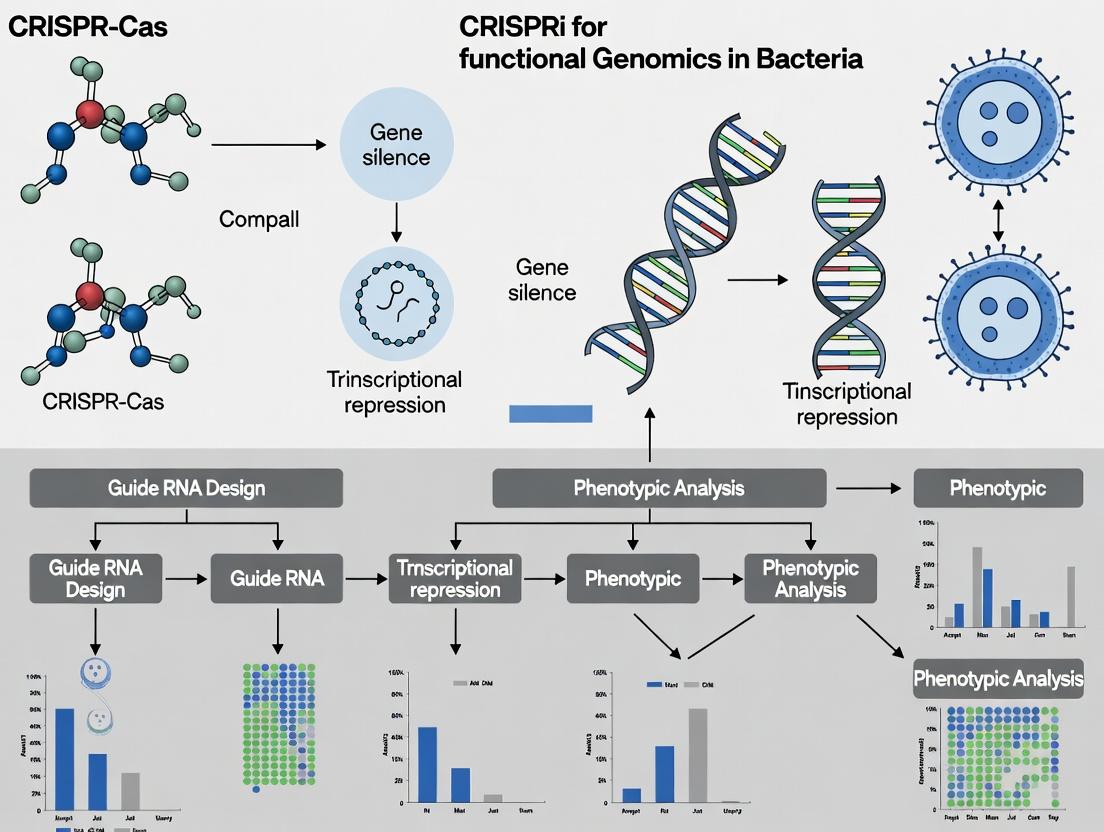
Diagrams
Title: CRISPRi Mechanism of dCas9-sgRNA Mediated Transcriptional Repression
Title: CRISPRi Experimental Workflow for Bacterial Gene Knockdown
The Scientist's Toolkit: Research Reagent Solutions
Table 3: Essential Materials for CRISPRi Experiments in Bacteria
| Item | Function & Critical Notes | Example Product/Catalog # |
|---|---|---|
| dCas9 Expression Plasmid | Constitutively or inducibly expresses catalytically dead Cas9. Requires compatibility with host and sgRNA plasmid. | pAN-3/dCas9 (Addgene #84832) for E. coli; pJV300 (Addgene #134127) for B. subtilis. |
| sgRNA Cloning Vector | Backbone for expressing sgRNA with a customizable 20-nt spacer. Contains terminator and selection marker. | pKDsgRNA (Addgene #134126) with BsaI sites for Golden Gate assembly. |
| High-Efficiency Competent Cells | For cloning and propagating plasmids. Essential for the research strain. | NEB 5-alpha (C2987H); Make chemically competent target strain as needed. |
| Golden Gate Assembly Kit | Efficient, one-pot digestion-ligation for sgRNA spacer insertion. | BsaI-HFv2 + T4 DNA Ligase (NEB, E1601). |
| Anhydrotetracycline (ATc) | A common, tightly-controlled inducer for Tet-regulated dCas9 systems. Use at low concentrations (e.g., 50-200 ng/mL). | Sigma-Aldrich, 37919. Prepare fresh in ethanol. |
| RNA Protect Reagent | Immediately stabilizes bacterial RNA at the point of sampling, ensuring accurate expression profiles. | Qiagen, 76526. |
| DNase I, RNase-free | Critical for complete removal of genomic DNA from RNA preps to prevent false positives in qPCR. | Thermo Scientific, EN0521. |
| SYBR Green qPCR Master Mix | For sensitive and specific detection of cDNA amplicons during quantification of gene knockdown. | PowerUp SYBR Green Master Mix (Thermo, A25742). |
| Flow Cytometer | For high-throughput measurement of repression/reversal kinetics using fluorescent protein reporters. | BD Accuri C6 or equivalent. |
Within a broader thesis investigating CRISPR interference (CRISPRi) for functional genomics in bacteria, a core mechanistic understanding is paramount. This application note details the fundamental distinction between CRISPRi’s transcriptional repression via steric hindrance and CRISPR-Cas9’s DNA cleavage. This comparison is critical for designing precise genetic perturbations in bacterial systems without introducing double-strand breaks (DSBs), enabling high-throughput gene knockdown studies, synthetic circuit tuning, and essential gene analysis.
Core Mechanism Comparison
CRISPRi (Steric Hindrance): Utilizes a catalytically "dead" Cas9 (dCas9) protein. When guided by a single-guide RNA (sgRNA) to a target DNA sequence, dCas9 binds but does not cut. By targeting the non-template strand within the promoter or the 5' early coding sequence (typically -50 to +300 relative to TSS), the bound dCas9 physically blocks the progression of RNA polymerase (RNAP), thus inhibiting transcription initiation or elongation.
CRISPR-Cas9 (DNA Cleavage): Employs wild-type Cas9, which, upon sgRNA-mediated target recognition and protospacer adjacent motif (PAM) binding, introduces a site-specific DSB. This triggers endogenous DNA repair pathways—error-prone non-homologous end joining (NHEJ) or homology-directed repair (HDR)—often leading to gene knockout.
Table 1: Quantitative & Functional Comparison of Core Mechanisms
| Parameter | CRISPRi (dCas9-SgRNA Complex) | CRISPR-Cas9 (Wild-Type) |
|---|---|---|
| Primary Action | Protein-DNA binding | DNA double-strand break |
| Catalytic Activity | Inactivated (D10A, H840A mutations in S. pyogenes Cas9) | Active RuvC & HNH nuclease domains |
| Typical Efficiency in E. coli | 95-99% knockdown (varies by target) | >90% knockout (with efficient repair) |
| Genetic Outcome | Reversible gene knockdown (transcriptional repression) | Permanent gene knockout (indel mutations) |
| Multiplexing Potential | High (via arrays of sgRNAs) | Moderate (DSB toxicity can limit multiplexing) |
| Off-Target Effects | Primarily binding-dependent; generally lower frequency & consequence | Cleavage-dependent; can cause genomic instability |
| Key Application in Functional Genomics | Essential gene analysis, fine-tuning expression, genome-scale screens | Gene deletion, library generation, allele replacement |
Experimental Protocols
Protocol 3.1: Implementing CRISPRi for Gene Knockdown inE. coli
Objective: To achieve targeted transcriptional repression of a gene of interest (GOI) in E. coli using a plasmid-based dCas9 and sgRNA system. Materials: See "Scientist's Toolkit" (Section 5).
Procedure:
- sgRNA Design: Design a 20-nt spacer sequence complementary to the non-template strand of the target gene. Optimal targeting region is from -50 to +300 relative to the transcription start site (TSS). Ensure an appropriate PAM (NGG for S. pyogenes dCas9) is present immediately downstream of the target sequence.
- Cloning: a. sgRNA Expression Vector: Order oligos encoding the spacer, anneal, and ligate into a plasmid containing the sgRNA scaffold under a constitutive promoter (e.g., J23119). b. dCas9 Expression Vector: Use a compatible plasmid expressing dCas9 (with D10A and H840A mutations) under an inducible promoter (e.g., anhydrotetracycline, aTc).
- Transformation: Co-transform both plasmids into your E. coli strain. Select on agar plates containing appropriate antibiotics for both plasmids.
- Induction & Culture: Inoculate a single colony into liquid media with antibiotics and inducer (e.g., 100 ng/mL aTc). Grow to desired OD~600~.
- Validation: a. Phenotypic Assay: Perform growth curves or specific functional assays relevant to the GOI. b. Transcript Quantification: Harvest cells, extract RNA, and perform RT-qPCR to quantify knockdown efficiency relative to a non-targeting sgRNA control.
Protocol 3.2: CRISPR-Cas9 Mediated Gene Knockout inE. coli
Objective: To generate a permanent deletion or mutation in a GOI. Procedure:
- Design: Design two sgRNAs flanking the region to delete, or a single sgRNA for point mutation if using an HDR template.
- Plasmid Assembly: Clone sgRNA(s) into a plasmid co-expressing wild-type Cas9 and the sgRNA(s). For HDR, a repair template oligo must be supplied.
- Transformation: Transform the Cas9-sgRNA plasmid (and repair oligo if needed) into an E. coli strain expressing recombinase proteins (e.g., λ Red system) to enhance HDR if desired.
- Selection & Screening: Plate transformations. The DSB is lethal unless repaired; survivors harbor mutations. Screen colonies by colony PCR and Sanger sequencing to identify indels or precise edits.
- Curing: Streak positive colonies on plates without antibiotic to cure the Cas9 plasmid.
Visualizations
Title: CRISPRi vs CRISPR-Cas9 Mechanism Comparison
Title: CRISPRi Experimental Protocol Flow
The Scientist's Toolkit
| Research Reagent / Material | Function & Brief Explanation |
|---|---|
| dCas9 Expression Plasmid | Plasmid encoding catalytically inactive Cas9 (e.g., with D10A/H840A mutations). Serves as the core effector protein for CRISPRi. |
| sgRNA Cloning Vector | Plasmid containing a constitutive promoter (e.g., J23119) upstream of a sgRNA scaffold. The spacer sequence is cloned into this scaffold. |
| Inducer (e.g., aTc) | Small molecule used to precisely control dCas9 expression from an inducible promoter (e.g., Ptet), allowing tunable knockdown. |
| Non-Targeting sgRNA Control | A sgRNA with a spacer that does not target the host genome. Critical negative control for distinguishing on-target effects. |
| RT-qPCR Kit | Reagents for reverse transcription quantitative PCR. Essential for quantifying transcript levels and measuring knockdown efficiency. |
| λ Red Recombinase System | For CRISPR-Cas9 knockout: Enhances HDR efficiency in E. coli when co-expressed with Cas9, facilitating precise edits using oligo templates. |
| Next-Generation Sequencing (NGS) Library Prep Kit | For genome-wide CRISPRi screens: Enables the quantification of sgRNA abundance pre- and post-selection to identify fitness genes. |
Application Notes
CRISPR interference (CRISPRi) has emerged as a premier tool for functional genomics in bacteria, enabling precise, programmable transcriptional repression. Its advantages are particularly transformative for bacterial research and antimicrobial drug target discovery.
Reversible Knockdown: Unlike CRISPR-Cas9 knockout, which creates permanent DNA breaks, CRISPRi uses a catalytically dead Cas9 (dCas9) to block transcription without altering the genome. Repression is titratable via inducer concentration and fully reversible upon removal of the sgRNA or inducer, allowing for dynamic studies of gene function and phenotypic rescue.
Essential Gene Study: Essential genes, whose loss is lethal, are prime targets for novel antibiotics but are intractable to traditional knockout screens. CRISPRi enables their systematic interrogation through potent, titratable knockdown, revealing phenotypes and genetic interactions without cell death at partial repression. This facilitates the identification and validation of essential gene function and vulnerability.
Reduced Off-Target Effects: CRISPRi exhibits significantly fewer off-target effects compared to RNAi (used in eukaryotes) or Cas9 nuclease activity. The dCas9-sgRNA complex binds with high specificity, and transcriptional repression has minimal nonspecific impact on the transcriptome. This increases the reliability of genotype-phenotype mappings in high-throughput screens.
Quantitative Data Summary:
Table 1: Comparison of Genetic Perturbation Methods in Bacteria
| Method | Mechanism | Reversible? | Suitable for Essential Genes? | Key Advantage | Reported Off-Target Rate |
|---|---|---|---|---|---|
| CRISPRi (dCas9) | Transcriptional repression | Yes | Yes | Tunable, reversible knockdown | < 1% significant off-target transcriptional changes |
| CRISPR-Cas9 Knockout | DNA cleavage & mutagenesis | No | No | Complete, permanent loss of function | 1-10% (due to sgRNA mismatch tolerance) |
| Transposon Mutagenesis | Random DNA insertion | No | No | Genome-wide saturation screening | N/A (random insertion) |
| Chemical Inducible Promoter | Transcriptional control | Yes | Yes | Tight, tunable control | Minimal (promoter-specific) |
Table 2: Performance Metrics in a Typical Essential Gene Screen (E. coli)
| Parameter | CRISPRi Performance | Notes |
|---|---|---|
| Repression Efficiency | 50-99% (gene-dependent) | Measured by qRT-PCR of target transcript |
| Growth Phenotype Detection Rate | >95% for known essentials | In pooled library screens |
| Screen False Discovery Rate | Typically <5% | Validated by follow-up assays |
| Reversibility (Recovery time) | 2-3 generations | After removal of inducer/sgRNA expression |
Protocols
Protocol 1: CRISPRi Knockdown for Essential Gene Phenotyping inE. coli
Objective: To observe the growth defect phenotype from knockdown of an essential gene.
Research Reagent Solutions Toolkit:
Table 3: Essential Reagents and Materials
| Item | Function |
|---|---|
| dCas9 Expression Plasmid | Constitutively expresses dCas9 protein (e.g., pAN-dCas9). |
| sgRNA Expression Plasmid | Contains inducible promoter driving sgRNA targeting gene of interest. |
| CRISPRi Bacterial Strain | E. coli strain harboring the chromosomal dCas9 expression system. |
| Anhydrotetracycline (aTc) | Inducer for the tet promoter controlling sgRNA expression. |
| LB Growth Media | Standard broth/agar for E. coli culture. |
| Spectrophotometer | For measuring optical density (OD600) to monitor growth. |
| qRT-PCR Reagents | To quantify knockdown efficiency at mRNA level. |
Methodology:
- Clone sgRNA: Design and clone a 20-nt spacer sequence specific to the non-template strand of the target gene's promoter or 5' coding region into the sgRNA expression vector.
- Transform: Co-transform the dCas9 plasmid (if not genomic) and the sgRNA plasmid into the target bacterial strain. Select with appropriate antibiotics.
- Culture & Induce: Inoculate 3 mL cultures in triplicate. At mid-exponential phase (OD600 ~0.3), add aTc to the experimental culture to induce sgRNA expression. Maintain an uninduced control.
- Monitor Growth: Measure OD600 every 30-60 minutes for 6-8 hours. Plot growth curves.
- Assess Knockdown: At a time point 1-2 hours post-induction, harvest cells for RNA extraction and perform qRT-PCR to quantify target mRNA levels relative to control.
- Reversibility Test: After induction and growth arrest, wash cells to remove inducer. Dilute and plate on non-inducing agar. Monitor colony formation compared to uninduced controls.
Protocol 2: Pooled CRISPRi Library Screen for Essential Gene Identification
Objective: To perform a genome-wide screen to identify genes essential for growth under a specific condition.
Methodology:
- Library Transformation: Transform the pooled genome-wide sgRNA library (e.g., E. coli CRISPRi Kohara library) at high coverage (>500x) into the expression strain carrying dCas9.
- Selection & Harvest: Plate the transformation on selective agar to create the "Input" pool. Harvest ~10^9 cells from these plates for genomic DNA (gDNA) extraction.
- Growth Passage: Inoculate the remainder of the pool into liquid media under the selective condition (e.g., antibiotic presence, nutrient limitation). Culture for ~15-20 generations, maintaining library representation.
- Harvest Output Pool: Collect cells from the final culture for gDNA extraction ("Output" pool).
- Amplify & Sequence sgRNA Barcodes: Perform PCR amplification of the sgRNA sequence region from both Input and Output gDNA pools. Submit for high-throughput sequencing.
- Data Analysis: Map sequencing reads to the sgRNA library. For each sgRNA, calculate the log2 fold change (Output/Input). Depleted sgRNAs in the Output pool indicate that their target gene is essential for the test condition.
Diagrams
CRISPRi Workflow from Induction to Reversal
Pooled CRISPRi Screen for Essential Genes
Within the broader thesis advocating for CRISPR interference (CRISPRi) as a superior platform for functional genomics in bacteria, it is critical to understand the limitations of its predecessor technologies. Traditional gene knockout (via homologous recombination) and RNA interference (RNAi) have been instrumental but possess significant constraints for systematic, large-scale studies in bacterial systems. This application note details these limitations with supporting data and protocols, providing context for the adoption of CRISPRi.
Limitations of Traditional Gene Knockouts
Complete gene knockout through homologous recombination is a cornerstone of bacterial genetics but is fraught with challenges for functional genomics.
Key Limitations:
- Time-Intensive and Laborious: The process requires multiple cloning and selection steps for each target.
- Lethality Bias: Essential genes cannot be studied as complete knockouts are inviable, creating a systematic gap in genomic coverage.
- Pleiotropic Effects: Secondary mutations or adaptive suppressors can arise during the construction process, confounding phenotypes.
- Low Throughput: Scaling to genome-wide libraries is exceptionally difficult in most bacterial species.
Quantitative Comparison of Construction Time: Table 1: Estimated Hands-on Time for Generating a Single Gene Knockout in E. coli K-12.
| Step | Process | Estimated Time |
|---|---|---|
| 1 | Primer Design, PCR of Resistance Cassette & Flanking Homology Arms | 4-6 hours |
| 2 | Cloning/Assembly & Transformation into Cloning Strain | 3-5 hours (plus 1-2 days incubation) |
| 3 | Plasmid Extraction & Verification | 2 hours (plus overnight culture) |
| 4 | Conjugation or Electroporation into Target Strain | 3-4 hours (plus 1-2 days selection) |
| 5 | Selection & Colony PCR Verification | 4-6 hours (plus 1-2 days growth) |
| 6 | Curing of Suicide Vector (if applicable) | 3-5 hours (plus 1-2 days counterselection) |
| Total Hands-on Time | ~19-30 hours |
Protocol: Traditional Knockout via Homologous Recombination Objective: Disrupt a target gene (geneX) in E. coli using a kanamycin resistance cassette. Materials: See "Research Reagent Solutions" (Table 3). Procedure:
- Design Homology Arms: Amplify ~500 bp sequences immediately upstream (UP) and downstream (DOWN) of geneX from genomic DNA.
- Amplify Resistance Cassette: PCR amplify the kanR gene from a template plasmid.
- Assemble Construct: Use overlap extension PCR or Gibson Assembly to fuse the UP-kanR-DOWN fragment.
- Clone into Suicide Vector: Ligate the assembled fragment into a temperature-sensitive origin suicide vector (e.g., pKO3).
- Transform into Cloning Host: Transform assembly into a standard E. coli cloning strain, select on ampicillin (vector resistance) and kanamycin.
- Conjugate/Electroporate: Mobilize the verified plasmid from the cloning strain into the target E. coli strain via conjugation or electroporation.
- First Crossover Selection: Plate on kanamycin at the permissive temperature (e.g., 30°C) to select for clones where the plasmid has integrated into the genome via homologous recombination.
- Second Crossover & Curing: Grow selected colonies at the restrictive temperature (e.g., 42°C) without selection to promote plasmid excision. Screen colonies for loss of vector marker (ampicillin sensitivity) and retention of kanR (kanamycin resistance).
- Verification: Confirm the knockout via PCR across the two junctions and Sanger sequencing.
Limitations of RNA Interference (RNAi) in Bacteria
RNAi is a potent gene silencing tool in eukaryotes but is largely ineffective in most prokaryotes due to the absence of the conserved RNAi machinery (Dicer, Argonaute proteins).
Key Limitations:
- Lack of Endogenous Machinery: Most bacteria do not possess the canonical RNAi pathway, making heterologous expression inefficient.
- High Off-Target Effects: Engineered expression of long double-stranded RNA (dsRNA) in artificial systems can trigger non-specific phenotypic effects.
- Variable and Incomplete Knockdown: Silencing efficiency is unpredictable and rarely reaches >90%, complicating phenotypic analysis.
- Toxicity and Fitness Cost: Constitutive expression of foreign dsRNA and necessary machinery components burdens bacterial growth.
Quantitative Data on Silencing Efficiency: Table 2: Reported Efficacy of Heterologous RNAi Systems in Bacteria.
| Bacterial Species | System/Vector | Max Knockdown Efficiency (%) | Key Caveat | Citation (Example) |
|---|---|---|---|---|
| E. coli | Heterologous Caenorhabditis elegans machinery | 50-70% | Severe growth defect, high variability | (Uhde et al., 2016) |
| Mycobacterium smegmatis | Plasmid-based antisense RNA | 60-80% | Strong target-dependent variation | (Engstrom et al., 2019) |
| Sinorhizobium meliloti | IPTG-inducible antisense RNA | ~70% | Incomplete repression, leaky expression | (Khan et al., 2018) |
Protocol: Attempted Gene Silencing via Heterologous RNAi in E. coli Objective: Express dsRNA targeting geneY using a heterologous system. Materials: See "Research Reagent Solutions" (Table 3). Procedure:
- Design dsRNA: Identify a 200-300 bp unique region of geneY. Clone this sense and antisense sequence, separated by a short intronic loop spacer, into an expression vector under a tight, inducible promoter (e.g., pL4440 derivative).
- Express RNAi Machinery: Co-transform the dsRNA vector with a second plasmid expressing the C. elegans Dicer (DCR-1) and Argonaute (ALG-1/2) genes under a constitutive promoter.
- Induce Silencing: Grow the double-transformant to mid-log phase and add inducer (e.g., IPTG) to express the dsRNA.
- Monitor Phenotype & Efficiency: Measure growth phenotype (OD600) over time compared to empty vector control.
- Assess Knockdown: Harvest cells 4-6 hours post-induction. Extract total RNA, perform reverse transcription, and quantify geneY mRNA levels via qRT-PCR using a housekeeping gene for normalization.
The Scientist's Toolkit
Table 3: Research Reagent Solutions for Traditional Bacterial Genetics.
| Item | Function & Application | Example Product/Catalog |
|---|---|---|
| Suicide Vector | Plasmid with temperature-sensitive origin; allows for chromosomal integration and subsequent curing. Essential for knockouts. | pKO3 (Temp-sensitive, sacB for counter-selection) |
| PCR Assembly Master Mix | Enzyme mix for seamless, Gibson Assembly-style cloning of homology arms and resistance cassettes. | NEBuilder HiFi DNA Assembly Master Mix |
| Counterselection Marker | Gene conferring sensitivity to a condition (e.g., sacB to sucrose), enabling selection for plasmid loss. | pRE112 (sacB, oriT for conjugation) |
| Antisense RNA Vector | Plasmid with strong, inducible promoter for expressing antisense or dsRNA for gene knockdown attempts. | pZA31 (Tet-inducible, low copy) |
| Broad-Host-Range Conjugation Helper | Strain providing trans-acting mobilization functions to transfer suicide vectors into target strains. | E. coli S17-1 λ pir |
| Cassette for Antibiotic Resistance | Selectable marker (e.g., kanR, cat) flanked by FRT or loxP sites for removal after knockout. | FRT-flanked kanR amplification template |
Title: Limitations of Knockouts & RNAi vs. CRISPRi Advantages
Title: Workflow Complexity: Traditional Knockout vs. CRISPRi
Within the broader thesis on CRISPR interference (CRISPRi) for functional genomics in bacteria, this application note details its transformative role in three critical areas. CRISPRi, utilizing a catalytically dead Cas9 (dCas9) to repress gene transcription, enables precise, programmable, and scalable functional genomics. This technology is pivotal for dissecting bacterial physiology, identifying genetic vulnerabilities, and accelerating antibacterial discovery, providing a robust framework for systematic genetic perturbation without permanent DNA cleavage.
Application Note 1: High-Throughput Genetic Screens
CRISPRi enables genome-wide or targeted arrayed/ pooled screens to identify genes essential for growth, stress response, or antibiotic susceptibility under defined conditions.
Table 1: Representative High-Throughput CRISPRi Screen Output (Model Organism: E. coli K-12)
| Screen Condition | Library Size (Guides) | Essential Genes Identified | Hit Rate (%) | Key Validation Method |
|---|---|---|---|---|
| Rich Medium (LB) | ~50,000 (genome-wide) | ~350 | ~0.7 | Individual knockdown & growth curve |
| Antibiotic (Sub-MIC) | ~10,000 (targeted) | ~150 (sensitizers) | ~1.5 | Checkerboard synergy assay |
| Biofilm Formation | ~5,000 (pathway-focused) | ~75 | ~1.5 | Microtiter plate crystal violet assay |
Detailed Protocol: Pooled CRISPRi Screen for Growth Essentials
Objective: To identify conditionally essential genes in E. coli using a pooled, genome-wide CRISPRi library.
Materials (Research Reagent Toolkit):
- dCas9 Expression Strain: E. coli strain harboring a chromosomally integrated, IPTG-inducible dCas9 (e.g., from plasmid pNDC-dCas9 integrated via λ-Red).
- CRISPRi Library: Pooled, cloned sgRNA library targeting all non-essential genes (e.g., ASAP library clone pool).
- Growth Medium: LB Lennox + appropriate antibiotics (e.g., Spectinomycin for library maintenance) + 100 µM IPTG.
- PCR Reagents: For amplifying sgRNA inserts for NGS.
- Sequencing Platform: Illumina MiSeq or NextSeq.
Procedure:
- Library Transformation: Electroporate the pooled sgRNA plasmid library into the induced dCas9 expression strain. Ensure high transformation efficiency (>10^7 CFU) to maintain library representation.
- Outgrowth & Selection: Recover cells in SOC medium for 1 hour, then inoculate into primary culture with antibiotic and IPTG. Grow for 6-8 hours (approx. 10 generations).
- Passaging & Bottlenecking: Dilute the primary culture 1:1000 into fresh, pre-warmed selective medium. Repeat this serial passaging for a total of ~15-20 generations. This enriches for cells carrying sgRNAs targeting non-essential genes.
- Sample Collection: Harvest cell pellets at generation 0 (T0) and at the final passage (Tend).
- sgRNA Amplification & Sequencing: Isolate plasmid DNA from T0 and Tend pellets. Amplify the sgRNA cassette via PCR using barcoded primers compatible with your sequencer. Pool and sequence amplicons.
- Data Analysis: Map sequencing reads to the sgRNA library reference. Calculate the fold-depletion of each sgRNA from T0 to Tend using read count normalization (e.g., DESeq2). sgRNAs targeting essential genes will be significantly depleted.
Workflow Diagram:
Title: Pooled CRISPRi Screen Workflow for Essential Genes
Application Note 2: Synthetic Lethality Screening
CRISPRi facilitates the discovery of synthetic lethal (SL) gene pairs, where repression of two genes is lethal while repression of either alone is not. This is powerful for identifying novel drug target combinations and understanding genetic networks.
Table 2: Example Synthetic Lethality Screen for Antibiotic Adjuvants
| Target Gene (Pathway) | Synergistic Partner Gene (Pathway) | Fitness Score (Double Knockdown) | Single Knockdown Fitness | Potential Therapeutic Use |
|---|---|---|---|---|
| folA (Folate synthesis) | purH (Purine synthesis) | -2.5 | ~0 (Neutral) | Dual-target antimicrobial |
| acrB (Efflux pump) | lpxC (LPS biosynthesis) | -3.1 | Mild Defect (-0.8) | Resensitization to antibiotics |
| gyrB (DNA gyrase) | topA (Topoisomerase I) | -4.0 | Essential | High-potency combination |
Detailed Protocol: Dual-guide CRISPRi for SL Identification
Objective: To systematically test pairwise gene repression for synthetic lethal interactions using a focused dual-guide CRISPRi system.
Materials (Research Reagent Toolkit):
- Dual-guide Vector: A plasmid expressing two sgRNAs from distinct promoters (e.g., pDUAL-ccdB with J23119 and J23100 promoters).
- Arrayed sgRNA Pairs: Pre-cloned pairs targeting candidate genes (e.g., genes in parallel pathways).
- Liquid Handling Robot: For high-density arrayed culture inoculation (e.g., 384-well plates).
- Plate Reader: For high-throughput OD600 measurements.
Procedure:
- Array Setup: In a 384-well plate, dispense growth medium + inducer (IPTG) into each well. Using automation, inoculate each well with the dCas9 strain harboring a unique dual-guide plasmid from the arrayed library. Include control wells with empty vector and single-target guides.
- Growth Kinetics: Incubate the plate with continuous shaking in a plate reader, measuring OD600 every 15-30 minutes for 16-24 hours.
- Data Extraction: Calculate the growth rate (μ) and/or maximum OD for each well.
- Interaction Scoring: Compute a genetic interaction score (ε) for each pair (A,B). A common method is: ε = μ(AB) - μ(A) - μ(B) + μ(empty). Strongly negative ε indicates a synthetic lethal/sick interaction.
- Validation: Retest top hits in biological triplicate and via individual knockdowns followed by complementary assays (e.g., viability staining).
Genetic Interaction Logic Diagram:
Title: Synthetic Lethality Interaction Logic Map
Application Note 3: Drug Target Discovery & Validation
CRISPRi is used to identify and validate novel antibacterial targets by linking gene repression to a desired phenotype (e.g., cell death, loss of virulence) and demonstrating correlation with drug action.
Table 3: CRISPRi-Based Prioritization of Novel Drug Targets
| Candidate Target Gene | CRISPRi Phenotype (Fitness Score) | Chemical Inhibitor Screen Hit? | MIC of Lead Compound (µg/mL) | Mammalian Cell Cytotoxicity (IC50, µM) |
|---|---|---|---|---|
| fabI (enoyl-ACP reductase) | -2.8 (Severe defect) | Yes (Triclosan analogs) | 0.5 | >50 |
| metK (S-adenosylmethionine synthetase) | -1.5 (Moderate defect) | Yes (Sinefungin analogs) | 8.0 | >100 |
| yjeQ (ribosome assembly GTPase) | -0.9 (Mild defect) | No | N/A | N/A |
Detailed Protocol: Chemical-Genetic Interaction Profiling
Objective: To validate a potential drug target by comparing the phenotypic footprint of genetic repression (CRISPRi) with treatment by a small-molecule inhibitor.
Materials (Research Reagent Toolkit):
- CRISPRi Strain Array: Arrayed strains with inducible dCas9 and sgRNAs targeting the candidate gene and essential/ non-essential controls.
- Compound Library: Array of putative inhibitors or a focused chemical library.
- Automated Imaging System: For endpoint viability assessment (e.g., via fluorescence).
Procedure:
- Strain Preparation: Grow overnight cultures of CRISPRi strains (with target gene sgRNA and control sgRNAs) with dCas9 induction.
- Chemical-Genetic Assay: In a 384-well plate, serially dilute the candidate drug compound. Add a uniform inoculum of each induced CRISPRi strain to separate compound plates. Include a no-drug control column.
- Phenotypic Readout: Incubate for 4-6 hours (approx. 5 generations). Add a viability stain (e.g., resazurin). Measure fluorescence after 1-2 hours.
- Data Integration: Calculate % inhibition for each strain at each drug concentration. Plot dose-response curves. A true target inhibitor will show hypersensitivity in the strain where the target gene is repressed by CRISPRi compared to a non-targeting control strain. This creates a characteristic "collateral sensitivity" profile.
- Mechanistic Follow-up: Use CRISPRi knockdown combined with sub-MIC drug to check for synergistic interaction, confirming on-target activity.
Chemical-Genetic Validation Pathway:
Title: CRISPRi-Chemical Screen Target Validation
Implementing CRISPRi: Step-by-Step Workflow from Design to Screening
Within the broader thesis on implementing CRISPR interference (CRISPRi) for functional genomics in bacterial research, the initial and critical step is the selection of an appropriate dCas9 protein and its expression system. This choice dictates the system's efficiency, specificity, orthogonality, and compatibility with the target bacterial host. This application note provides a current, comparative analysis of key dCas9 orthologs and vector considerations, along with detailed protocols for initial validation.
Comparative Analysis of dCas9 Orthologs for Bacterial CRISPRi
The optimal dCas9 ortholog balances high binding affinity, minimal off-target effects, and compatibility with the host's cellular environment (e.g., codon usage, temperature). The following table summarizes key characteristics of the most utilized dCas9 variants.
Table 1: Comparison of Common dCas9 Orthologs for Bacterial CRISPRi
| Ortholog (Species Source) | Size (aa) | Optimal PAM Sequence | Working Temperature | Key Advantages | Primary Considerations |
|---|---|---|---|---|---|
| dCas9 (Streptococcus pyogenes) | 1368 | 5'-NGG-3' | 37°C | Most well-characterized; extensive sgRNA design tools; high activity. | Large size may burden some cells; prevalent in synthetic circuits may cause crosstalk. |
| dCas9 (Staphylococcus aureus) | 1053 | 5'-NNGRRT-3' | 37°C | Smaller size, easier delivery; different PAM expands targeting range. | Slightly lower binding affinity in some reports; fewer validated sgRNAs. |
| dCas9 (Streptococcus thermophilus) | 1121 | 5'-NNAGAAW-3' | 30-42°C | Good for thermophiles or mesophiles; orthogonal to Sp-dCas9. | Less characterized; toolbox of parts is smaller. |
| dCas9 (Neisseria meningitidis) | 1082 | 5'-NNNNGATT-3' | 37°C | Long PAM allows for highly specific targeting; orthogonal. | Very restricted targeting range due to long PAM. |
| dCas9 (Campylobacter jejuni) | 984 | 5'-NNNNRYAC-3' | 37°C (C. jejuni grows at 42°C) | Smallest common ortholog; useful for targeting AT-rich genomes. | Optimal activity may require host-specific adaptations. |
Vector System Considerations
The expression vector must be tailored to the host bacterium and experimental goals (inducible vs. constitutive repression).
Table 2: Key Vector Features for dCas9 Expression
| Feature | Options | Recommendation |
|---|---|---|
| Origin of Replication | High-copy (ColE1), Medium-copy (p15A), Low-copy (SC101, F-plasmid) | Use low-copy for toxicity concerns; medium-copy for standard applications. |
| Selection Marker | Antibiotic resistance (KanR, AmpR, CmR), Auxotrophic complementation | Choose marker compatible with host and downstream assays. |
| Promoter for dCas9 | Constitutive (J23100, Pveg), Inducible (Ptet, ParaBAD, PLtetO-1) | Strongly recommend inducible promoters to mitigate fitness cost and allow control of repression timing. |
| sgRNA Expression | Constitutive promoter (e.g., J23119) with terminator (e.g., T1). | Use a separate, constitutive promoter for sgRNA. Multiplexing requires array with processing elements (tRNA, Csy4). |
| Additional Features | MCS for sgRNA cloning, RBS library for tuning dCas9 expression, Transcriptional terminators. | Include a strong double terminator after dCas9 to prevent read-through. |
Protocols
Protocol 4.1: Cloning dCas9 Ortholog into an Inducible Expression Vector
Objective: Clone a chosen dCas9 gene into a medium-copy plasmid under the control of an inducible promoter (e.g., Ptet).
Materials:
- Destination vector with inducible promoter and appropriate selection.
- PCR-amplified dCas9 gene (codon-optimized for host) with flanking homology arms.
- High-fidelity DNA polymerase, DpnI.
- Gibson Assembly or In-Fusion cloning mix.
- Competent E. coli cloning strain (DH5α).
- LB agar plates with appropriate antibiotic.
- Induction agent (e.g., anhydrotetracycline, aTc).
Procedure:
- PCR Amplification: Amplify the dCas9 gene from source DNA using primers that add 20-30 bp overlaps homologous to the vector sequence immediately downstream of the promoter and upstream of the terminator.
- Vector Preparation: Linearize the destination vector by PCR or restriction digest. If using a restriction enzyme, dephosphorylate the ends.
- Assembly: Mix ~50 ng of linearized vector with a 2:1 molar ratio of the dCas9 insert. Add assembly master mix. Incubate per manufacturer's instructions (typically 50°C for 15-60 min).
- Transformation: Transform 2-5 µl of the assembly reaction into chemically competent E. coli DH5α. Recover in SOC medium for 1 hour at 37°C.
- Plating and Screening: Plate on LB agar with the appropriate antibiotic. Incubate overnight at 37°C. Screen colonies by colony PCR using primers flanking the insertion site.
- Validation: Sequence-confirmed clones should be validated by inducing with the appropriate agent and performing a western blot for dCas9 (using a FLAG or HA tag engineered at the C-terminus).
Protocol 4.2: Functional Validation of CRISPRi Repression Efficiency
Objective: Quantify the knockdown efficiency of a selected dCas9-sgRNA system on a reporter gene (e.g., GFP).
Materials:
- Validated dCas9 expression plasmid.
- sgRNA expression plasmid (or a single plasmid with both).
- Reporter plasmid with GFP under a constitutive promoter.
- Target bacterial strain.
- Microplate reader with fluorescence capability.
- Sterile 96-well plates.
Procedure:
- Strain Construction: Co-transform or sequentially transform the target bacterium with three plasmids: (1) dCas9 expression plasmid, (2) sgRNA plasmid targeting the GFP RBS or early coding sequence, (3) GFP reporter plasmid. Include controls lacking dCas9 or sgRNA.
- Culture Conditions: Inoculate triplicate wells of a 96-well deep-well plate with 500 µl of medium containing all necessary antibiotics and the dCas9 inducer (if applicable).
- Growth and Measurement: Grow cultures with shaking at the appropriate temperature. Monitor OD600 and fluorescence (Ex: 485 nm, Em: 520 nm) in a plate reader every 30-60 minutes.
- Data Analysis: Normalize fluorescence to OD600 for each time point. Calculate repression efficiency as:
[1 - (Fluor/OD)sample / (Fluor/OD)control] * 100%. The control is the strain with non-targeting sgRNA.
Visualization
Diagram 1: CRISPRi System Component Workflow
Diagram 2: dCas9-sgRNA Mechanism of Transcriptional Interference
The Scientist's Toolkit
Table 3: Essential Research Reagents for CRISPRi System Selection & Validation
| Reagent / Material | Function / Purpose |
|---|---|
| Codon-Optimized dCas9 Genes | Gene fragments optimized for expression in your target host (e.g., E. coli, B. subtilis, Pseudomonas). |
| Modular Cloning Vectors | Plasmids with standardized parts (promoters, RBS, terminators) for easy assembly of dCas9 and sgRNA expression cassettes. (e.g., MoClo, Golden Gate systems). |
| Induction Agents | Small molecules for precise control of dCas9 expression (e.g., aTc for Ptet, Arabinose for ParaBAD). |
| Anti-FLAG/HA Antibody | For western blot validation of tagged dCas9 protein expression levels. |
| Fluorescent Reporter Plasmids | Plasmids expressing GFP/mCherry under constitutive promoters to serve as knockdown targets for quantitative validation. |
| High-Efficiency Competent Cells | For both cloning strains (DH5α) and target experimental strains. Crucial for multi-plasmid transformations. |
| Next-Gen Sequencing Library Prep Kit | For preparing sequencing libraries from genome-wide CRISPRi screens to identify essential genes. |
| sgRNA Design Software | Tools like CHOPCHOP, Benchling, or species-specific design tools to predict efficient sgRNAs with minimal off-targets. |
Application Notes
Within a broader thesis exploring CRISPR interference (CRISPRi) for functional genomics in bacteria, strategic gRNA design is paramount. Optimal design ensures potent, specific transcriptional repression, enabling high-quality genetic screens and target validation in drug discovery.
Recent literature and database analyses (2023-2024) confirm two cardinal rules for maximizing CRISPRi efficiency in bacteria:
- Target the Non-Template (NT) Strand: gRNAs complementary to the non-template strand of the target gene consistently yield higher repression efficiency. This is attributed to the stable R-loop formation when dCas9 binds to the NT strand, creating a more effective steric block for RNA polymerase.
- Proximal Promoter Targeting: gRNAs targeting regions from -50 to +300 nucleotides relative to the transcription start site (TSS) show the strongest repression. The region immediately downstream of the TSS is particularly effective.
Table 1: Quantitative Impact of gRNA Positioning on CRISPRi Efficiency
| Target Region (relative to TSS) | Median Repression Efficiency (%) | Key Rationale |
|---|---|---|
| -50 to +1 (Promoter) | 85-95% | Blocks RNAP binding or initial unwinding. |
| +1 to +50 (Early 5' CDS) | 90-99% | Optimal steric blocking of elongating RNAP. |
| +50 to +150 (Early CDS) | 75-90% | High efficiency, but can decline with distance. |
| +150 to +300 (Mid CDS) | 50-75% | Moderate efficiency; subject to sequence effects. |
| > +300 (Distal CDS) | < 50% | Generally low and unreliable repression. |
Table 2: Strand Selection Impact in Model Bacteria
| Organism | Non-Template Strand Efficiency | Template Strand Efficiency | Efficiency Ratio (NT/T) |
|---|---|---|---|
| E. coli | 92% ± 5% | 68% ± 12% | ~1.35 |
| B. subtilis | 88% ± 7% | 60% ± 15% | ~1.47 |
| M. tuberculosis | 85% ± 10% | 55% ± 18% | ~1.55 |
Experimental Protocols
Protocol 1:In SilicogRNA Design and Selection
Objective: To design and prioritize high-efficacy gRNAs for a bacterial target gene.
Materials:
- Bacterial genome sequence (FASTA).
- Confirmed Transcription Start Site (TSS) data for target gene. (If unknown, use translation start site (ATG) as proxy, assuming -35/-10 promoter).
- gRNA design software (e.g., CHOPCHOP, Benchling).
Procedure:
- Define Target Window: Identify the sequence from -50 to +300 nucleotides relative to the TSS.
- Extract Sequence: Retrieve both template and non-template strand sequences for the target window.
- Identify PAM Sites: Scan the non-template strand for NGG (or other Cas9 variant-specific PAM, e.g., NGG for Sp-dCas9) sequences. The PAM is located 3' of the target sequence on the non-template strand.
- Generate gRNA Sequences: For each NGG, extract the 20-nt genomic sequence immediately upstream (5') of the PAM. This 20-nt sequence is your prospective gRNA spacer.
- Filter and Rank: a. Eliminate spacers with significant off-target homology (>12-nt contiguous match) using BLAST against the host genome. b. Prioritize spacers within the +1 to +50 region. c. Select 3-5 top candidate gRNAs for empirical testing.
Protocol 2: Empirical Validation of gRNA Efficiency
Objective: To measure the transcriptional repression efficiency of designed gRNAs in vivo.
Materials:
- Bacterial strain with integrated, inducible dCas9 expression system (e.g., E. coli MG1655 with pZA-dCas9).
- Cloning vectors for gRNA expression (e.g., pZS-sgRNA).
- qRT-PCR reagents (SYBR Green, primers for target and reference gene).
- Spectrophotometer and qPCR instrument.
Procedure:
- Clone gRNA Candidates: Clone each 20-nt spacer sequence into the gRNA expression vector via inverse PCR or Golden Gate assembly.
- Co-transform: Transform the dCas9-expressing strain with each gRNA plasmid and an empty gRNA vector control.
- Induction: Grow triplicate cultures to mid-log phase (OD600 ~0.3-0.5) and induce dCas9 and gRNA expression with appropriate inducers (e.g., aTc, IPTG).
- Harvest RNA: After 2-3 hours of induction, harvest cells and extract total RNA. Treat with DNase I.
- Quantify mRNA: Perform qRT-PCR. Use primers amplifying a 100-150 bp region ~100 bp downstream of the gRNA target site. Include a housekeeping gene (e.g., rpoB) for normalization.
- Calculate Efficiency: Use the ΔΔCt method. Repression efficiency = (1 - 2^(-ΔΔCt)) * 100%, where ΔΔCt compares induced gRNA samples to the induced empty vector control.
Diagrams
gRNA Design & Validation Workflow (95 chars)
Optimal CRISPRi gRNA Binding Mechanism (94 chars)
The Scientist's Toolkit
Table 3: Essential Research Reagent Solutions for CRISPRi gRNA Design & Validation
| Reagent/Material | Function/Application | Example/Notes |
|---|---|---|
| dCas9 Expression System | Constitutively or inducibly expresses catalytically dead Cas9 protein. | Plasmid: pZA-dCas9 (aTc inducible). Integrated into genome for stability. |
| gRNA Scaffold Vector | Allows easy cloning of 20-nt spacer sequences upstream of the invariant gRNA scaffold. | Plasmid: pZS-sgRNA (IP inducible). Contains SapI or BsaI sites for Golden Gate cloning. |
| High-Fidelity DNA Polymerase | For inverse PCR to linearize the gRNA vector for spacer insertion. | Q5 or Phusion Polymerase for error-free amplification. |
| Golden Gate Assembly Mix | Efficient, one-pot modular cloning of spacer sequences into the gRNA scaffold. | Esp3I or BsaI-HF enzyme with T4 DNA Ligase. |
| Chemically Competent Cells | For transformation of constructed plasmids into the dCas9-expressing bacterial strain. | E. coli MG1655 with pZA-dCas9 made competent via CaCl2 or TSS method. |
| RNA Protect Reagent | Immediately stabilizes bacterial mRNA profiles at time of harvest. | Qiagen RNAprotect Bacteria Reagent. |
| DNase I (RNase-free) | Removes genomic DNA contamination from RNA preps, critical for accurate qRT-PCR. | |
| Reverse Transcriptase | Synthesizes cDNA from the purified mRNA template for downstream qPCR. | M-MLV or similar, with random hexamers/gene-specific primers. |
| SYBR Green qPCR Master Mix | For quantitative real-time PCR to measure relative transcript levels. | Contains hot-start Taq polymerase, dNTPs, buffer, and SYBR Green dye. |
| Validated qPCR Primers | Amplify a short fragment (~100 bp) of the target gene and a housekeeping control. | Design to amplicon region ~100 bp downstream of gRNA target site. |
Within a thesis investigating CRISPR interference (CRISPRi) for functional genomics in bacteria, the construction of the sgRNA library is a pivotal step. The choice between pooled and arrayed formats dictates experimental scale, screening methodology, and downstream analysis. This application note details the considerations, protocols, and reagents for both approaches in bacterial systems.
Comparative Analysis: Pooled vs. Arrayed Screening
Table 1: Key Characteristics of Pooled vs. Arrayed Screening Formats
| Feature | Pooled Screening | Arrayed Screening |
|---|---|---|
| Library Format | All sgRNA plasmids are cloned and maintained in a single, complex mixture. | Each sgRNA clone is maintained individually in a separate well (e.g., 96- or 384-well plate). |
| Primary Application | Positive and negative selection screens (e.g., survival under antibiotic pressure). | Phenotypic screens requiring individual strain analysis (e.g., microscopy, growth kinetics, biofilm formation). |
| Throughput | Extremely high (can assay entire library in 1-2 culture flasks). | Lower, limited by plate-based assays. |
| Phenotypic Readout | Bulk population fitness, assessed by NGS of sgRNA abundance over time. | Multidimensional, per-strain measurements (OD600, fluorescence, enzymatic activity). |
| Cost per Datapoint | Very low. | High. |
| Key Instrumentation | Next-Generation Sequencer, PCR thermocycler. | Liquid handler, plate reader, automated microscopy. |
| Data Complexity | High (requires statistical modeling of NGS counts). | Simpler, often direct measurement per well. |
| Typical Library Size | 10^3 – 10^5 sgRNAs. | 10^2 – 10^3 sgRNAs. |
| CRISPRi Context | Ideal for genome-wide identification of genes essential for growth or stress tolerance. | Ideal for targeted, mechanistic follow-up on pathways of interest. |
Table 2: Quantitative Comparison of Workflow Steps
| Workflow Step | Pooled Format Duration (Days) | Arrayed Format Duration (Days) |
|---|---|---|
| Library Cloning & Validation | 7-10 | 10-14 |
| Transformation into Bacterial Cells | 1 (bulk electroporation) | 3-5 (arrayed transformation or spotting) |
| Library Amplification & Selection | 2-3 (outgrowth with antibiotic) | 5-7 (colony picking, inoculating plates) |
| Screening Experiment | 5-10 (passaging under selection) | 1-3 (plate-based assay) |
| Sample Prep for Readout | 3-4 (PCR amplicon prep for NGS) | 0 (direct measurement) |
| Data Acquisition & Analysis | 2-3 (NGS run) + 2-4 (bioinformatics) | 1-2 (plate reader) + 1-2 (analysis) |
Detailed Protocols
Protocol 1: Construction of a Pooled CRISPRi sgRNA Library for Bacteria
Objective: To generate a complex plasmid pool targeting every non-essential gene in the bacterial genome. Materials: See "Scientist's Toolkit" below.
- sgRNA Library Design: Using a bioinformatics tool (e.g., CHOPCHOP), design 3-5 sgRNAs per gene target, focusing on the 5' region of the coding sequence. Include positive and negative control sgRNAs. Synthesize the oligo pool commercially.
- Pool Cloning (Golden Gate Assembly): a. Amplify the oligo pool in a 50 µL PCR reaction using primers that add the requisite overhangs for the destination CRISPRi plasmid (e.g., pCRISPRi-v2). b. Purify the PCR product using a spin column. c. Set up a Golden Gate assembly reaction: 50 ng digested backbone, 20 ng PCR insert, 1 µL T4 DNA Ligase, 1 µL Type IIs restriction enzyme (e.g., BsaI), in 1X T4 Ligase Buffer. Cycle: (37°C for 5 min, 16°C for 5 min) x 25 cycles; then 60°C for 10 min.
- Library Transformation & Amplification: a. Desalt the assembly reaction and electroporate into high-efficiency E. coli cloning strain (e.g., NEB 10-beta) in 5-10 reactions. Recover in SOC for 1 hour. b. Pool all recoveries, plate a dilution series to assess library coverage (>500 colonies per sgRNA), and incubate the remainder in liquid culture with antibiotic overnight to amplify the plasmid library. c. Perform maxiprep to obtain the high-quality pooled plasmid library. Validate complexity by NGS of the sgRNA cassette region.
Protocol 2: Arrayed Screening Using a CRISPRi Bacterial Library
Objective: To assay phenotypic responses of individual CRISPRi knockdown strains in a multi-well format. Materials: See "Scientist's Toolkit" below.
- Arrayed Library Generation: a. Starting from the pooled plasmid library (Protocol 1, Step 3b) or from individually cloned sgRNAs, perform a series of 96-well plate transformations into your bacterial strain of interest using chemical transformation or electroporation. b. Spot each transformation on individual wells of a 96-well agar plate containing antibiotic. Incubate. c. Using a 96-pin replicator, inoculate from the agar plate into a 96-well deep-well block containing 1 mL of medium + antibiotic per well. Grow overnight. d. Add glycerol to 15% final concentration to create the master stock plate. This is your arrayed library.
- Phenotypic Screening (Example: Growth Curves): a. Using a liquid handler, inoculate 5 µL from the master stock into 195 µL of fresh medium (+ inducer for CRISPRi) in a 96-well optical plate. Include control wells (non-targeting sgRNA, empty vector). b. Load the plate into a plate reader. Set protocol: Shake continuously, measure OD600 every 15 minutes for 24 hours, 37°C. c. Export data and calculate per-well metrics: lag time, maximum growth rate, and final OD.
Visualizations
Workflow for Pooled and Arrayed CRISPRi Screens
Decision Guide for Screening Format Selection
The Scientist's Toolkit: Key Research Reagent Solutions
Table 3: Essential Materials for CRISPRi Library Construction & Screening
| Item | Function/Description | Example Product/Catalog |
|---|---|---|
| CRISPRi Vector Backbone | Inducible dCas9 expression, sgRNA scaffold, bacterial origin, selection marker. | pCRISPRi-v2 (Addgene #125597) |
| Pooled sgRNA Oligo Library | Custom-designed, synthesized oligo pool containing all sgRNA sequences. | Twist Bioscience Custom Pooled Oligo Library |
| High-Efficiency Cloning Strain | E. coli strain for maximizing library transformation efficiency and representation. | NEB 10-beta Electrocompetent E. coli (C3020K) |
| Type IIs Restriction Enzyme | Enzyme for Golden Gate assembly (creates unique, directional overhangs). | BsaI-HFv2 (NEB, R3733) |
| Electrocompetent Target Bacteria | Competent cells of the bacterial species under study for library delivery. | Species-specific preparation required. |
| Next-Generation Sequencing Kit | For preparing sgRNA amplicon libraries from genomic DNA of pooled screens. | Illumina Nextera XT DNA Library Prep Kit (FC-131-1096) |
| Automated Liquid Handler | For accurate, high-throughput reagent dispensing in arrayed screens. | Beckman Coulter Biomek i7 |
| Multimode Plate Reader | For kinetic growth and absorbance/fluorescence measurements in arrayed screens. | Tecan Spark or BioTek Synergy H1 |
| 96-/384-Well Deep Well Blocks | For growing and maintaining the arrayed library culture stocks. | Axygen P-DW-20-C-S |
| Bioinformatics Pipeline | Software for analyzing NGS count data from pooled screens. | MAGeCK (Li et al., 2014) |
Within a CRISPRi functional genomics workflow, efficient and broad delivery of the CRISPRi machinery (dCas9 and sgRNA expression constructs) into diverse bacterial strains is the critical gateway to large-scale genetic perturbation. This step dictates the experimental scope, throughput, and applicability across complex microbial communities. The choice of delivery method balances transformation efficiency, host range, cargo capacity, and labor intensity.
The three primary delivery modalities are:
- Transformation: Direct introduction of naked DNA, optimal for tractable, laboratory-adapted strains.
- Conjugation: Broad-host-range DNA transfer via bacterial mating, essential for non-model and genetically recalcitrant bacteria.
- Phage Delivery: Highly efficient, strain-specific delivery using engineered bacteriophages, enabling targeted editing within complex consortia.
The selection criteria are summarized in Table 1.
Table 1: Quantitative Comparison of CRISPRi Delivery Methods
| Method | Typical Efficiency (CFU/µg DNA or Transconjugant/Donor) | Max Cargo Capacity (kb) | Key Bacterial Targets | Throughput | Key Limitation |
|---|---|---|---|---|---|
| Electroporation | 10⁶ – 10¹⁰ CFU/µg | 10 – >100 | Electrotrophic lab strains (E. coli, Salmonella) | High | Restricted to competent strains. |
| Chemical Transformation | 10⁵ – 10⁷ CFU/µg | 1 – 10 | Naturally competent or chemically treated strains. | High | Low efficiency for many species. |
| Conjugation | 10⁻⁵ – 10⁻¹ (per donor) | 10 – >100 | Gram-negative & many Gram-positive bacteria. | Medium | Requires filter plating, donor removal. |
| Phage Delivery (Transduction) | 10⁻³ – 10⁻¹ (PFU/transductant) | ~5 – 10 (λ phage) | Phage-specific hosts (e.g., E. coli, B. subtilis). | Medium-High | Narrow host range, cargo limit. |
Detailed Experimental Protocols
Protocol 2.1: High-Efficiency Electroporation for Plasmid Delivery
Objective: Introduce CRISPRi plasmid(s) into electrocompetent E. coli or similar Gammaproteobacteria. Reagents: Target strain, CRISPRi plasmid DNA (100-500 ng/µL, in sterile water or TE buffer), 1 mM HEPES or 10% glycerol (ice-cold), recovery medium (e.g., SOC). Equipment: Electroporator, 1 mm gap cuvettes, temperature-controlled shaker. Procedure:
- Prepare Electrocompetent Cells: Grow target strain to mid-log phase (OD₆₀₀ ~0.5-0.6). Chill culture on ice 30 min. Pellet cells (4°C, 5000 x g, 10 min). Wash pellet gently 3x with 1 volume of ice-cold 10% glycerol or 1 mM HEPES. Resuspend final pellet in 1/1000ᵗʰ original volume of wash buffer.
- Electroporation: Mix 50 µL cells with 1 µL plasmid DNA in pre-chilled cuvette. Pulse with standard parameters (e.g., 1.8 kV, 200 Ω, 25 µF for E. coli). Immediately add 950 µL pre-warmed SOC.
- Recovery & Selection: Incubate at 37°C with shaking for 60-90 min. Plate serial dilutions on selective agar. Incubate 16-24h.
Protocol 2.2: Triparental Conjugation for Broad-Host-Range Delivery
Objective: Deliver a CRISPRi plasmid from an E. coli donor to a non-model recipient bacterium via a helper plasmid providing mobilization (tra) functions. Reagents: Donor E. coli (carrying CRISPRi plasmid), Recipient strain, Helper E. coli (carrying pRK2013 or similar mobilizing plasmid), LB agar with/without selective antibiotics, sterile 0.22 µm filters or non-selective agar plates. Procedure:
- Grow Cultures: Grow donor, helper, and recipient strains to late-log phase (OD₆₀₀ ~0.8-1.0).
- Mix & Mate: Combine 100 µL of each culture. Pellet (5000 x g, 2 min). Resuspend in 30 µL LB. Spot onto a sterile filter placed on a non-selective agar plate, OR plate directly onto a non-selective agar plate. Incubate 6-12h at 30°C or recipient's permissive temperature.
- Select Transconjugants: Resuspend mating mixture in 1 mL saline. Plate serial dilutions onto agar containing antibiotics that select for the CRISPRi plasmid and counter-select against the E. coli donor (e.g., antibiotic resistance of recipient, or lack of nutrient required by donor). Incubate until transconjugant colonies appear (24-72h).
Protocol 2.3: Phage λ Transduction of CRISPRi Constructs
Objective: Package and deliver a CRISPRi construct integrated into a phage λ genome to an E. coli recipient. Reagents: E. coli donor strain with CRISPRi construct in λ attB site, E. coli recipient strain, λ packaging lysate, Lambda Dilution Buffer (LDB: 10 mM Tris-HCl pH 7.5, 5 mM MgSO₄), CaCl₂ (10 mM), LB agar/broth. Procedure:
- Prepare Phage Lysate: Infect donor strain (OD₆₀₀ ~0.3) with λ phage at MOI ~0.1. Incubate with shaking until lysis (3-5h). Centrifuge debris (10,000 x g, 10 min), filter supernatant (0.45 µm). Titer lysate.
- Infect Recipient: Grow recipient to OD₆₀₀ ~0.5. Centrifuge 1 mL, resuspend in 1 mL 10 mM CaCl₂. Mix 100 µL cells with 100 µL diluted phage lysate (~10⁶ PFU/mL). Incubate 30 min at 37°C without shaking.
- Select Transductants: Add 1 mL LB, recover 60 min. Pellet, resuspend in 100 µL saline, plate on selective agar. Incubate overnight.
Visualization of Method Selection & Workflow
The Scientist's Toolkit: Key Research Reagent Solutions
Table 2: Essential Reagents for CRISPRi Delivery
| Reagent / Solution | Function & Application | Key Consideration |
|---|---|---|
| High-Efficiency Electrocompetent Cells | Ready-to-use cells for electroporation, maximizing transformation efficiency for common lab strains. | Strain genotype (e.g., DH10B, MG1655) crucial for library applications. |
| Broad-Host-Range Cloning Vectors (e.g., pBBR1, RSF1010 origins) | Plasmid backbones with replication origins functional in diverse Gram-negative bacteria. | Copy number and compatibility with other vectors must be verified. |
| Mobilizable Helper Plasmids (e.g., pRK2013, pUX-BF13) | Provide trans-acting tra functions to mobilize non-conjugative plasmids during conjugation. | Requires tri-parental mating setup; helper should not be maintained in transconjugants. |
| Phage λ Packaging Lysates | Commercial in vitro packaging extracts to transduce genetic material into E. coli. | For delivering large constructs; efficiency depends on insert size. |
| CRISPRi-dCas9 Plasmid Libraries | Pre-cloned, arrayed or pooled libraries of sgRNA expression constructs for genome-wide screens. | Ensure compatibility of promoter/terminator with host strain. |
| Counterselection Antibiotics (e.g., Streptomycin, Nalidixic Acid) | Used in conjugation to select against the E. coli donor strain based on intrinsic resistance of recipient. | Must pre-determine recipient's antibiotic resistance profile. |
| SOC Outgrowth Medium | Nutrient-rich recovery medium post-electroporation to maximize cell viability and transformation yield. | Critical for obtaining high colony counts; prepare fresh. |
Application Notes
Phenotypic screening is the critical translational step in a CRISPRi functional genomics pipeline, moving from a list of candidate genes to a validated set with clear functional roles. By coupling targeted gene repression with high-throughput phenotypic assays, researchers can systematically decipher gene function in contexts relevant to infection and treatment.
Key Quantitative Phenotypes in Bacterial Research
| Phenotypic Category | Specific Assay | Readout Method | Typical Experimental Timeframe | Key Data Output |
|---|---|---|---|---|
| Growth & Fitness | Growth Curve Analysis | Optical Density (OD600) | 12-24 hours | Growth Rate (μ), Maximum OD, Lag Time |
| Competitive Growth | Barcode Sequencing (BarSeq) | 12-48 hours | Fitness Score (log2 fold change) | |
| Virulence-Associated | Invasion/Intracellular Survival | Gentamicin Protection Assay | 4-24 hours | % Invasion or CFU Count |
| Biofilm Formation | Crystal Violet Staining | 24-72 hours | Absorbance (A570-600) | |
| Toxin Production | ELISA or Reporter Assay | 6-18 hours | Concentration or Fluorescence Units | |
| Drug Resistance | Minimum Inhibitory Concentration (MIC) | Broth Microdilution | 16-24 hours | MIC Value (μg/mL) |
| Time-Kill Kinetics | CFU Enumeration | 0-24 hours | Log10 Reduction in CFU/mL | |
| Synergistic Screening (Checkerboard) | Fractional Inhibitory Concentration Index (FICI) | 16-24 hours | FICI Score (Synergy: ≤0.5) |
Experimental Protocols
Protocol 1: High-Throughput Fitness Screening via Pooled CRISPRi Libraries Objective: To identify essential and conditionally essential genes under a specific stress (e.g., antibiotic sub-MIC).
- Library Preparation: Transform a pooled, barcoded CRISPRi knockdown library (e.g., containing 10 sgRNAs/gene) into your target bacterium expressing dCas9.
- Inoculation & Passaging: Dilute the transformed pool to OD600 ~0.05 in media ± stressor. Grow with shaking, passaging into fresh media daily to maintain mid-log growth for 3-5 population doublings.
- Sample Collection: At T0 (inoculation) and Tfinal, collect 1 mL of culture. Centrifuge, pellet, and store at -80°C.
- Genomic DNA (gDNA) Extraction & Barcode Amplification: Isolate gDNA from all pellets. Amplify the barcode regions via PCR using primers containing Illumina adapters and sample indices.
- Sequencing & Analysis: Pool PCR products for high-throughput sequencing (Illumina MiSeq). Count barcode reads. Calculate a fitness defect for each sgRNA: Fitness Score = log2(ReadssgRNATfinal / ReadssgRNAT0) for test condition, normalized to a non-targeting control pool.
Protocol 2: Linking Gene Repression to Virulence via Biofilm Assay Objective: To quantify the impact of gene repression on biofilm formation.
- Strain Preparation: Individually array strains from your CRISPRi library (each with a unique sgRNA) in a 96-well plate containing appropriate inducer (aTc/IPTG) for dCas9-sgRNA expression.
- Biofilm Growth: Incubate plate statically at relevant temperature (e.g., 37°C) for 24-48 hours.
- Biofilm Staining: Carefully aspirate planktonic culture. Wash wells gently with 200 μL PBS. Add 125 μL of 0.1% crystal violet solution for 15 minutes.
- Destaining & Quantification: Aspirate stain, wash thoroughly with water. Add 125 μL of 30% acetic acid to solubilize stain. Incubate 15 min. Transfer 100 μL to a new plate.
- Data Acquisition: Measure absorbance at 550-600 nm. Normalize data: Biofilm Index = (Abssample - Absmediablank) / Absnon-targeting_control.
Protocol 3: Determining Impact on Drug Resistance (MIC) Objective: To assess if repression of a specific gene alters the Minimum Inhibitory Concentration of an antibiotic.
- Inoculum Prep: Grow CRISPRi knockdown strain and a non-targeting control with inducer to mid-log phase. Dilute to ~5 x 10^5 CFU/mL in broth containing inducer.
- Broth Microdilution: In a 96-well plate, perform 2-fold serial dilutions of the antibiotic in broth + inducer (100 μL final volume). Add 100 μL of bacterial inoculum to each well. Include growth (no drug) and sterility (no inoculum) controls.
- Incubation & Reading: Incubate plate 16-20 hours at 37°C. The MIC is the lowest concentration of antibiotic that completely inhibits visible growth. Confirm by plating 10 μL from clear wells on non-selective agar.
- Analysis: Compare the MIC of the knockdown strain to the non-targeting control. A ≥4-fold decrease in MIC indicates the repressed gene contributes to intrinsic resistance.
Diagrams
Title: Pooled CRISPRi Phenotypic Screening Workflow
Title: Gene Repression to Phenotype Logic
The Scientist's Toolkit
| Research Reagent / Material | Function / Application |
|---|---|
| Pooled, Barcoded CRISPRi sgRNA Library | Enables simultaneous screening of knockdowns for all target genes in a single experiment. Barcodes allow tracking via sequencing. |
| dCas9 Expression Strain | Constitutive or inducible bacterial strain expressing a catalytically dead Cas9 protein for targeted repression. |
| Inducer (aTc or IPTG) | Small molecule to precisely control the timing and level of dCas9 or sgRNA expression. |
| 96/384-Well Cell Culture Plates | Platform for high-throughput arrayed phenotypic assays (growth, biofilm, MIC). |
| Automated Liquid Handler | Enables precise, high-throughput pipetting for library handling, assay setup, and serial dilutions. |
| Plate Reader (Absorbance/Fluorescence) | Quantifies optical density (growth), fluorescence from reporters, or absorbance from colorimetric assays (e.g., crystal violet). |
| Next-Generation Sequencer (e.g., Illumina MiSeq) | Decodes barcode abundance from pooled screens to calculate fitness scores. |
| Crystal Violet Solution (0.1%) | Stain used to quantify adherent biofilm biomass. |
| Cation-Adjusted Mueller Hinton Broth (CAMHB) | Standardized medium for antimicrobial susceptibility testing (MIC). |
| Automated Colony Picker | Facilitates rapid transfer of individual CRISPRi strains from libraries to assay plates for arrayed screens. |
Troubleshooting CRISPRi: Solving Common Problems and Optimizing Repression
Within a CRISPR interference (CRISPRi) functional genomics screen in bacteria, a weak or absent phenotypic readout following targeted gene repression is a common challenge. This issue complicates the interpretation of gene essentiality and function. This application note details systematic strategies to enhance dCas9-mediated repression efficiency, ensuring robust phenotypic outputs in bacterial studies.
Key Challenges and Diagnostic Framework
Ineffective repression can stem from multiple factors. A structured diagnostic approach is required to identify and rectify the underlying cause.
Diagram Title: Diagnostic Framework for Weak CRISPRi Phenotypes
Strategy 1: Optimizing sgRNA Design and Validation
The sgRNA sequence is the primary determinant of targeting efficiency. Key parameters are summarized below.
Table 1: Quantitative Parameters for High-Efficiency sgRNA Design in Bacteria
| Parameter | Optimal Target | Quantitative Measure | Recommended Tool/Resource |
|---|---|---|---|
| On-Target Score | > 80% | Predicts binding/repression efficiency | CHOPCHOP, Benchling |
| Target Region | -35 to +10 bp from TSS | Distance to Transcription Start Site (TSS) | TSS database (e.g., RegulonDB) |
| GC Content | 40-60% | Percent of Guanine and Cytosine bases | Manual calculation |
| Off-Target Potential | Zero mismatches in seed region (PAM proximal 10-12 bp) | Number of genomic sites with ≤3 mismatches | BLAST against host genome |
| Secondary Structure | ΔG > -5 kcal/mol | Free energy of sgRNA scaffold folding | NUPACK, RNAfold |
Protocol 1.1: Empirical Validation of sgRNA Efficiency by RT-qPCR Objective: Quantify knockdown efficiency at the mRNA level for candidate sgRNAs. Materials:
- Bacterial strains expressing dCas9 and sgRNA.
- Control: Non-targeting sgRNA strain.
- RNA extraction kit (e.g., hot phenol-chloroform or column-based).
- DNase I (RNase-free).
- Reverse transcription kit.
- qPCR master mix, gene-specific primers. Procedure:
- Culture & Induction: Grow biological triplicates of sgRNA strains to mid-log phase. Induce dCas9 and sgRNA expression with optimal concentrations of anhydrotetracycline (aTc) or IPTG as required.
- RNA Extraction: Harvest 1-5 mL cells by centrifugation. Extract total RNA following kit protocol. Treat with DNase I. Verify RNA integrity (A260/A280 ~2.0) and absence of DNA contamination by PCR.
- cDNA Synthesis: Synthesize cDNA from 1 µg total RNA using random hexamers.
- qPCR: Perform qPCR in triplicate using primers for the target gene and a stable reference gene (e.g., rpoD, gyrB). Use a 2-∆∆Ct method for analysis.
- Analysis: Repression efficiency = (1 - 2^(-∆∆Ct)) * 100%. Select sgRNAs yielding >80% mRNA knockdown.
Strategy 2: Enhancing dCas9 Expression and Activity
Maximizing the intracellular concentration and functionality of the dCas9 protein is critical.
Table 2: Strategies to Enhance dCas9 System Potency
| Component | Enhancement Strategy | Typical Efficiency Gain (Fold) | Key Consideration |
|---|---|---|---|
| dCas9 Promoter | Use strong, tunable promoters (e.g., P_tet, P_LtetO-1, P_trc) over constitutive ones. | 2-10x (in repression) | Leaky expression can cause toxicity; inducible systems are preferred. |
| RBS Optimization | Utilize strong, predicted RBS (e.g., from RBS Calculator). | 1.5-5x (in protein level) | Must balance with plasmid copy number and cell health. |
| dCas9 Variant | Use S. pyogenes dCas9 with recoded, codon-optimized sequence for the host. Consider higher-fidelity or engineered variants (e.g., dCas9*). | Up to 2x (in specific activity) | Ensure compatibility with sgRNA scaffold. |
| Multiplexing | Co-express multiple sgRNAs targeting the same gene (tandem array on plasmid). | Additive/Synergistic | Increases likelihood of blocking RNA polymerase. |
Protocol 2.1: Titration of dCas9/sgRNA Expression for Optimal Repression Objective: Identify the inducer concentration that maximizes repression while minimizing dCas9-related fitness costs. Materials:
- Strain with inducible dCas9 and sgRNA expression systems.
- Appropriate inducer (e.g., aTc, IPTG).
- Plate reader or flow cytometer (if using fluorescent reporter).
- RT-qPCR reagents (from Protocol 1.1). Procedure:
- Inducer Gradient: Inoculate cultures in a 96-well deep-well plate with a gradient of inducer concentration (e.g., 0, 1, 10, 50, 100, 500 ng/mL aTc).
- Growth Kinetics: Monitor OD600 over 12-24 hours. Calculate growth rate (µ) and final biomass.
- Repression Assessment: At mid-log phase, sample cells for RT-qPCR (Protocol 1.1) or measure fluorescence of a target-GFP fusion.
- Optimal Point: Plot repression efficiency and relative growth rate vs. inducer concentration. The optimal point is the lowest inducer concentration yielding near-maximal repression with <20% growth defect.
Strategy 3: Addressing Biological Context and Redundancy
Some genes are inherently resistant to knockdown due to biological factors.
Diagram Title: Strategies to Overcome Biological Resistance to CRISPRi
Protocol 3.1: Combinatorial CRISPRi Knockdown of Paralogous Genes Objective: Phenotype a gene within a redundant family by simultaneously repressing multiple members. Materials:
- Array of sgRNA expression plasmids or a single plasmid with a tandem sgRNA array.
- dCas9-expressing strain.
- Electrocompetent cells and electroporator. Procedure:
- Identify Paralogs: Use bioinformatics (BLAST, COG) to identify genes with high sequence homology or functional overlap.
- Design sgRNAs: Design high-efficiency sgRNAs for each target paralog (see Strategy 1).
- Construct Array: Clone up to 3 sgRNA expression cassettes (each with its own promoter or as a tRNA-gRNA array) into a single, compatible plasmid.
- Transformation: Co-transform the sgRNA array plasmid and the dCas9 plasmid (or transform array into genome-integrated dCas9 strain).
- Phenotyping: Conduct the functional assay (e.g., growth curve, antibiotic sensitivity, metabolite profiling) comparing the multiplex strain to single knockdowns and non-targeting control.
The Scientist's Toolkit: Research Reagent Solutions
Table 3: Essential Materials for Enhancing CRISPRi Repression
| Item | Function in Protocol | Example Product/Catalog Number | Key Notes |
|---|---|---|---|
| Tunable Induction System | Controlled expression of dCas9 and sgRNA to balance efficacy and toxicity. | pET system (IPTG), pLtetO-1 (aTc), pBAD (arabinose). | Low-leakiness and linear dose-response are critical. |
| Strong, Codon-Optimized dCas9 | Provides the foundational repression machinery with high activity in the host. | Addgene #44249 (E. coli codon-optimized dCas9). | Ensure the PAM specificity matches sgRNA design. |
| High-Efficiency sgRNA Cloning Kit | Rapid and reliable construction of sgRNA expression vectors. | Golden Gate Assembly Kit (BsaI), Site-Directed Mutagenesis Kit. | Enables parallel construction of sgRNA libraries. |
| Robust RNA Extraction Kit | Provides high-quality, DNA-free RNA for RT-qPCR validation. | Macherey-Nagel NucleoSpin RNA, TRIzol reagent. | On-column DNase treatment is recommended. |
| One-Step RT-qPCR Master Mix | Streamlines quantification of target mRNA knockdown. | Bio-Rad iTaq Universal SYBR Green One-Step Kit. | Includes reverse transcriptase and hot-start Taq. |
| Fluorescent Transcriptional Reporter Plasmid | Enables rapid, indirect assessment of repression efficiency via flow cytometry. | Target gene promoter fused to GFP/mCherry on a medium-copy plasmid. | Normalize fluorescence to cell size (FSC) or control fluorophore. |
| M9 Minimal Media Kit | For stringent control of growth conditions during sensitive phenotypic assays. | Prepared M9 salts, glucose, MEM vitamins, and casamino acids. | Removes confounding factors from rich media. |
Within the broader thesis exploring CRISPR interference (CRISPRi) for high-throughput functional genomics in bacteria, controlling specificity is paramount. Off-target effects, where dCas9-sgRNA complexes bind and repress non-cognate genomic sites, can lead to misleading phenotypic data and confound genetic assignment. This document outlines an integrated computational and experimental pipeline for predicting, validating, and mitigating off-target effects in bacterial CRISPRi screens, ensuring robust functional annotations.
Table 1: Comparison of Major CRISPR Off-Target Prediction Tools for Bacterial Genomes
| Tool Name | Primary Algorithm | Input Requirements | Key Output Metrics | Best For |
|---|---|---|---|---|
| CRISPOR (v4.8) | FlashFry, Doench '16 rules | sgRNA seq (20nt), GenBank/FASTA | Off-target list, CFD score, MIT specificity | Comprehensive scoring, usability |
| CHOPCHOP (v3) | Bowtie, Hsu rules | sgRNA seq, genome FASTA | Off-target sites, mismatch count, efficiency | Quick screening, multiple genomes |
| CRISPRater | Integrated biochemical rules | sgRNA seq, genome FASTA | On-target efficacy & off-target propensity | Combined on/off-target analysis |
| BLAST (Custom) | gapped alignment | sgRNA seq (extended), genome FASTA | Mismatch, bulge locations, E-value | User-defined, PAM-flexible searches |
Table 2: Typical Experimental Validation Outcomes from NGS-based Off-Target Profiling (e.g., CIRCLE-seq adapted for E. coli)
| Assay | Target sgRNA(s) Tested | Total Off-Target Sites Identified (CFD<0.2) | Sites with Significant Repression (>1.5 log₂FC) | Key Mitigation Strategy Success Rate |
|---|---|---|---|---|
| CIRCLE-seq | 10 (essential genes) | 3-15 per sgRNA | 10-30% of identified sites | Truncated sgRNAs (17-18nt): ~70% reduction |
| ChIP-seq (dCas9) | 5 (high-expression) | 5-12 per sgRNA | 15-40% of identified sites | Enhanced specificity dCas9 (eSpCas9): ~85% reduction |
| RNA-seq (Perturb-seq) | Pooled library (100 sgRNAs) | NA (phenotypic readout) | 2-5% of sgRNAs show confounding phenotypes | Combined in silico filtering: >90% specificity |
Experimental Protocols
Protocol 3.1: In Silico Off-Target Prediction for Bacterial sgRNA Design
- Objective: Identify putative off-target sites for candidate sgRNAs.
- Materials: sgRNA sequence (20nt+NGG PAM), annotated genome FASTA file of target bacterium.
- Procedure:
- Access the CRISPOR web tool (http://crispor.tefor.net).
- Paste the target organism's genome sequence in FASTA format or select from database.
- Input the candidate sgRNA sequence (including PAM).
- Execute the search. Use default parameters (allow up to 4 mismatches, consider DNA bulge).
- Export the list of predicted off-target sites ranked by CFD (Cutting Frequency Determination) score. A CFD score >0.2 for a potential off-target site warrants caution.
- Cross-validate using a second tool (e.g., run local BLAST with the sgRNA sequence against the genome, permitting gaps).
Protocol 3.2: Experimental Validation Using CIRCLE-seq (Adapted for Bacteria)
- Objective: Empirically identify genome-wide off-target binding sites for a given dCas9-sgRNA complex.
- Materials: Purified genomic DNA (gDNA) from target strain, sgRNA expression plasmid, dCas9 protein (or expressing strain), Phi29 polymerase, T4 PNK, T4 Ligase, NGS library prep kit.
- Procedure:
- Circularize gDNA: Fragment 5 µg gDNA (~300bp), end-repair, and intramolecularly ligate under dilute conditions to form circles.
- Remove Linear DNA: Treat with Plasmid-Safe ATP-Dependent DNase to digest linear fragments.
- In Vitro Cleavage Reaction: Incubate circularized DNA with recombinant dCas9-sgRNA ribonucleoprotein (RNP) complex. Note: For CRISPRi validation, use catalytically active Cas9 (not dCas9) in this step to create double-strand breaks at binding sites, or use dCas9 fused to a nonspecific nuclease domain.
- Linearize Off-Target Cleaved DNA: Treat reaction with exonuclease to degrade DNA not protected by the RNP complex, leaving only linearized fragments from cleavage sites.
- Amplify & Sequence: Purify fragments, amplify with Phi29 polymerase, and prepare an NGS library. Sequence on an Illumina platform.
- Bioinformatic Analysis: Map reads to the reference genome. Sites with significant read start clusters represent potential off-target binding sites.
Protocol 3.3: Phenotypic Confirmation via RT-qPCR
- Objective: Validate transcriptional repression at predicted off-target genes.
- Materials: Bacterial strains with integrated CRISPRi system + target sgRNA, RNA extraction kit, DNase I, cDNA synthesis kit, SYBR Green qPCR master mix, primers for off-target and control genes.
- Procedure:
- Culture triplicate samples of the CRISPRi strain and a non-targeting sgRNA control to mid-log phase.
- Induce sgRNA/dCas9 expression if using an inducible system.
- Harvest cells, extract total RNA, and treat rigorously with DNase I.
- Synthesize cDNA from equal amounts of RNA.
- Perform qPCR using primers specific for the putative off-target gene(s) and at least two stable reference genes (e.g., rpoD, gyrB).
- Calculate fold-change using the 2^(-ΔΔCt) method. A significant reduction (>1.5 log₂ fold) indicates functional off-target repression.
Mandatory Visualizations
The Scientist's Toolkit: Research Reagent Solutions
| Item | Function & Application | Example/Supplier Consideration |
|---|---|---|
| High-Fidelity dCas9 Protein | Purified protein for in vitro binding assays (e.g., modified CIRCLE-seq) and biochemical characterization of off-target binding kinetics. | His-tagged S. pyogenes dCas9, recombinant. |
| Enhanced Specificity dCas9 Variants | Expression plasmids encoding eSpCas9 or HypaCas9 for in vivo use. Reduce off-target binding while maintaining on-target efficacy. | Addgene plasmids #71814, #72247. |
| Truncated sgRNA (tru-gRNA) Scaffold | Backbone vectors for expressing sgRNAs with shortened spacer sequences (17-18nt), increasing specificity but potentially reducing on-target potency. | Addgene plasmid #60954. |
| CIRCLE-seq Kit (Bacterial Adapted) | Optimized enzyme mix and buffers for streamlined, high-sensitivity in vitro off-target profiling from bacterial genomic DNA. | Requires adaptation from commercial mammalian kits (e.g., IDT). |
| dCas9-Specific ChIP-grade Antibody | Essential for in vivo binding site mapping via ChIP-seq in bacterial cells, validating computational predictions. | Anti-FLAG, Anti-HA, or direct dCas9 antibodies. |
| Stable Reference Gene Primers (Bacterial) | Validated primer sets for RT-qPCR normalization during off-target transcriptional validation. Target genes like rpoD, gyrB, recA. | Must be validated for your specific strain/growth condition. |
| Next-Generation Sequencing Service | For deep sequencing of CIRCLE-seq, ChIP-seq, or RNA-seq libraries to genome-wide identification/confirmation of off-target effects. | Providers: Novogene, GENEWIZ, in-house MiSeq. |
| CRISPOR Web Tool / CLI | Free, comprehensive web tool and command-line interface for sgRNA design and off-target prediction across thousands of genomes. | http://crispor.tefor.net |
Within the broader thesis exploring CRISPR interference (CRISPRi) for functional genomics in bacterial systems, a critical challenge is achieving predictable and tunable gene knockdown. Unlike complete knockout, knockdown requires precise control over the expression levels of the two core components: a catalytically dead Cas9 (dCas9) and a single guide RNA (gRNA). This application note details protocols and strategies for modulating these expression levels to generate variable, titratable repression of target bacterial genes, enabling sophisticated studies of essential genes, genetic interactions, and metabolic pathways in functional genomics and drug target validation.
The level of gene repression is influenced by several controllable factors. The data below summarizes findings from recent literature on the impact of these variables.
Table 1: Factors Influencing CRISPRi Knockdown Efficiency
| Factor | Variable | Typical Range | Effect on Repression | Key Notes |
|---|---|---|---|---|
| dCas9 Expression | Promoter Strength | Weak (e.g., tetA) to Strong (e.g., PLtetO-1) | ~5% to >99% repression | Tunable via inducer concentration (aTc for tet systems). |
| gRNA Expression | Promoter Strength | Constitutive (e.g., J23119) to Titratable (e.g., PBAD) | Directly affects complex formation | Stronger promoters increase repression but may cause toxicity. |
| Copy Number | Plasmid Origin | Low (SC101) to High (pUC) copy | Higher copy increases dCas9/gRNA availability | Low-copy systems are often less toxic and more tunable. |
| gRNA Design | Spacer Position | +25 to -50 relative to TSS | Optimal within -50 to +10 of TSS | Repression >90% typically achieved within this window. |
| Inducer Titration | aTc (for tet) or Arabinose (for PBAD) | 0-100 ng/mL aTc; 0-0.2% Ara | Linear or sigmoidal dose-response | Enables fine-tuning of repression levels. |
Table 2: Example Titration Results for an Essential Gene (acpP) in E. coli
| dCas9 Promoter | aTc (ng/mL) | gRNA Promoter | Arabinose (%) | Relative Growth Rate (%) | Estimated Repression (%) |
|---|---|---|---|---|---|
| PLtetO-1 | 0 | J23119 | N/A | 100 | <10 |
| PLtetO-1 | 10 | J23119 | N/A | 85 | ~40 |
| PLtetO-1 | 50 | J23119 | N/A | 45 | ~80 |
| PLtetO-1 | 100 | J23119 | N/A | 10 | >95 |
| PLtetO-1 | 100 | PBAD | 0.002 | 60 | ~65 |
Experimental Protocols
Protocol 1: Constructing a Titratable dCas9 System
Objective: Clone dCas9 under the control of the anhydrotetracycline (aTc)-inducible PLtetO-1 promoter on a low-copy plasmid.
Materials:
- Low-copy plasmid backbone with SC101 origin and Kanamycin resistance.
- PLtetO-1 promoter and tetR gene fragment.
- dCas9 gene (from S. pyogenes, codon-optimized for host).
- Gibson Assembly or Golden Gate Assembly reagents.
- E. coli DH5α competent cells.
Method:
- Perform a one-step isothermal assembly reaction to combine the linearized backbone, PLtetO-1-tetR module, and the dCas9 gene fragment.
- Transform the assembly into E. coli DH5α, plate on LB-Kanamycin, and incubate overnight at 37°C.
- Screen colonies by colony PCR and sequence-validate the final construct (pSC101-PLtetO-1-dCas9).
- For precise quantification, measure dCas9 protein levels via Western blot (anti-FLAG, if tagged) across a range of aTc concentrations (0, 2, 10, 50, 100 ng/mL).
Protocol 2: Titrating gRNA Expression Using a Tunable Promoter
Objective: Assemble a plasmid expressing a target-specific gRNA from the arabinose-inducible PBAD promoter.
Materials:
- Medium-copy plasmid with p15A origin and Chloramphenicol resistance.
- PBAD promoter and araC regulator fragment.
- gRNA scaffold sequence and oligonucleotides for target spacer (e.g., targeting acpP TSS).
- Restriction enzymes (Bsal for Golden Gate), T4 DNA Ligase.
Method:
- Design and anneal oligonucleotides encoding a 20-nt spacer sequence complementary to the target site near the Transcription Start Site (TSS).
- Perform a Golden Gate assembly to clone the spacer into the BsaI-digested gRNA scaffold vector containing the PBAD promoter.
- Transform into E. coli containing the pSC101-PLtetO-1-dCas9 plasmid, selecting on LB-Kan+Chloramphenicol.
- To titrate, grow strains in media with a fixed, saturating aTc level (100 ng/mL) and varying arabinose (0%, 0.0002%, 0.002%, 0.02%, 0.2%). Measure growth (OD600) and target mRNA levels (via qRT-PCR).
Protocol 3: Measuring Variable Knockdown Phenotypes
Objective: Quantify the functional consequence of titrated repression on bacterial growth and gene expression.
Materials:
- Bacterial strains harboring the tunable dCas9 and gRNA plasmids.
- 96-well deep-well plates and plate reader.
- RNA extraction kit, cDNA synthesis kit, qPCR reagents.
- Primers for target gene and a housekeeping control (e.g., rpoD).
Method:
- Inoculate strains in triplicate into 1 mL of media with the desired combination of inducers (aTc and arabinose).
- Grow in a 96-deep well plate at 37°C with shaking, monitoring OD600 every 30 minutes for 12-16 hours.
- Fit growth curves to calculate maximum growth rate.
- At mid-log phase (OD600 ~0.5), harvest 500 µL of culture for RNA extraction.
- Perform qRT-PCR to calculate relative expression of the target gene using the ΔΔCt method. Correlate expression levels with growth rates and inducer concentrations.
Visualizations
The Scientist's Toolkit: Key Research Reagent Solutions
Table 3: Essential Materials for Variable Knockdown Experiments
| Item | Function in Protocol | Example Product/Catalog |
|---|---|---|
| Tunable Promoter Plasmid Kit | Provides modular vectors with inducible promoters (Tet, Bad, etc.) for dCas9/gRNA cloning. | Addgene Kit #1000000059 (CRISPRi induction plasmids). |
| dCas9 Expression Plasmid | Source of codon-optimized, catalytically dead Cas9 for repression. | Addgene Plasmid #44249 (pKdSG-dCas9). |
| Golden Gate Assembly Kit | Enables rapid, modular cloning of gRNA spacer sequences into expression vectors. | NEB Golden Gate Assembly Kit (BsaI-HF v2). |
| aTc (Anhydrotetracycline) | High-purity inducer for the Tet promoter system; allows fine control of dCas9 expression. | Cayman Chemical #10009542. |
| L-Arabinose | Inducer for the P_BAD promoter; allows titration of gRNA expression. | Sigma-Aldrich A3256. |
| Chromogenic β-galactosidase Substrate | Used in reporter assays (e.g., lacZ) to quantitatively measure repression efficiency. | MilliporeSigma ONPG (N1127). |
| qRT-PCR Master Mix | For sensitive quantification of target mRNA levels following knockdown. | Bio-Rad iTaq Universal SYBR Green One-Step Kit. |
| Anti-FLAG M2 Antibody | For Western blot detection of FLAG-tagged dCas9 to confirm protein expression levels. | Sigma-Aldrich F1804. |
Within the broader thesis on developing next-generation CRISPR interference (CRISPRi) platforms for functional genomics in bacteria, a core challenge is achieving predictable, strong, and tunable gene silencing. Traditional CRISPRi utilizes a deactivated Cas9 (dCas9) fused to a single repressor domain (e.g., KRAB). This application note details the optimization of synergistic, titratable silencing by engineering dCas9 fusions with multiple copies of potent repressor domains, such as the Max-interacting protein 1 (Mxi1) domain. This approach is particularly valuable for bacterial research where fine-control over essential gene expression is required for target validation in drug development.
Core Principle: Synergistic Repression with Mxi1
Mxi1 is a mammalian transcriptional repressor that functions as part of the Mad-Max complex, recruiting histone deacetylases (HDACs) to compact chromatin. While bacteria lack histones, the Mxi1 domain retains strong, generic repression activity when targeted to bacterial promoters by dCas9. Fusing multiple Mxi1 domains in tandem to dCas9 creates a synergistic repressive effect, dramatically increasing silencing efficacy beyond additive single-domain effects. Titration is achieved by modulating the expression level of the dCas9-Mxi1(n) construct or the sgRNA.
Table 1: Comparison of Repressor Domain Efficacy in E. coli
| dCas9 Fusion Construct | Repressor Domain(s) | Silencing Efficacy (GFP Reporter) | Dynamic Range (Fold-Repression) | Leakiness (% of Baseline) |
|---|---|---|---|---|
| dCas9-only | None | <10% | 1.5x | >90% |
| dCas9-KRAB | Single KRAB | ~65% | 10x | ~15% |
| dCas9-Mxi1 | Single Mxi1 | ~80% | 25x | ~8% |
| dCas9-Mxi1-Mxi1 | Tandem Mxi1 (2x) | ~95% | 100x | ~2% |
| dCas9-Mxi1-Mxi1-Mxi1 | Tandem Mxi1 (3x) | ~98% | 250x | <1% |
Table 2: Titration Parameters for dCas9-Mxi1(3x) System
| Induction Parameter | Control Method | Expression Range (AU) | Repression Range (Target Gene) |
|---|---|---|---|
| aTc (dCas9 vector) | Tetracycline Promoter | 0 - 1000 | 100% - 5% |
| IPTG (sgRNA vector) | lac Promoter | 0 - 1.0 mM | 100% - 2% |
| Arabinose (sgRNA) | araBAD Promoter | 0 - 0.2% | 100% - <1% |
Experimental Protocols
Protocol 4.1: Cloning Tandem Mxi1-dCas9 Fusions
Objective: Construct a bacterial expression vector with dCas9 C-terminally fused to 3x tandem Mxi1 repressor domains. Materials:
- pND3-dCas9 (or similar bacterial dCas9 backbone with inducible promoter, e.g., Ptet).
- Mxi1 domain gene blocks (codon-optimized for host, e.g., E. coli).
- Restriction enzymes (BsaI, AarI) for Golden Gate assembly.
- T4 DNA Ligase.
- Chemically competent E. coli DH5α.
Method:
- Design oligonucleotides encoding the Mxi1 domain (~90 aa) with 5' and 3' overhangs for seamless fusion. Include flexible glycine-serine linkers (e.g., (GGS)5) between domains.
- Perform a one-pot Golden Gate assembly: Mix 50 ng linearized pND3-dCas9 vector with 20 fmol of each Mxi1 gene block fragment, 1 µL BsaI-HFv2, 1 µL T4 DNA Ligase, and 1x Ligase Buffer. Cycle 25 times (37°C for 2 min, 16°C for 5 min), then 50°C for 5 min, 80°C for 10 min.
- Transform 2 µL of the assembly reaction into DH5α cells, plate on selective antibiotic, and incubate overnight.
- Screen colonies by colony PCR and confirm assembly by Sanger sequencing across all fusion junctions.
Protocol 4.2: Evaluating Silencing Titration in a Reporter Strain
Objective: Quantify the dose-responsive silencing of a GFP reporter gene using the dCas9-Mxi1(3x) system. Materials:
- E. coli reporter strain with chromosomally integrated Ptet-GFP.
- Constructed pND3-dCas9-Mxi1(3x) vector.
- sgRNA vector targeting the GFP promoter (constitutive expression).
- Microplate reader (fluorescence capable).
- Inducer molecules: anhydrotetracycline (aTc), IPTG.
Method:
- Co-transform the reporter strain with the pND3-dCas9-Mxi1(3x) vector and the GFP-targeting sgRNA vector. Select with appropriate antibiotics.
- Inoculate 3 mL cultures (LB + antibiotics) and grow overnight.
- Dilute cultures 1:100 into fresh medium in a 96-well deep-well block. Set up a matrix of inducer concentrations: aTc (for dCas9 expression) from 0-100 ng/mL and IPTG (for sgRNA expression, if applicable) from 0-1 mM.
- Grow cultures at 37°C with shaking for 6 hours to mid-log phase.
- Transfer 200 µL to a black-walled, clear-bottom 96-well assay plate. Measure OD600 and GFP fluorescence (excitation 485 nm, emission 520 nm).
- Normalize fluorescence to OD600. Plot normalized fluorescence vs. inducer concentration to generate titration curves for each parameter.
Protocol 4.3: Genome-Scale CRISPRi Screening with Synergistic Repressor
Objective: Perform a pooled fitness screen to identify essential genes using a dCas9-Mxi1(3x) library. Materials:
- Genome-scale sgRNA library (e.g., ~10 sgRNAs/gene) cloned in a high-copy plasmid.
- E. coli strain expressing dCas9-Mxi1(3x) from a chromosomal locus.
- Next-generation sequencing (NGS) platform.
- M9 minimal glucose media.
Method:
- Transform the pooled sgRNA library into the dCas9-Mxi1(3x) expression strain at high coverage (>500x per sgRNA).
- Plate transformations on large LB agar plates with antibiotic selection. Scrape all colonies to create the "T0" pool. Harvest genomic DNA (gDNA).
- Inoculate the "T0" pool into competitive culture in M9 minimal medium (more stringent than rich media). Propagate for ~15 generations.
- Harvest cells from the final "T_end" pool and extract gDNA.
- Amplify the sgRNA cassette from all gDNA samples (T0 and T_end) using barcoded PCR primers compatible with NGS.
- Sequence the amplicons. Align reads to the sgRNA library and count abundances.
- Calculate depletion scores (e.g., log2 fold-change T_end/T0) for each sgRNA and gene. Essential genes are identified by significant depletion of targeting sgRNAs compared to non-targeting controls.
Diagrams
Title: Mechanism of Titratable Silencing by dCas9-Mxi1
Title: Experimental Workflow for Titration Profiling
Title: Genome-Scale CRISPRi Screen Workflow
The Scientist's Toolkit: Research Reagent Solutions
Table 3: Essential Materials for dCas9-Mxi1 CRISPRi Experiments
| Reagent / Solution | Function & Application | Example Product / Source |
|---|---|---|
| dCas9 Backbone Vector | Provides inducible expression of dCas9 protein scaffold for fusion. | pND3 (Addgene #136167) or pDusk (Addgene #136169) |
| Mxi1 Domain Gene Fragment | Building block for constructing tandem repressor fusions. | Codon-optimized gBlock (Integrated DNA Technologies) |
| Golden Gate Assembly Mix | Enzymatic kit for efficient, scarless assembly of multiple DNA fragments. | NEBridge Golden Gate Assembly Kit (BsaI-HFv2) |
| Inducer Molecules | Small molecules for titrating expression of dCas9 or sgRNA components. | Anhydrotetracycline (aTc), Isopropyl β-d-1-thiogalactopyranoside (IPTG) |
| Fluorescent Reporter Strain | Validates silencing efficiency and enables quantitative titration studies. | E. coli MG1655 with chromosomally integrated Ptet-GFP |
| Genome-Scale sgRNA Library | Pooled guide RNAs for high-throughput functional genomics screens. | E. coli CRISPRi Knockout Library (Mo et al., 2017, available from Addgene) |
| Next-Gen Sequencing Kit | For quantifying sgRNA abundance pre- and post-selection in screens. | Illumina DNA Prep Kit |
| sgRNA Cloning Vector | High-copy plasmid for constitutive or inducible sgRNA expression. | pGuide (Addgene #136169) or pZA31-sgRNA |
Within the broader thesis on applying CRISPR interference (CRISPRi) for functional genomics in bacterial research, precise temporal control of gene repression is paramount. Inducible promoters enable dynamic, time-sensitive studies, allowing researchers to initiate repression at specific time points. This facilitates the investigation of essential genes, metabolic shifts, and adaptive responses, moving beyond static knockout models to observe real-time functional consequences.
Key Inducible Systems for Bacterial CRISPRi: Quantitative Comparison
The selection of an inducible system depends on kinetics, dynamic range, and compatibility with the bacterial host. Below is a comparison of the most current, widely used systems.
Table 1: Quantitative Comparison of Major Inducible Promoter Systems for Bacterial CRISPRi Studies
| Promoter System | Inducer Molecule(s) | Typical Induction Range (Fold-Change) | Time to Half-Maximal Induction (approx.) | Key Advantages | Key Limitations |
|---|---|---|---|---|---|
| anhydrotetracycline (aTc)-inducible (Tet system) | aTc, Doxycycline | 100 - 500x | 20 - 40 min | Low basal expression, high dynamic range, works in many Gram-negatives. | Slow reversibility, potential pleiotropic effects of tetracyclines. |
| Isopropyl β-D-1-thiogalactopyranoside (IPTG)-inducible (Lac system) | IPTG | 10 - 1000x (strain dependent) | 10 - 30 min | Fast on/off kinetics, well-characterized, inexpensive inducer. | High basal expression in some setups, can affect metabolism. |
| Arabinose-inducible (PBAD) | L-Arabinose | 50 - 1000x | 5 - 20 min | Very low basal level, tight regulation, fast induction. | Catabolite repression by glucose, metabolized by host. |
| Rhamnose-inducible (PrhaBAD) | L-Rhamnose | 50 - 200x | 15 - 30 min | Tight regulation, low cost inducer, low basal expression. | Slower kinetics than PBAD, less studied in some species. |
| Cumate-inducible (CymR system) | Cumate | 100 - 1000x | 20 - 60 min | Extremely tight repression, non-metabolized inducer. | Newer system, less orthogonal in some hosts, inducer cost. |
| Light-inducible (Optogenetic) | Blue Light (e.g., 450 nm) | 10 - 50x | Seconds to minutes | Unparalleled temporal precision ( | Requires specialized equipment, potential phototoxicity, lower dynamic range. |
Detailed Protocols
Protocol 1: Establishing a Dynamic CRISPRi Knockdown Time-Course Using an aTc-Inducible dCas9
Objective: To repress a target gene at defined time points during bacterial growth and sample for downstream phenotypic (e.g., transcriptomic, growth) analysis.
Materials:
- Bacterial strain harboring integrated, aTc-inducible dCas9 and sgRNA expression constructs.
- Appropriate rich and minimal media.
- Anhydrotetracycline (aTc) stock solution (100 ng/µL in 70% ethanol).
- ˚70% ethanol (for sterile aTc preparation).
- Sterile culture tubes/flasks.
- Spectrophotometer or plate reader for OD600 measurement.
- Microcentrifuge.
Method:
- Pre-culture & Dilution: Inoculate a single colony into 2-5 mL of appropriate medium containing selective antibiotics. Grow overnight at required temperature with shaking.
- Experimental Culture Initiation: Dilute the overnight culture to an OD600 of 0.05 in fresh, pre-warmed medium (without inducer) in at least two separate flasks (one uninduced control, one for induction).
- Growth Monitoring: Grow cultures with shaking, monitoring OD600 every 30-60 minutes.
- Time-Point Induction: When the experimental culture reaches the target OD600 (e.g., mid-log phase, OD600 ~0.3-0.5), add aTc to the induction flask at a pre-optimized final concentration (e.g., 100 ng/mL). Immediately remove a sample (T=0) from both induced and uninduced cultures for analysis.
- Time-Course Sampling: Continue incubation. Remove equal-volume samples from both cultures at defined post-induction intervals (e.g., T=15, 30, 60, 90, 120 min). For each sample:
- Immediately place on ice.
- If analyzing RNA, add a stop solution (e.g., RNAprotect) per manufacturer's instructions, then pellet cells.
- If analyzing protein or growth, pellet cells (4°C, 3 min, max speed) and snap-freeze pellet or proceed directly to assay.
- For growth curves, continue measuring OD600 of the bulk cultures.
- Analysis: Process samples for qRT-PCR (to measure transcript knockdown kinetics), western blotting (for protein depletion), or phenotypic assays.
Protocol 2: Rapid-Fire Induction Using an Optogenetic CRISPRi System
Objective: To achieve ultra-fast, reversible gene repression for studying immediate-early transcriptional responses.
Materials:
- E. coli strain expressing light-sensitive dCas9-EL222 fusion and sgRNA under constitutive promoter.
- Customizable LED array or blue light box (450 nm).
- Transparent-bottomed, clear 96-well plates or culture tubes.
- Plate reader capable of maintaining temperature and controlling LED illumination, or a dedicated light chamber.
- Appropriate media and antibiotics.
Method:
- Culture Preparation: Grow overnight culture as in Protocol 1. Dilute to OD600 ~0.05 in fresh medium in a light-protected container (e.g., wrapped in foil).
- Plate Setup: Aliquot 200 µL of diluted culture per well into a clear 96-well plate. Include multiple replicates for light and dark conditions.
- Pre-Adaptation: Load the plate into a pre-warmed (37°C) plate reader. Allow cultures to equilibrate with continuous shaking for 30-60 minutes in the dark.
- Precision Illumination Program: Program the plate reader or external light source.
- Dark Control Wells: Maintain in darkness for the entire experiment.
- Light-Induced Wells: Subject to cycles of illumination (e.g., 450 nm light at 10-50 µW/mm² for 1-5 min) followed by dark periods (e.g., 10 min). The specific regimen is experiment-dependent.
- High-Resolution Sampling: Immediately before and after each light pulse, use the plate reader to record OD600 (growth) and, if equipped, fluorescence (if using a transcriptional reporter). For endpoint molecular analysis, rapidly transfer the entire content of specific wells at precise time points to pre-chilled, light-protected tubes containing stop solution, then process as in Protocol 1.
- Reversibility Test: To test reversal of repression, induce with a single light pulse, then return culture to dark conditions and sample over time to observe recovery of gene expression.
Visualization: Workflows and Pathways
The Scientist's Toolkit: Research Reagent Solutions
Table 2: Essential Materials for Inducible Promoter CRISPRi Studies
| Item | Function & Rationale |
|---|---|
| Tunable Inducer Stocks (e.g., aTc, IPTG, Arabinose) | High-purity, filter-sterilized stocks at defined concentrations enable precise and reproducible titration of dCas9 expression levels. |
| CRISPRi-Compatible Bacterial Strains (e.g., E. coli MG1655 with genomic landing pad) | Strains engineered for stable, single-copy integration of inducible dCas9 and sgRNA arrays minimize experimental variability. |
| Validated, Positive-Control sgRNA Plasmids | sgRNAs targeting essential genes (e.g., fabI, dnaN) provide a benchmark for maximum repression efficiency and kinetics of the inducible system. |
| Rapid RNA Stabilization Solution (e.g., RNAprotect Bacteria Reagent) | Immediately halts transcription and degrades RNases upon sampling, capturing accurate snapshots of transcript levels at each time point. |
| dCas9 Protein Antibody | For western blot analysis to correlate inducer concentration and timing with intracellular dCas9 protein levels, verifying system performance. |
| Optogenetic Hardware (Programmable LED array, light-proof culture vessels) | Enables millisecond-to-minute precision for induction studies using light-sensitive CRISPRi systems, such as those based on EL222 or Cry2. |
| High-Throughput Culture Monitoring System (Microplate reader with shaking & induction control) | Allows parallel growth (OD600) and fluorescence (reporter) monitoring of dozens of induction conditions or time points in a single experiment. |
Validating CRISPRi Data and Comparing It to Alternative Genomics Tools
Within a thesis on CRISPR interference (CRISPRi) for functional genomics in bacterial research, validation of target gene knockdown and its direct link to an observed phenotype is paramount. This protocol details two essential validation methodologies: RT-qPCR for quantifying transcript knockdown and phenotypic rescue experiments to confirm on-target effects. These techniques are critical for differentiating specific knockdown effects from off-target artifacts, especially when screening for novel antibacterial targets.
Application Notes
The Role of Validation in a CRISPRi Workflow
CRISPRi utilizes a deactivated Cas9 (dCas9) protein and a guide RNA (gRNA) to repress transcription of a target gene. After observing a phenotype of interest (e.g., growth defect, loss of virulence) following CRISPRi knockdown, essential validation steps must follow:
- Transcript-Level Validation (RT-qPCR): Confirms the repression of mRNA for the targeted gene. A successful knockdown typically shows >70% reduction in transcript levels.
- Phenotypic Rescue: Confirms the direct link between the knockdown of the specific gene and the observed phenotype. Restoration of the wild-type phenotype upon expression of an orthogonal, CRISPRi-resistant copy of the gene is the gold standard for establishing on-target causality.
Key Considerations
- Normalization for RT-qPCR: Use at least two stable reference genes (e.g., rpoB, gyrA in E. coli) validated under your experimental conditions.
- Design of Rescue Constructs: The rescue gene must be expressed from a different genomic locus or a plasmid, using a promoter native to the species. It must contain silent mutations in the PAM/protospacer region to prevent targeting by the original gRNA.
- Controls: Always include non-targeting gRNA controls and, if possible, a non-essential gene knockdown control (e.g., lacZ) for phenotypic assays.
Protocols
Protocol 1: RT-qPCR for Quantifying Transcript Knockdown
Objective: To quantitatively measure the reduction in target gene mRNA levels following CRISPRi induction.
Materials & Reagents:
- Bacterial cultures with induced CRISPRi knockdown and appropriate controls.
- RNA stabilization reagent (e.g., RNAprotect Bacteria Reagent).
- Lysozyme, Proteinase K.
- Commercial bacterial RNA extraction kit with DNase I treatment.
- Reverse transcription kit (using random hexamers).
- qPCR master mix (SYBR Green or probe-based).
- Primers for target gene and reference genes.
Procedure:
- Sample Harvest: Grow bacterial strains with and without CRISPRi inducer to mid-log phase. Mix 1 mL culture with 2 mL RNAprotect reagent. Incubate 5 min at room temp, pellet cells.
- RNA Extraction: Lyse cell pellet using lysozyme/Proteinase K. Complete extraction using the RNA kit, including on-column DNase I digestion. Elute RNA in nuclease-free water.
- RNA Quantification & Quality Check: Measure RNA concentration via spectrophotometry (e.g., Nanodrop). Assess integrity by agarose gel electrophoresis (sharp 16S/23S rRNA bands).
- cDNA Synthesis: Using 500 ng - 1 µg total RNA, perform reverse transcription with random hexamers according to the kit protocol. Include a no-reverse transcriptase (-RT) control for each sample to detect genomic DNA contamination.
- qPCR Setup: Dilute cDNA 1:10-1:20. Prepare reactions in triplicate for each gene/sample. Use a 10-20 µL reaction volume containing master mix, primers (200-500 nM final), and cDNA template.
- qPCR Run: Use a standard two-step cycling protocol (e.g., 95°C for 10 min, then 40 cycles of 95°C for 15 sec and 60°C for 1 min) with a melt curve analysis for SYBR Green assays.
- Data Analysis: Calculate average Cq values for triplicates. Use the comparative ΔΔCq method. Normalize target gene Cq to the geometric mean of reference gene Cqs for each sample. Calculate fold change (2^-ΔΔCq) of the induced sample relative to the uninduced control.
Table 1: Representative RT-qPCR Data for CRISPRi Knockdown Validation
| Target Gene | Condition (Inducer) | Mean Cq (Target) | Mean Cq (Ref GeoMean) | ΔCq | ΔΔCq | % Transcript Remaining |
|---|---|---|---|---|---|---|
| essentialA | - | 22.1 | 17.5 | 4.6 | 0.0 | 100.0 |
| essentialA | + | 26.8 | 17.6 | 9.2 | 4.6 | 4.2 |
| controlGene | - | 20.5 | 17.5 | 3.0 | 0.0 | 100.0 |
| controlGene | + | 20.8 | 17.6 | 3.2 | 0.2 | 87.1 |
Protocol 2: Phenotypic Rescue Experiment
Objective: To confirm that the observed phenotype from CRISPRi knockdown is specifically due to repression of the target gene.
Materials & Reagents:
- CRISPRi knockdown strain.
- Rescue construct: Plasmid or genomically integrated copy of the target gene with silent mutations in the gRNA-binding site, expressed from a constitutive or native promoter.
- Empty vector control: Identical backbone without the rescue gene.
- Phenotypic assay materials (e.g., broth for growth curves, plates for colony formation, specific substrate for enzymatic assay).
Procedure:
- Strain Construction: Transform the CRISPRi knockdown strain with either the rescue construct or the empty vector control plasmid. Select on appropriate antibiotics.
- Experimental Setup: Inoculate triplicate cultures for four conditions:
- a. Knockdown strain + empty vector, no inducer.
- b. Knockdown strain + empty vector, with inducer.
- c. Knockdown strain + rescue construct, no inducer.
- d. Knockdown strain + rescue construct, with inducer.
- Phenotypic Assay: Perform the relevant phenotypic assay (e.g., measure OD600 over time for growth, plate for CFU counts, measure reporter activity).
- Growth Curve Example: Dilute overnight cultures and grow in a plate reader with and without inducer. Measure OD600 every 15-30 minutes.
- Data Analysis: Plot phenotype (e.g., OD600, CFU/mL) vs. time or condition. Successful rescue is demonstrated when the phenotype in Condition d (inducer + rescue) is restored to resemble Condition a (no inducer), while Condition b (inducer + empty vector) shows the defective phenotype.
Table 2: Phenotypic Rescue Data for Growth Defect
| Strain (Knockdown of essentialA) | Plasmid | Inducer Added | Doubling Time (min) | Maximum OD600 |
|---|---|---|---|---|
| 1 | Empty Vector | - | 45 ± 3 | 1.05 ± 0.04 |
| 2 | Empty Vector | + | 120 ± 15 | 0.25 ± 0.02 |
| 3 | Rescue (essentialA-R) | - | 48 ± 4 | 1.02 ± 0.03 |
| 4 | Rescue (essentialA-R) | + | 50 ± 5 | 0.98 ± 0.05 |
The Scientist's Toolkit: Research Reagent Solutions
Table 3: Essential Materials for CRISPRi Validation Experiments
| Item | Function & Relevance |
|---|---|
| RNAprotect Bacteria Reagent | Immediately stabilizes bacterial RNA transcripts upon mixing, preserving the in vivo expression levels critical for accurate RT-qPCR. |
| DNase I (RNase-free) | Essential for removing genomic DNA contamination during RNA purification, preventing false positive signals in qPCR. |
| Reverse Transcriptase with Random Hexamers | Ensures comprehensive cDNA synthesis from all mRNA sequences, not just those with poly-A tails (which bacteria lack). |
| SYBR Green qPCR Master Mix | Cost-effective dye for monitoring amplicon accumulation in real-time. Requires post-run melt curve analysis to confirm amplicon specificity. |
| TaqMan Probe qPCR Master Mix | Provides higher specificity through a sequence-specific fluorescent probe, ideal for multiplexing or when primer-dimer is a concern. |
| Validated Reference Gene Primers | Primers for stable housekeeping genes (e.g., rpoB, gyrA, recA) are necessary for reliable normalization of qPCR data. |
| Phusion or Q5 High-Fidelity DNA Polymerase | Used for generating rescue constructs with silent mutations, due to its ultra-high accuracy to prevent unwanted coding changes. |
| CRISPRi-Inducible Expression Vector | Plasmid or genomic system for controlled expression of dCas9 and the target-specific gRNA (e.g., using anhydrotetracycline (aTc)-inducible promoter). |
Visualizations
CRISPRi Validation Workflow
Phenotypic Rescue Logic
Within the framework of a thesis investigating CRISPR interference (CRISPRi) for functional genomics in bacteria, selecting the appropriate CRISPR tool is paramount. This analysis contrasts CRISPR-Cas9 knockout with CRISPRi, focusing on their applications, advantages, and limitations in bacterial research and drug target discovery.
CRISPR-Cas9 Knockout creates permanent, irreversible gene disruption via double-strand breaks (DSBs) repaired by error-prone non-homologous end joining (NHEJ) or homologous recombination (HR) in bacteria with recombineering systems. CRISPRi, typically using a catalytically "dead" Cas9 (dCas9), binds DNA without cleavage, sterically blocking transcription initiation or elongation, resulting in potent, reversible gene repression.
Diagram 1: Core mechanistic pathways of CRISPR-KO vs. CRISPRi.
Comparative Quantitative Analysis
Table 1: Core Characteristics Comparison
| Feature | CRISPR-Cas9 Knockout | CRISPRi (dCas9-based) |
|---|---|---|
| Genetic Outcome | Permanent deletion/disruption | Reversible transcriptional repression |
| Effect on Protein | Complete absence | Reduced levels (typically 10-95% knockdown) |
| Reversibility | Irreversible | Reversible (via inducer depletion/sgRNA loss) |
| Multiplexing Ease | Moderate (risk of genomic rearrangements) | High (multiple sgRNAs + single dCas9) |
| Tunability | Binary (on/off) | Tunable via sgRNA design/expression level |
| Primary Risk | Off-target cleavage, toxicity from DSBs | Off-target binding, incomplete repression |
| Best for | Essential gene validation, creating stable mutants | Essential gene studies, phenotypic screening, dynamic processes |
Table 2: Performance Metrics in Model Bacteria (E. coli)
| Metric | CRISPR-Cas9 Knockout | CRISPRi | Notes |
|---|---|---|---|
| Efficiency | >90% (with recombineering) | 95-99% repression (for essential genes) | KO efficiency depends on repair pathway. |
| Off-target Effects | Lower in AT-rich genomes due to specific PAM (NGG) | Higher potential for binding off-targets (no cleavage) | CRISPRi specificity enhanced by truncated sgRNAs. |
| Toxicity/Cellular Burden | High (DSB toxicity) | Low to Moderate | dCas9 expression can burden growth in some strains. |
| Time to Phenotype | Longer (requires repair/selection) | Rapid (hours post-induction) | |
| Screening Suitability | Lower for essential genes | High (enables knockdown of essentials) |
Application-Specific Protocols
Protocol 1: CRISPRi for Essential Gene Phenotypic Screening inE. coli
Goal: Identify growth-defect phenotypes from knockdown of essential gene targets.
Research Reagent Solutions:
| Reagent | Function |
|---|---|
| dCas9 Expression Plasmid (e.g., pRH2501, araBAD promoter) | Expresses optimized dCas9 protein for bacterial repression. |
| sgRNA Library Plasmid (e.g., pRH2522, constitutive expression) | Expresses target-specific sgRNAs (targeting -35 to +50 region relative to TSS). |
| IPTG or Arabinose | Inducer for dCas9 and/or sgRNA expression (enables tunability). |
| CRISPRI-optimized sgRNA Design Tool (e.g., CHOPCHOP, EuPaGDT) | Designs sgRNAs with high on-target binding efficiency. |
| Next-Generation Sequencing (NGS) Reagents | For pool screening and hit identification via sgRNA abundance changes. |
Workflow:
- Design & Clone: Design 3-5 sgRNAs per target gene focusing on the non-template strand near the 5' region. Clone arrayed or pooled sgRNA library into the expression vector.
- Transform: Co-transform the dCas9 plasmid and sgRNA library plasmid into the target bacterial strain. Include non-targeting sgRNA controls.
- Induce Repression: Plate transformed cells on selective media containing inducer (e.g., 0.2% L-arabinose) to activate dCas9/sgRNA.
- Phenotype Assay: Incubate and measure colony size, optical density, or use a chromogenic assay over 12-48 hours. For pooled screens, inoculate library in liquid culture, induce, and sample over time.
- Hit Identification: For arrayed screens, compare growth to control. For pooled screens, extract genomic DNA, PCR-amplify sgRNA regions, and sequence via NGS to quantify sgRNA abundance depletion.
- Validation: Retest hits with individual sgRNAs and dose-response induction.
Diagram 2: CRISPRi workflow for essential gene screening.
Protocol 2: CRISPR-Cas9 Knockout for Creating Isogenic Mutants inB. subtilis
Goal: Generate clean, markerless gene deletions for comparative physiology.
Research Reagent Solutions:
| Reagent | Function |
|---|---|
| Temperature-sensitive Cas9 Plasmid (e.g., pJOE8999) | Allows Cas9 expression at permissive temperature, facilitates curing. |
| Editing Template (ssDNA or dsDNA) | Homology-directed repair (HDR) template with desired deletion/flanking homology arms. |
| PAM-compatible sgRNA Plasmid | Targets sequence (NGG) within the gene to be deleted. |
| Sucrose Counter-Selection Marker | Used in systems with sacB for efficient plasmid curing post-editing. |
Workflow:
- Design: Design sgRNA targeting the early region of the gene. Synthesize an HDR template (dsDNA) with 500-800 bp homology arms flanking the desired deletion.
- Transform: Co-transform the Cas9 plasmid, sgRNA plasmid, and the HDR repair template into competent cells.
- Selection & Editing: Plate on selective media at the permissive temperature. Colonies have undergone DSB and HDR.
- Cure Plasmids: Streak colonies onto non-selective media (or media with sucrose if using sacB) at the restrictive temperature to lose the Cas9/sgRNA plasmids.
- Verification: Screen colonies via colony PCR and Sanger sequencing across the edited locus to confirm clean deletion.
Strategic Selection Guide
Diagram 3: Decision logic for selecting CRISPR-KO or CRISPRi.
CRISPRi is not merely a complementary technique but a transformative tool for bacterial functional genomics, particularly within a thesis framework. It enables systematic, genome-scale interrogation of essential genes and gene networks under dynamic control—a feat difficult with traditional knockout approaches. While CRISPR-Cas9 knockout remains the gold standard for constructing stable, defined mutants, CRISPRi excels in high-resolution, reversible perturbation studies. The choice is goal-dependent: use knockout for definitive genetic ablation and mutant construction; employ CRISPRi for functional screening, essential gene analysis, and studying nuanced phenotypic consequences of knockdowns, thereby providing a more complete systems-level understanding of bacterial physiology and potential drug targets.
Within the broader thesis on CRISPRi for functional genomics in bacterial research, the accurate mapping of essential genes is a cornerstone for understanding core physiology and identifying novel drug targets. This analysis compares two powerful functional genomics approaches: CRISPR interference (CRISPRi) and Transposon Mutagenesis coupled with deep sequencing (Tn-Seq). CRISPRi offers precise, titratable transcriptional repression, while Tn-Seq provides a genome-wide, stochastic disruption screen. This application note details their methodologies, data output, and applications in essential gene identification.
Table 1: Core Characteristics of CRISPRi and Tn-Seq for Essential Gene Mapping
| Feature | CRISPRi | Tn-Seq (Random Transposon Mutagenesis) |
|---|---|---|
| Primary Mechanism | Targeted transcriptional repression via dCas9 binding. | Random genomic insertion disrupting gene function. |
| Type of Screen | Targeted, hypothesis-driven or genome-scale but predefined. | Untargeted, genome-wide, stochastic. |
| Genetic Outcome | Knockdown (reversible, titratable). | Knockout (permanent disruption). |
| Essential Gene Call | Based on fitness defect from repression. | Based on statistical absence of insertions in essential genes. |
| Resolution | Gene-level (can target specific domains/TSs). | Gene-level (insertions map functional domains). |
| Key Quantitative Output | Fold-change in gene expression; Growth defect (fitness score). | Read counts per insertion site; Gene essentiality statistic (e.g., q-value). |
| False Positives | Off-target repression; Poor sgRNA efficiency. | Read-through transcription; "Tolerant" domains; suppressor mutations. |
| False Negatives | Inefficient sgRNA/dCas9 delivery/expression. | Insufficient library saturation; essential genes with "permissive" sites. |
| Typical Turnaround Time | Longer (library cloning, induction optimization). | Shorter (random insertion, direct selection). |
| Best For | Conditional essentiality; Tunable knockdown; Hypomorphic alleles; High-throughput genetics. | Definitive essentiality; Non-coding regions; Genome saturation maps; Minimal genome definition. |
Table 2: Typical Quantitative Data Output Comparison
| Metric | CRISPRi Experiment | Tn-Seq Experiment |
|---|---|---|
| Library Size | 10³–10⁵ predefined sgRNAs. | 10⁵–10⁶ unique transposon insertion mutants. |
| Coverage | 3-10 sgRNAs per gene. | Aim for insertion every 10-50 bp (saturation). |
| Fitness Metric | Log₂(Fold Change) in sgRNA abundance over time. | Log₂(Insertion Index) or normalized read count. |
| Essentiality Threshold | Common cutoff: Fitness score < -1.0; p-value < 0.05. | Common cutoff: q-value < 0.05; Essentiality Index > 0. |
| Reproducibility (R²) | High between technical replicates (0.85-0.98). | High between replicates for essential calls (0.8-0.95). |
| Typical Essential Genes Identified | 200-500 in model bacteria (e.g., E. coli, B. subtilis). | 300-600 in model bacteria. |
Detailed Experimental Protocols
Protocol 1: CRISPRi Essentiality Screen in Bacteria
Objective: To identify essential genes via dCas9-mediated transcriptional repression and growth phenotype measurement.
Materials: See "The Scientist's Toolkit" below.
Procedure:
- sgRNA Library Design & Cloning: Design 5-10 sgRNAs per gene targeting the non-template strand near the transcription start site (TSS). Clone oligonucleotide pools into a plasmid containing a dCas9 expression system (e.g., pCRISPRi) via Golden Gate assembly. Transform the pooled library into an electrocompetent E. coli strain expressing dCas9 from a chromosomal locus or compatible plasmid.
- Library Expansion & Baseline Sampling: Plate transformed cells on selective agar and scrape all colonies to create the "Time Zero" library plasmid pool. Isinate plasmid DNA (input library).
- Outgrowth & Selection: Dilute the pooled library into liquid medium with appropriate inducers (for dCas9/sgRNA expression) and antibiotics. Culture for ~15-20 generations, ensuring library representation is maintained (>>10x library size).
- Harvest Endpoint Samples: Pellet cells from the endpoint culture and isolate plasmid DNA.
- Sequencing Library Prep: Amplify the sgRNA cassette from input and endpoint plasmid DNA using barcoded primers compatible with Illumina sequencing. Use a high-fidelity polymerase and limit PCR cycles (≤18) to minimize bias.
- Sequencing & Data Analysis: Sequence on an Illumina MiSeq or HiSeq platform (single-end, 75 bp+). Align reads to the sgRNA reference library. Calculate the fold-change (FC) for each sgRNA as (Endpoint reads / Input reads), normalized to total reads. Compute a gene-level fitness score (e.g., robust z-score of log₂(FC) for all targeting sgRNAs). Genes with significantly negative fitness scores are classified as essential.
Protocol 2: Tn-Seq (Transposon Sequencing) for Essential Gene Mapping
Objective: To identify essential genes by quantifying the relative abundance of transposon insertions across the genome after selection.
Materials: See "The Scientist's Toolkit" below.
Procedure:
- Transposon Mutant Library Generation: Perform in vitro or in vivo transposition. For in vitro: Mix purified transposase (e.g., Himar1 MarC9) with genomic DNA, then transform into competent cells. For in vivo: Conjugate or transform a mariner-based transposon plasmid (e.g., pSAM_Bt) into the target strain, allowing transposition onto the chromosome. Plate on selective media to select for transposon insertions.
- Pooling & Selection: Scrape ≥200,000 colonies to create a master mutant pool. This is your "input pool." Use this pool to inoculate experimental conditions (e.g., rich medium for essential gene mapping). Grow for several generations.
- Genomic DNA Extraction & Fragmentation: Harvest cells from input and selected pools. Isolate high-molecular-weight genomic DNA. Fragment the DNA by sonication or enzymatic digestion to ~300 bp.
- Transposon Junction Enrichment: Enrich fragments containing the transposon end. Common methods include: (a) MmeI digestion & ligation: Ligate an adapter to the fragmented DNA, digest with MmeI (cuts 20/18 bp away from its recognition site, capturing genomic sequence), and circularize. (b) PCR-based enrichment: Use a primer specific to the transposon end and a primer for a ligated adapter.
- Sequencing Library Amplification: Amplify the enriched fragments with primers adding Illumina flow cell adapters and sample barcodes.
- Sequencing & Data Analysis: Perform paired-end Illumina sequencing. Map the genomic read to the reference genome to identify the precise insertion site. Count the number of reads per insertion site for input and selected pools. Use bioinformatics pipelines (e.g., TRANSIT, ESSENTIALS, or ARTIST) to statistically compare insertion densities per gene. Genes with a statistically significant lack of insertions (after correcting for local TA site density and other biases) are classified as essential.
Visualizations
CRISPRi Screen Workflow (98 chars)
Tn-Seq Essential Gene Mapping Workflow (96 chars)
CRISPRi vs Tn-Seq Logical Basis (85 chars)
The Scientist's Toolkit: Research Reagent Solutions
Table 3: Key Reagents and Materials
| Reagent/Material | Function in Experiment | Example Product/System |
|---|---|---|
| dCas9 Expression Plasmid | Expresses catalytically dead Cas9 for targeted DNA binding without cleavage. Required for CRISPRi. | pCRISPRi (Addgene #140249); pdCas9-bacteria. |
| sgRNA Library Cloning Vector | Backbone for cloning and expressing pools of sgRNAs. Often combined with dCas9 plasmid. | pGuide (Addgene #140250); pACYA sgRNA array vectors. |
| Himar1 MarC9 Transposase | Purified hyperactive mutant of Himar1 mariner transposase for efficient in vitro transposition. | Commercial kits (e.g., Thermo Fisher Scientific MuA Transposase). |
| Mariner-based Transposon Donor Plasmid | Plasmid carrying a mariner transposon with selectable marker (e.g., kanR) for in vivo delivery. | pSAM_Bt; pKMW7 (Tn-seq optimized). |
| High-Efficiency Electrocompetent Cells | Essential for high-efficiency transformation of pooled sgRNA or transposon libraries. | Commercial E. coli strains (e.g., MegaX, 10-GOLD). |
| Next-Gen Sequencing Kit | For preparing Illumina-compatible libraries from sgRNA amplicons or transposon junctions. | Illumina Nextera XT; NEBNext Ultra II DNA Library Prep. |
| Transposon Junction Enrichment Enzymes | Enzymes for specific capture of transposon-genome junctions (e.g., MmeI, Tn5). | MmeI (NEB); Custom Tn5 loaded with sequencing adapters. |
| Bioinformatics Pipeline Software | Essential for processing sequencing data and calculating fitness/essentiality statistics. | CRISPRi: MAGeCK, PinAPL-Py. Tn-Seq: TRANSIT, ESSENTIALS, ARTIST. |
| Inducer for dCas9/sgRNA | Chemical to titrate dCas9/sgRNA expression (e.g., aTc for Tet systems). | Anhydrotetracycline (aTc); Isopropyl β-d-1-thiogalactopyranoside (IPTG). |
1. Introduction Within the broader thesis on applying CRISPRi for functional genomics in bacterial research, target validation remains a critical step. Two predominant methodologies are CRISPR interference (CRISPRi) and small-molecule chemical inhibitors. This application note provides a comparative analysis and detailed protocols for both approaches, focusing on their utility, limitations, and experimental workflows for validating essential genes in bacterial pathogens.
2. Comparative Analysis: Key Parameters
Table 1: Comparative Summary of CRISPRi vs. Chemical Inhibitors
| Parameter | CRISPRi | Chemical Inhibitors |
|---|---|---|
| Mechanism of Action | Sequence-specific, transcriptional repression via dCas9 binding. | Biochemical inhibition of protein function; often competitive or allosteric. |
| Target Specificity | High (DNA sequence-dependent). Can have off-target binding with minimal repression. | Variable. High risk of off-target effects due to promiscuous binding. |
| Development Timeline | Slow (design and clone gRNAs, construct strains). | Fast (commercial availability). |
| Reversibility | Fully reversible (inducible systems). | Reversible or irreversible, compound-dependent. |
| Tunability | High (promoter strength, gRNA design). | Limited (fixed potency, EC50). |
| Cost per Experiment | Low post-construction. | High (compound purchase). |
| Applicability | Typically essential genes; requires protospacer adjacent motif (PAM) site. | Requires a druggable, often enzymatic, active site. |
| Resistance Development | Rare. | Common (single-point mutations). |
| Primary Use Case | Functional genomics, validation of genetic essentiality. | Early-stage drug discovery, pharmacodynamic studies. |
Table 2: Quantitative Performance Metrics in *E. coli Model Studies*
| Metric | CRISPRi (Targeting fabI) | Chemical Inhibitor (Triclosan vs. fabI) |
|---|---|---|
| Repression/Inhibition Efficiency | 95% ± 3% mRNA knockdown | 99% enzyme inhibition (IC50 = 2 nM) |
| Onset of Phenotype (Growth Arrest) | 2-3 generations post-induction | < 1 generation |
| Off-Target Effects (Genome-wide) | < 5 genes differentially expressed | > 50 proteins bound in chemoproteomic screens |
| Minimum Inhibitory Concentration (MIC) Correlation | Excellent correlation with genetic essentiality | Can be misleading due to efflux/ permeability |
| Experimental Variability (CV) | 8-12% (strain-dependent) | 15-25% (batch-dependent) |
3. Detailed Protocols
Protocol 3.1: CRISPRi Target Validation in E. coli Objective: To validate gene essentiality by inducible, sequence-specific knockdown. Materials: See "Scientist's Toolkit" below. Procedure:
- gRNA Design & Cloning: Design two 20-nt guide sequences targeting the non-template strand of the target gene's 5' region. Clone into a CRISPRi plasmid (e.g., pCRISPRi) using BsaI Golden Gate assembly.
- Strain Construction: Transform the constructed plasmid into your E. coli target strain expressing chromosomal dCas9 (from a separate construct). Select on appropriate antibiotics.
- Knockdown Induction: Inoculate 3 biological replicate colonies into media with sub-inhibitory antibiotic. At OD600 ~0.3, induce CRISPRi with anhydrotetracycline (aTc, 100 ng/mL). Maintain an uninduced control.
- Phenotypic Analysis: Monitor growth (OD600) for 8-16 hours. Calculate doubling times in exponential phase.
- Validation: Extract RNA 60 min post-induction. Perform qRT-PCR to quantify mRNA knockdown relative to uninduced control and a housekeeping gene.
Protocol 3.2: Validation Using a Chemical Inhibitor Objective: To assess target vulnerability using a small-molecule inhibitor. Materials: Target-specific chemical inhibitor (e.g., Triclosan for FabI), DMSO vehicle control. Procedure:
- Dose-Response Curve: Prepare a 2-fold serial dilution of the inhibitor in a 96-well microtiter plate, covering a range from 0.5x to 64x the reported MIC. Include DMSO-only wells.
- Inoculation: Dilute mid-log phase bacterial culture to ~5 x 10^5 CFU/mL and add to each well.
- Growth Measurement: Incubate with shaking (if possible) at 37°C for 16-20 hours. Measure OD600 hourly in a plate reader.
- Data Analysis: Calculate percent inhibition relative to DMSO control at each concentration. Determine the IC50 (concentration causing 50% growth inhibition) using a four-parameter logistic curve fit.
- Specificity Check (Optional): Perform whole-genome sequencing on spontaneously resistant mutants to identify target-site mutations, confirming on-target activity.
4. Visualization: Workflows and Pathways
Title: CRISPRi Experimental Workflow
Title: Chemical Inhibitor Mode of Action
5. The Scientist's Toolkit
Table 3: Key Research Reagent Solutions
| Reagent/Material | Function in Experiment |
|---|---|
| dCas9 Expression Strain | Provides the catalytically dead Cas9 protein for CRISPRi repression. |
| CRISPRi Plasmid Backbone (e.g., pCRISPRi) | Carries inducible promoter and scaffold for gRNA cloning. |
| Golden Gate Assembly Kit (BsaI) | Enables rapid, seamless cloning of gRNA sequences. |
| Anhydrotetracycline (aTc) | Inducer for tet-promoter driven dCas9/gRNA expression. |
| Target-Specific Chemical Inhibitor | Small molecule for direct protein inhibition. |
| RNAprotect Bacteria Reagent | Stabilizes bacterial RNA instantly for accurate qRT-PCR. |
| SYBR Green qRT-PCR Master Mix | For one-step quantification of target mRNA levels. |
| AlamarBlue/CellTiter-Fluor | Cell viability assays for dose-response profiling. |
Application Notes
CRISPR interference (CRISPRi) has become a cornerstone for functional genomics in bacteria, enabling precise, programmable knockdown of gene expression without permanent genetic alteration. When integrated with multi-omics readouts—transcriptomics and proteomics—it facilitates a systems-level understanding of gene function, regulatory networks, and adaptive responses. This integrated approach is central to a thesis on CRISPRi's role in elucidating bacterial physiology, identifying novel drug targets, and understanding mechanisms of antibiotic resistance and persistence.
Key Applications:
- Functional Gene Validation & Network Mapping: CRISPRi-mediated knockdown followed by RNA-seq and LC-MS/MS proteomics identifies direct transcriptional effects and downstream proteomic adaptations, revealing compensatory pathways and network robustness.
- Mode-of-Action (MoA) Studies for Antimicrobials: Combining CRISPRi sensitization (e.g., knockdown of efflux pumps) with omics profiling of treated bacterial cells helps deconvolute drug mechanisms and identify resistance signatures.
- Discovery of Essential Gene Interactions: Dual-gene knockdown strategies combined with proteomic profiling can uncover synthetic lethal interactions, highlighting potential combination drug targets.
- Metabolic Engineering: Tuning pathway enzyme levels via CRISPRi with concurrent metabolomic and proteomic analysis optimizes microbial cell factories for compound production.
Table 1: Representative Multi-Omic Studies Integrating CRISPRi in Bacteria
| Study Focus (Bacteria) | CRISPRi Target Class | Omics Layers Used | Key Quantitative Findings | Reference (Year) |
|---|---|---|---|---|
| Antibiotic Persistence (E. coli) | Toxin-Antitoxin Modules | Transcriptomics (RNA-seq), Proteomics (TMT-MS) | Knockdown of tisB reduced persister cells by 85%; 322 transcripts and 45 proteins differentially expressed (>2-fold, p<0.01) in persistent state. | (2023) |
| Cell Wall Biogenesis (B. subtilis) | Penicillin-Binding Proteins (PBPs) | Transcriptomics, Proteomics (Label-free) | dCas9-sgRNA targeting pbp2b reduced growth rate by 70%; Proteomics revealed 15 cell envelope stress response proteins upregulated >5-fold. | (2022) |
| Metabolic Flux (C. glutamicum) | Glycolytic Enzymes | Transcriptomics, Proteomics (SILAC), Metabolomics | pfkA knockdown reduced fructose-1,6-BP pool by 90%; Proteomic shifts indicated redirection of carbon to pentose phosphate pathway (5 enzymes upregulated 3-8 fold). | (2023) |
| Stress Response (P. aeruginosa) | Sigma Factors (rpoS) | Dual RNA-seq (Host-Pathogen), Proteomics | rpoS knockdown attenuated infection in model, reducing bacterial load 10-fold; Host immune response transcripts (e.g., IL-1β) decreased 50%. | (2024) |
Detailed Experimental Protocols
Protocol 1: CRISPRi Knockdown Followed by Dual Omics Profiling (RNA-seq & Proteomics)
Aim: To characterize the systems-level response to targeted gene knockdown in Escherichia coli.
Part A: CRISPRi Strain Construction and Knockdown
- Design sgRNAs: For your target gene, design two sgRNAs targeting the non-template strand within -50 to +300 relative to the TSS. Use a validated design tool (e.g., CHOPCHOP).
- Clone sgRNA: Clone oligonucleotide pairs into a CRISPRi plasmid (e.g., pKD-sgRNA) containing a dCas9 gene and anhydrotetracycline (aTc)-inducible promoter.
- Transform & Induce: Transform plasmid into your bacterial strain. Grow biological triplicates in appropriate media to mid-log phase (OD600 ~0.5). Induce dCas9-sgRNA expression with 100 ng/mL aTc for 2-4 hours.
- Validate Knockdown: Harvest 1 mL of culture for RNA extraction and qRT-PCR to confirm target gene knockdown (expected >70% reduction).
Part B: RNA-seq for Transcriptomics
- RNA Extraction: Harvest 10^9 cells by centrifugation. Extract total RNA using a column-based kit with on-column DNase I digestion. Assess integrity (RIN >9.0).
- Library Prep: Deplete ribosomal RNA using a species-specific kit. Prepare stranded cDNA libraries with a standard kit (e.g., Illumina Stranded Total RNA Prep).
- Sequencing & Analysis: Sequence on an Illumina platform (≥10 million 150bp paired-end reads per sample). Align reads to reference genome (e.g., with STAR). Perform differential expression analysis (e.g., with DESeq2, cutoff: |log2FC|>1, adj. p-value <0.05).
Part C: LC-MS/MS for Proteomics
- Protein Extraction: Pellet remaining cells from the same induced culture. Lyse cells in urea lysis buffer (8M urea, 50mM Tris-HCl pH8.0) via sonication. Clear lysate by centrifugation.
- Digestion & TMT Labeling: Quantify protein. Take 50 µg per sample, reduce, alkylate, and digest with trypsin (1:50 w/w) overnight. Label resulting peptides with tandem mass tag (TMT) reagents (e.g., 11-plex) as per manufacturer's protocol. Pool labeled samples.
- Fractionation & MS: Desalt pooled peptides. Perform basic pH reversed-phase fractionation into 8-12 fractions. Analyze each fraction by LC-MS/MS on an Orbitrap Eclipse Tribrid mass spectrometer coupled to a nanoLC.
- Data Analysis: Identify and quantify proteins using a search engine (e.g., Sequest HT in Proteome Discoverer 3.0). Normalize to internal standards. Differential analysis (cutoff: |log2FC|>0.58, p-value <0.05).
Protocol 2: Integrating CRISPRi Fitness Screens with Proteomics (Perturb-seq/Proteotype)
Aim: To link gene essentiality data from pooled CRISPRi screens to proteomic consequences.
- Pooled CRISPRi Library Screening: Perform a standard pooled CRISPRi knockout screen in your bacterium using a genome-wide sgRNA library. Sequence the sgRNA barcodes pre- and post-selection (e.g., under antibiotic stress) to calculate fitness scores.
- Hit Validation & Parallel Cultures: Select hits (e.g., top 20 sensitizing genes). For each, create an arrayed, inducible CRISPRi strain as in Protocol 1A.
- Multiplexed Proteomic Sample Prep: Grow and induce all 20 strains + controls in biological triplicate. Harvest cells. Process each sample individually through protein extraction and digestion (Protocol 1C, steps 1-2).
- Barcoding with Isobaric Carriers: Instead of TMT, label each sample's peptides with a unique isobaric carrier tag (e.g., TMTpro 16-plex). Pool all samples into one single tube.
- Single-Run Deep Proteomics: Analyze the pooled sample using an advanced LC-MS/MS method with real-time search (RTS) and multi-notch MS3 quantification to maximize depth and accuracy from a single experiment.
- Data Integration: Correlate proteomic signatures (clustered by hierarchical clustering) with fitness scores from the initial screen to identify functional protein modules critical under the tested condition.
Visualizations
Title: CRISPRi Multi-Omic Workflow for Systems Biology
Title: CRISPRi Mechanism & Multi-Omic Measurement Links
The Scientist's Toolkit
Table 2: Essential Research Reagents & Materials for CRISPRi-Omics Integration
| Item | Function & Application | Example Product/Kit |
|---|---|---|
| dCas9 Expression Plasmid | Constitutively or inducibly expresses catalytically dead Cas9 protein for targeted repression. | Addgene #44249 (pDG1663-dCas9, inducible) |
| sgRNA Cloning Vector | Backbone for synthesizing and expressing target-specific sgRNA sequences. | Addgene #44251 (pDG1661-sgRNA) |
| Genome-wide sgRNA Library | Pooled library targeting all non-essential genes for large-scale fitness screens. | Custom-designed (e.g., Calgary or whole-genome) |
| Anhydrotetracycline (aTc) | Inducer for TetR-regulated promoters controlling dCas9/sgRNA expression. | Sigma-Aldrich, 37919 |
| RiboZero rRNA Depletion Kit | Removes bacterial ribosomal RNA prior to RNA-seq library prep for enhanced mRNA coverage. | Illumina, MRZMB126 |
| Stranded RNA Library Prep Kit | Prepares sequencing libraries that preserve strand-of-origin information for accurate transcript mapping. | Illumina Stranded Total RNA Prep |
| Tandem Mass Tag (TMT) Reagents | Isobaric chemical tags for multiplexed quantitative proteomics (e.g., 6-plex, 11-plex, 16-plex). | Thermo Fisher Scientific, A34808 (TMTpro 16-plex) |
| MS-Grade Trypsin/Lys-C | Protease for digesting proteins into peptides for bottom-up proteomics. | Promega, V5073 |
| High-pH Reversed-Phase Peptide Fractionation Kit | Fractionates complex peptide mixtures to increase proteome depth prior to LC-MS/MS. | Thermo Fisher, 84868 |
| Proteome Discoverer Software | Primary software suite for processing, searching, and quantifying LC-MS/MS proteomics data. | Thermo Fisher Scientific |
| DESeq2 R Package | Statistical analysis package for determining differential expression from RNA-seq count data. | Bioconductor |
Conclusion
CRISPRi has emerged as a transformative tool for functional genomics in bacteria, offering unparalleled precision and scalability for gene function discovery. Its core strength lies in the ability to conduct reversible, titratable knockdowns—particularly vital for probing essential genes and synthetic lethal interactions that are intractable with traditional knockouts. As outlined, successful implementation hinges on meticulous design, systematic troubleshooting, and rigorous validation against complementary methods like Tn-Seq. For the biomedical and clinical research community, CRISPRi's application accelerates the identification and validation of novel antibacterial drug targets and resistance mechanisms. Future directions will likely involve the integration of CRISPRi with single-cell technologies and machine learning for predictive genomics, as well as its expansion into complex microbial communities. By providing a robust framework for genetic interrogation, CRISPRi is poised to remain a cornerstone technology in the ongoing fight against antimicrobial resistance and in the fundamental understanding of bacterial physiology.