COG Database Explained: A Guide to Functional Categories for Biomedical Research and Drug Discovery
This comprehensive guide explains the Clusters of Orthologous Groups (COG) database and its functional categories, designed for researchers and drug development professionals.
COG Database Explained: A Guide to Functional Categories for Biomedical Research and Drug Discovery
Abstract
This comprehensive guide explains the Clusters of Orthologous Groups (COG) database and its functional categories, designed for researchers and drug development professionals. It covers foundational knowledge of COGs and their classification system, practical applications in genomic annotation and comparative analyses, common pitfalls and strategies for optimizing their use, and methods for validating COG-based findings. The article provides a complete resource for leveraging this essential bioinformatics tool to drive hypothesis generation, functional prediction, and target identification in biomedical research.
What is the COG Database? Demystifying Functional Categories for New Users
Historical Development
The Clusters of Orthologous Genes (COG) database was initiated in 1997 at the National Center for Biotechnology Information (NCBI). Its creation was driven by the rapid influx of fully sequenced genomes, which necessitated a systematic framework for functional annotation and evolutionary classification of gene products. The project was spearheaded by Roman L. Tatusov, Michael Y. Galperin, and Eugene V. Koonin. The core innovation was the move from analyzing individual sequences to comparing entire genomes, allowing for the identification of orthologs—genes in different species that evolved from a common ancestral gene by speciation.
Key historical milestones are summarized below:
| Year | Milestone | Significance |
|---|---|---|
| 1997 | Publication of the first COG paper and database. | Introduced the concept of genome-wide orthology detection. |
| 2000 | COGs expanded to 43 complete genomes. | Demonstrated scalability and utility for comparative genomics. |
| 2003 | Major update with the "clusters of orthologous groups" method refined. | Inclusion of prokaryotic and eukaryotic genomes. |
| 2014+ | Integration into the NCBI's Conserved Domain Database (CDD) and maintenance as part of the "eggnog" expanded resources. | Transition from a standalone resource to a component of larger annotation pipelines. |
Purpose and Core Principles
The primary purpose of the COG database is to provide a phylogenetic classification of proteins encoded in complete genomes. This classification serves as a foundation for:
- Functional Annotation: Predicting functions of novel proteins by association with well-characterized orthologs.
- Evolutionary Studies: Tracing the evolutionary history of genes and genomes.
- Genome Analysis: Identifying conserved core genes, lineage-specific gene losses, and horizontal gene transfer events.
- Pathway Reconstruction: Facilitating the reconstruction of metabolic and signaling pathways across organisms.
The core operational principles are:
- Orthology as the Primary Criterion: Classification is based on inferred orthology, not simple sequence similarity (paralogy).
- Genome-Centric Approach: Triangles of best hits (BeTs) across multiple complete genomes are used to define clusters, minimizing false assignments from paralogs.
- Functional Consistency: Proteins within a COG are assumed to share a common general function, though specifics may diverge.
- Hierarchical Structure: The system includes COGs (for entire protein), domains (functional modules), and superfamilies.
COG Construction Methodology (Experimental Protocol)
The classic protocol for constructing COGs is detailed below.
Protocol Title: Construction of Clusters of Orthologous Genes (COGs) Objective: To systematically identify and cluster orthologous proteins from complete genomes.
Materials & Software:
- Input Data: Complete proteomes (all protein sequences) from a set of genomes.
- Algorithm: All-against-all protein sequence comparison (e.g., using BLASTP).
- Thresholds: Predefined E-value and alignment coverage cutoffs.
Procedure:
- All-against-all BLAST: Perform a reciprocal BLAST search for every protein in every genome against every other genome.
- Identify Best Hits (BeTs): For each protein (A) in genome 1, identify its best match (B) in genome 2, based on highest alignment score.
- Form Triangles of Reciprocal Best Hits: A cluster is seeded when a triangle of BeTs is formed among three genes from three different genomes (e.g., Gene A1 in Genome 1, A2 in Genome 2, and A3 in Genome 3 are all mutual best hits).
- Cluster Merging and Expansion: Initial triangles are merged if they share a common side (protein). The cluster is then expanded to include orthologs from other genomes that are BeTs to any member of the growing cluster.
- Manual Curation (Historical): Early COGs involved expert review to split fused clusters (containing paralogs) and assign functional categories.
- Functional Category Assignment: Each finalized COG is assigned one or more of the 26 functional categories (e.g., [J] Translation, [K] Transcription).
Analysis: The resulting set of COGs provides a map of orthologous relationships. Quantitative metrics include the number of core COGs (present in all genomes), variable COGs, and lineage-specific COGs.
The following table summarizes key quantitative aspects of the classic COG database as a reference resource, alongside its modern extended counterpart.
| Metric | Classic COG (NCBI) | eggNOG (Extended Framework) |
|---|---|---|
| Number of Clusters | ~4,800 COGs | Over 5.7 million orthologous groups (OGs) |
| Functional Categories | 26 broad categories | Inherits and extends the 26 COG categories |
| Coverage of Genomes | Primarily prokaryotes & some unicellular eukaryotes | > 12,000 organisms (prokaryotes & eukaryotes) |
| Update Status | Static reference (maintained in CDD) | Regularly updated (eggNOG 6.0, 2023) |
| Primary Use Case | Foundational classification, teaching, core genome analysis | Large-scale automated annotation, metagenomics |
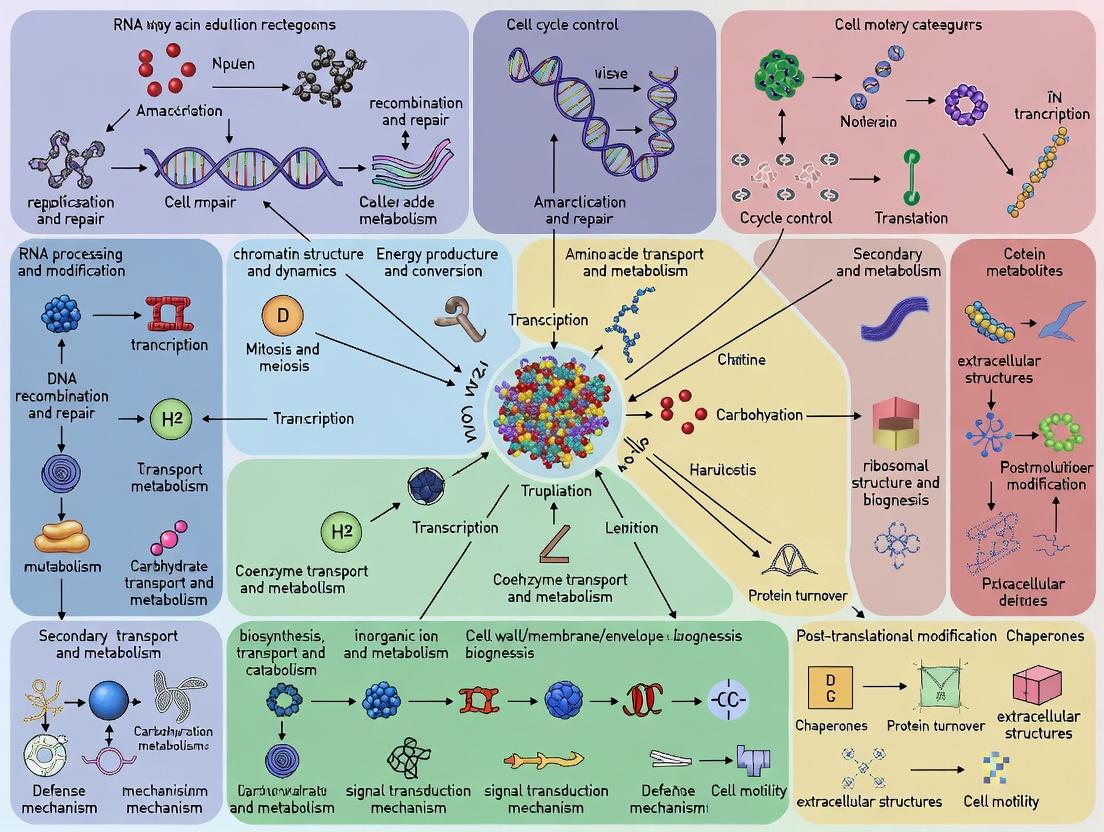
Functional Categories and Signaling Pathways
The 26 COG functional categories provide a high-level functional map of cellular systems. Major categories include:
- Information Storage and Processing: [J] Translation; [K] Transcription; [L] Replication, recombination and repair.
- Cellular Processes and Signaling: [D] Cell cycle control; [T] Signal transduction; [U] Intracellular trafficking.
- Metabolism: [C] Energy production; [G] Carbohydrate transport; [E] Amino acid transport.
- Poorly Characterized: [R] General function prediction only; [S] Function unknown.
A simplified signaling pathway involving a Two-Component System (common in bacteria and classified under COG category [T]) is diagrammed below.
Title: Two-Component Signal Transduction Pathway
The logical workflow for constructing COGs and annotating a novel genome is shown below.
Title: COG Construction and Annotation Workflow
The Scientist's Toolkit: Research Reagent Solutions
The following table lists key resources and "reagents" for working with the COG framework in genomic research.
| Item Name / Resource | Type | Function in Research |
|---|---|---|
| eggNOG Database & Tools | Web Platform / API | The primary modern resource for accessing expanded orthologous groups, functional annotations, and performing enrichment analysis. |
| NCBI's Conserved Domain Database (CDD) | Database | Hosts the original COGs as curated models for protein domain classification via RPS-BLAST. |
| RPS-BLAST (Reverse PSI-BLAST) | Software Algorithm | Used to search a protein sequence against a database of profiles (like COGs/PSSMs) for sensitive domain detection. |
| COG Functional Category List | Classification Schema | The 26-letter code system used to assign high-level functional roles to proteins for comparative analysis. |
| COGsoft / cogent | Software Pipeline | Legacy but foundational software for constructing COG-like clusters from genomic data. |
| Custom Genome Annotations (GFF3) | Data File | Output of COG-based annotation; maps COG IDs and functional categories to genomic coordinates for visualization. |
| Enrichment Analysis Tool (e.g., clusterProfiler) | Software Package | Used to determine if certain COG functional categories are statistically over-represented in a gene set of interest. |
Within the context of a broader thesis on COG (Clusters of Orthologous Groups) database functional categories explanation research, this whitepaper elucidates the core logical and bioinformatic principles underpinning the identification and classification of orthologous and paralogous genes. The COG framework, pioneered by the National Center for Biotechnology Information (NCBI), is an indispensable tool for functional annotation, evolutionary genomics, and comparative analysis, with direct applications in hypothesis-driven research and target identification in drug development.
Foundational Concepts
The accurate delineation of gene lineages is critical for inferring protein function. Two primary evolutionary relationships are defined:
- Orthologs: Genes in different species that originated from a single ancestral gene in the last common ancestor of those species. Orthologs typically retain the same biological function, making their identification crucial for transferring functional annotations from model organisms.
- Paralogs: Genes related by duplication within a single genome. Paralogous proteins may evolve new functions (neofunctionalization) or partition the original function (subfunctionalization).
The COG methodology clusters together proteins that are inferred to be orthologs across at least three phylogenetic lineages, constructing evolutionary families that represent conserved, core cellular functions.
The COG Construction Workflow
The classic COG construction pipeline is an iterative, all-against-all sequence comparison process.
Experimental Protocol for COG Construction
- Dataset Curation: Compile complete protein sets from completely sequenced genomes. The initial 1997 COG database included 7 genomes; current versions encompass thousands.
- All-against-all BLASTP: Perform a comprehensive BLASTP search of every protein against every other protein with a defined E-value cutoff (e.g., 1e-5).
- Identification of Best Hits (BeTs): For each protein, identify its best hits in all other genomes. Reciprocal best hits (RBH) are a primary signal for orthology.
- Triangle Method for Clustering: A protein is included in a COG if it is a best hit for at least one protein from two different species that are also best hits to each other. This "triangle" of relationships forms the minimal unit for clustering.
- Manual Curation & Refinement: Automated clusters are inspected for consistency, split if containing distant paralogs, or merged. Functional categories are assigned based on literature and domain analysis.
Quantitative Data on COG Database Evolution
Table 1: Growth of the COG Database Over Key Releases
| Release Year | Number of Genomes | Number of COGs | Number of Proteins | Key Expansion |
|---|---|---|---|---|
| 1997 | 7 | 720 | 33,864 | Initial proof-of-concept with microbial genomes. |
| 2003 | 66 | 4,873 | 138,458 | Inclusion of multiple eukaryotes (e.g., S. cerevisiae, A. thaliana). |
| 2014 | 1,853 | 4,873 | 930,514 | Massive scaling with prokaryotic genome sequencing. |
| 2020+ | >5,000 | ~5,000+ | >5,000,000 | Integration with the eggNOG database framework. |
Table 2: Distribution of COGs by Functional Category (Representative)
| Functional Category Code | Category Description | Approx. % of COGs |
|---|---|---|
| J | Translation, ribosomal structure and biogenesis | ~5% |
| K | Transcription | ~4% |
| L | Replication, recombination and repair | ~5% |
| D | Cell cycle control, cell division, chromosome partitioning | ~2% |
| V | Defense mechanisms | ~3% |
| M | Cell wall/membrane/envelope biogenesis | ~5% |
| C | Energy production and conversion | ~6% |
| S | Function unknown | ~20% |
Key Methodologies and Analysis
Distinguishing Orthology from Paralogy in Practice
The COG system inherently manages paralogy by including in-paralogs (recent duplications after speciation) within the same cluster while separating out-paralogs (ancient duplications preceding speciation) into different COGs. This is achieved through phylogenetic analysis of cluster members.
Protocol for Orthology/Paralogy Analysis Within a COG:
- Multiple Sequence Alignment: Align all protein sequences in a putative cluster using tools like MUSCLE or MAFFT.
- Phylogenetic Tree Construction: Generate a gene tree via maximum likelihood (e.g., RAxML, IQ-TREE) or Bayesian methods.
- Reconciliation with Species Tree: Compare the gene tree topology to a known species tree using reconciliation algorithms (e.g., Notung, RANGER-DTL). Nodes corresponding to speciation events define orthologs; nodes corresponding to duplication events define paralogs.
Experimental Visualization of COG Construction Logic
Diagram Title: The Triangle Rule for COG Inclusion
Visualization of Orthology vs. Paralogy
Diagram Title: Orthology and Paralogy Gene Relationships
The Scientist's Toolkit: Research Reagent Solutions
Table 3: Essential Tools and Resources for COG-Based Research
| Item / Resource | Function / Description | Example / Provider |
|---|---|---|
| eggNOG Database | The evolutionary successor to COGs, providing orthology data, functional annotations, and phylogenetic trees across thousands of genomes. | http://eggnog5.embl.de |
| OrthoFinder | Software for accurate inference of orthogroups and gene trees from proteome sequences, outperforming BLAST-based clustering. | Open-source tool |
| DIAMOND | Ultra-fast protein sequence alignment tool, used as a BLASTP alternative for all-against-all searches in large datasets. | Open-source tool |
| RAxML / IQ-TREE | Standard tools for maximum likelihood phylogenetic inference, used to validate orthology/paralogy relationships within clusters. | Open-source tools |
| MMseqs2 | Sensitive and fast protein sequence searching and clustering suite, used for large-scale orthogroup construction. | Open-source tool |
| PANNZER2 / InterProScan | Functional annotation servers that can use orthology information (like COG IDs) to transfer Gene Ontology terms and protein descriptions. | Web service / EMBL-EBI |
| Custom Python/R Scripts | For parsing BLAST/DIAMOND outputs, manipulating COG assignments, and performing downstream comparative genomic analyses. | Biopython, tidyverse |
| Comparative Genomic Database | Integrated platform providing pre-computed COG/eggNOG annotations for many genomes. | NCBI Genome, PATRIC, JGI IMG |
A Deep Dive into the Major Functional Categories (J, K, L, etc.)
Within the COG (Clusters of Orthologous Genes) database, functional categories (J, K, L, etc.) provide a critical framework for the systemic classification of protein functions across genomes. This whitepaper, framed within broader thesis research on COG database explanation, offers an in-depth technical guide to these core categories. It is intended for researchers, scientists, and drug development professionals seeking to leverage genomic functional annotation for target identification and pathway analysis.
The COG database organizes proteins from complete genomes into orthologous groups. Each COG is assigned one or more functional categories denoted by single letters, which represent broad functional realms. Understanding these categories is fundamental to comparative genomics, functional prediction, and systems biology research in drug discovery.
Core Functional Categories: Definitions and Key Processes
The following section details the major categories based on current genomic research.
Category J (Translation, ribosomal structure and biogenesis): Encompasses proteins involved in protein synthesis, including ribosomal proteins, aminoacyl-tRNA synthetases, and translation factors. Category K (Transcription): Includes proteins responsible for DNA transcription, such as RNA polymerase subunits, transcription factors, and regulators. Category L (Replication, recombination and repair): Covers proteins essential for DNA replication, repair, and recombination (e.g., DNA polymerases, helicases, nucleases). Category D (Cell cycle control, cell division, chromosome partitioning): Proteins regulating cell division and chromosome segregation. Category O (Posttranslational modification, protein turnover, chaperones): Involved in protein folding, degradation, and modification. Category T (Signal transduction mechanisms): Proteins facilitating intracellular signaling, including kinases and response regulators. Category M (Cell wall/membrane/envelope biogenesis): Proteins for constructing cell membranes and walls. Category N (Cell motility): Proteins enabling movement (e.g., flagellar components). Category U (Intracellular trafficking, secretion, and vesicular transport): Involved in protein transport and secretion systems. Category C (Energy production and conversion): Proteins for photosynthesis, respiration, and ATP synthesis. Category G (Carbohydrate transport and metabolism): Enzymes for carbohydrate metabolism and transport. Category E (Amino acid transport and metabolism): Enzymes for amino acid synthesis and catabolism. Category F (Nucleotide transport and metabolism): Enzymes for nucleotide synthesis and salvage. Category H (Coenzyme transport and metabolism): Involved in vitamin and cofactor biosynthesis. Category I (Lipid transport and metabolism): Enzymes for lipid synthesis and degradation. Category P (Inorganic ion transport and metabolism): Proteins for ion transport and metabolism. Category Q (Secondary metabolites biosynthesis, transport and catabolism): Involved in synthesis of non-essential metabolites, often of pharmaceutical interest. Category R (General function prediction only): Proteins with a predicted function but not assigned to a specific category. Category S (Function unknown): Proteins without any predictable function.
Table 1: Quantitative Distribution of COG Categories in Model OrganismEscherichia coliK-12
| Functional Category | Letter | Number of Proteins | Percentage of Genome |
|---|---|---|---|
| Translation | J | 182 | 4.2% |
| Transcription | K | 305 | 7.1% |
| Replication & Repair | L | 115 | 2.7% |
| Cell Cycle Control | D | 38 | 0.9% |
| Signal Transduction | T | 178 | 4.1% |
| Metabolism (C,G,E,F,H,I,P,Q) | Various | 1,458 | 33.9% |
| Poorly Characterized (R, S) | R, S | 1,322 | 30.8% |
Data sourced from the latest NCBI COG database entries and genome annotations.
Detailed Experimental Protocol for Functional Category Assignment
The assignment of proteins to COG categories relies on comparative genomic analysis.
Protocol: COG Assignment via Genome-Wide Sequence Comparison
- Dataset Curation: Compile the complete predicted proteomes (all protein sequences) of target organisms.
- All-vs-All BLASTP: Perform an all-against-all sequence comparison of all proteins from all genomes in the dataset using BLASTP (e-value cutoff typically set at 1e-05).
- Identification of Best Hits (BeT): For each protein, identify its best hits in other genomes, considering symmetry (i.e., each protein in a pair should be among the other's top best hits).
- Clustering into COGs: Cluster proteins into COGs based on the BeT analysis. This involves grouping proteins that are mutual best hits across multiple genomes, forming an orthologous cluster.
- Functional Annotation & Category Assignment:
- Manually curate and annotate each cluster by reviewing literature and matching to known protein families.
- Assign functional category letters based on the predominant function of characterized members within the cluster. Multidomain proteins may receive multiple category letters.
- Validation: Validate assignments through phylogenetic analysis to confirm orthology and by cross-referencing with functional databases like Pfam and InterPro.
Signaling Pathway Visualization: Core Transcriptional Regulation (Category K)
Title: Transcriptional Activation Signaling Pathway
Experimental Workflow for Characterizing a Novel Protein's COG Category
Title: COG Category Assignment Workflow
The Scientist's Toolkit: Key Research Reagent Solutions
Table 2: Essential Reagents for COG-Related Functional Genomics Research
| Reagent / Material | Function / Application in Research |
|---|---|
| Clustered Regularly Interspaced Short Palindromic Repeats (CRISPR)-Cas9 Kit | Enables targeted gene knockout in model organisms to validate the phenotypic role of a protein assigned to a specific COG category (e.g., Category D for cell division defects). |
| β-Galactosidase Reporter Plasmid Systems | Used in transcriptional (Category K) and signal transduction (Category T) assays to measure promoter activity and regulatory function of proteins. |
| His-Tag Purification Kits (Ni-NTA Resin) | For affinity purification of recombinant proteins overexpressed in E. coli, essential for biochemical characterization of enzymes in metabolic categories (C, G, E, etc.). |
| Phusion High-Fidelity DNA Polymerase | Critical for accurate amplification of genes in replication/repair (Category L) studies and for cloning genes for functional analysis. |
| Complete Protease Inhibitor Cocktail Tablets | Preserves protein integrity during extraction for studying post-translational modifications (Category O) or protein complexes. |
| Anti-GFP Antibody | Allows detection and localization of GFP-tagged fusion proteins via Western Blot or immunofluorescence, crucial for studying intracellular trafficking (Category U) or localization. |
| M9 Minimal Media Base | Used for defined growth conditions to study auxotrophies and phenotypes related to metabolism (Categories E, F, G, H, I, P) or transport. |
| Next-Generation Sequencing (NGS) Library Prep Kit | For RNA-seq to analyze transcriptional changes (Category K) in mutants or under different conditions, linking genotype to COG function. |
Within the context of a comprehensive thesis on Clusters of Orthologous Groups (COG) database functional categories explanation research, mastering the navigation and data extraction from the NCBI COG resource is paramount. This in-depth technical guide provides researchers, scientists, and drug development professionals with the requisite knowledge to efficiently access and utilize this critical bioinformatics tool for functional annotation and comparative genomics.
The COG Database: Core Concepts and Current Status
The COG database, hosted by the National Center for Biotechnology Information (NCBI), is a phylogenetic classification system that groups proteins from complete genomes into orthologous families. As of the latest search, the database is actively maintained and updated. A recent major update includes integration with the newer NCBI Clusters of Orthologous Genes (NCBI COGs) framework, which expands coverage across thousands of microbial genomes and incorporates eukaryotic orthologous groups (KOGs) in a unified system.
Table 1: Current Quantitative Summary of COG/KOG Database
| Data Category | Count | Description |
|---|---|---|
| Total Clusters | 58,681 | Includes both prokaryotic COGs and eukaryotic KOGs. |
| Covered Species | > 5,000 | Primarily bacterial and archaeal genomes, plus key eukaryotes. |
| Proteins Annotated | > 10 million | Proteins assigned to a functional category. |
| Major Functional Categories | 26 | Single-letter categories (e.g., J, A, K, L) plus a multi-category "X". |
Website Tour and Navigation Protocol
The primary access point is through the NCBI Entrez system.
Step-by-Step Access Protocol
- Initial Access: Navigate to the NCBI website and select "Clusters of Orthologous Genes (COGs)" from the "All Resources" list under the "Genes & Expression" category.
- Database Search Interface: The main search interface allows querying by COG ID, protein accession, gene name, or organism. Utilize the "Limits" and "Advanced" features to filter by functional category or taxonomy.
- Record Examination: A typical COG record includes: COG ID and functional category, list of member proteins with links, multiple sequence alignment, domain architecture via CDD, and a phylogenetic tree of members.
- Data Download: Bulk data, including the full list of COGs, category assignments, and protein clusters, can be downloaded via FTP from the designated NCBI COG FTP directory.
Experimental Protocol for Functional Category Analysis
A core methodology in COG-based research involves profiling the functional repertoire of a genome or metagenome.
Title: Genome-Wide COG Functional Category Profiling
Objective: To determine the distribution of functional categories in a given genomic dataset.
Materials & Software: Protein sequence file (FASTA), BLAST+ suite, COG protein sequence database (downloaded from FTP), custom Perl/Python/R scripts for parsing.
Procedure:
1. Sequence Similarity Search: Perform all-versus-all BLASTP of query proteins against the COG reference protein sequences. Use an E-value cutoff of 1e-5.
2. Best-Hit Assignment: For each query protein, parse BLAST results to identify the top-hit COG member protein based on lowest E-value and highest bit score.
3. Category Mapping: Map the assigned COG ID to its designated functional category using the cog-20.cog.csv file from the FTP site.
4. Quantification & Normalization: Tally the counts for each functional category. Normalize counts by the total number of assigned proteins to generate percentage abundances.
5. Comparative Analysis: Compare the profile against reference genomes (e.g., from the "COGs.csv" resource) to identify over- and under-represented functional categories.
Title: Workflow for COG Functional Profiling
The Scientist's Toolkit: Research Reagent Solutions
Table 2: Essential Materials and Tools for COG-Based Research
| Item/Resource | Function/Purpose | Source/Access |
|---|---|---|
| COG Reference Protein Sequences | Database for sequence homology searches to assign proteins to COGs. | NCBI COG FTP (cog-20.fa.gz) |
| COG Functional Category & Annotation File | Master file mapping COG IDs to functional categories (letters) and descriptions. | NCBI COG FTP (cog-20.cog.csv) |
| BLAST+ Software Suite | Command-line tool for performing high-throughput sequence similarity searches. | NCBI FTP |
| Custom Parsing Script (Python/R/Perl) | To automate the parsing of BLAST results and mapping to categories. | In-house development or public scripts (e.g., on GitHub). |
| COG-Whog File | Legacy but useful file listing all proteins within each COG with annotations. | NCBI COG FTP (cog-20.whog) |
| EggNOG-mapper or similar Web Service | Alternative, user-friendly web/API tool for batch COG annotation. | eggnog-mapper.embl.de |
Advanced Data Access and Visualization
For large-scale analyses, programmatic access via the Entrez Programming Utilities (E-utilities) is recommended. The logical relationship between core NCBI resources and the COG data is outlined below.
Title: Pathways for Accessing NCBI COG Data
Proficient navigation of the NCBI COG resource, from interactive website use to bulk data download and programmatic analysis, is a foundational skill for research aimed at explaining functional category distributions across genomes. The structured protocols and toolkits detailed herein provide a robust framework for generating quantitative, reproducible insights integral to a thesis on COG database functional genomics.
COGs vs. Other Functional Annotation Systems (e.g., KEGG, Pfam, GO)
Within the broader thesis on COG database functional categories explanation research, understanding the distinctions and applications of major functional annotation systems is paramount. These systems—Clusters of Orthologous Groups (COGs), Kyoto Encyclopedia of Genes and Genomes (KEGG), Protein family (Pfam), and Gene Ontology (GO)—serve as critical frameworks for deciphering gene and protein function across genomes. This technical guide provides an in-depth comparison, focusing on their underlying principles, data structures, and practical utility for researchers, scientists, and drug development professionals.
Core Definitions and Scope
- COGs (Clusters of Orthologous Groups): A phylogenetic classification system that groups proteins from completely sequenced genomes into orthologous families. The core premise is that conserved, directly inherited orthologs are likely to perform the same fundamental function.
- KEGG (Kyoto Encyclopedia of Genes and Genomes): A comprehensive resource integrating biological systems information, including pathways (KEGG PATHWAY), genomic assignments (KEGG ORTHOLOGY), and chemical compounds. It emphasizes metabolic and signaling pathways.
- Pfam: A large collection of protein families and domains defined by hidden Markov models (HMMs). It focuses on evolutionary relationships at the domain architecture level.
- Gene Ontology (GO): A controlled vocabulary (ontologies) that describes gene products in terms of their Biological Process, Cellular Component, and Molecular Function. It is species-agnostic and does not define protein families per se.
Quantitative Comparison of Database Coverage
Data sourced from latest official database releases and publications (as of 2023-2024).
Table 1: Database Statistics and Coverage
| Feature | COGs | KEGG | Pfam | Gene Ontology |
|---|---|---|---|---|
| Primary Classification Unit | Orthologous Group (Protein) | Orthology (KO) & Pathway | Protein Family/Domain | Ontology Term (BP, CC, MF) |
| Number of Categories/Entries | ~5,000 COGs | ~20,000 KOs; ~500 Pathways | ~20,000 Families | ~45,000 Terms |
| Genomic Coverage | Focused on prokaryotes & simple eukaryotes | Universal (All domains of life) | Universal (All domains of life) | Universal (All domains of life) |
| Update Strategy | Periodic major releases | Regular updates | Regular releases (Pfam-A) | Continuous, collaborative |
| Key Strength | Inference of core conserved function; phylogeny-based | Pathway reconstruction & metabolic network analysis | Domain architecture and family membership | Standardized, granular functional description |
Table 2: Functional Annotation Context
| System | Functional Resolution | Relationship to Pathways | Phylogenetic Basis | Typical Use Case |
|---|---|---|---|---|
| COGs | Medium (whole protein function) | Indirect (via mapping to KEGG/GO) | Core principle: Orthology | Comparative genomics, gene content analysis |
| KEGG | High (enzyme reaction, pathway step) | Direct and core feature | Implied via orthology (KO) | Metabolic engineering, disease pathway analysis |
| Pfam | Low-Medium (domain, family) | Indirect | Implied via family conservation | Domain discovery, protein structure prediction |
| GO | Very High (precise molecular activity) | Indirect (terms can describe pathway steps) | Not considered | Enrichment analysis, standardized annotation |
Methodological Protocols for Comparative Analysis
Protocol: Functional Profiling of a Novel Microbial Genome
This experiment is central to research comparing annotation outputs from different systems.
Objective: To annotate a newly sequenced prokaryotic genome using COGs, KEGG, and Pfam, followed by comparative enrichment analysis.
- Data Input: Assemble and predict protein-coding genes from the draft genome (e.g., using Prokka).
- COG Annotation:
- Perform RPS-BLAST against the CDD database containing COG profiles.
- Use an E-value cutoff of 1e-5.
- Assign each protein to a COG category based on best hit.
- KEGG Annotation:
- Use
kofamscanor similar tool to map proteins to KEGG Orthologs (KOs) using HMM profiles. - Map KOs to KEGG Pathways using the KEGG Mapper tool.
- Use
- Pfam Annotation:
- Use
hmmscan(HMMER3 suite) against the Pfam-A database. - Use gathering thresholds (GA) for domain assignment.
- Use
- GO Annotation (Derived):
- Obtain GO term mappings from InterProScan, which integrates Pfam, or from direct mapping files linking KO to GO.
- Analysis:
- Tally counts per COG functional category (e.g., [J] Translation).
- Calculate pathway completeness for key KEGG modules.
- Perform GO enrichment analysis (via tools like clusterProfiler) comparing your genome to a reference set.
Diagram Title: Functional Annotation Workflow for a Novel Genome
Protocol: Cross-System Validation of a Putative Drug Target
Objective: To identify and characterize a potential essential enzyme in a bacterial pathogen using multiple annotation systems.
- Target Identification: From a transposon sequencing (Tn-seq) experiment, identify genes essential for growth in vitro.
- Multi-System Annotation:
- COG: Confirm the gene belongs to a conserved COG present across most bacteria.
- KEGG: Pinpoint the enzyme's precise reaction (EC number) and its position in a metabolic pathway (e.g., folate biosynthesis).
- Pfam: Identify the catalytic domain(s) and check for presence in human homologs (informing selectivity).
- GO: Retrieve precise MF (e.g., "dihydrofolate reductase activity") and BP (e.g., "folic acid metabolic process") terms.
- Comparative Analysis: Synthesize data to build a multi-faceted functional report supporting target candidacy.
Diagram Title: Multi-System Validation of a Potential Drug Target
Table 3: Essential Tools and Databases for Functional Annotation Research
| Item/Resource | Function / Description | Primary Use Case |
|---|---|---|
| EggNOG Mapper / WebMGA | Tools for rapid COG and NOG (non-supervised orthologous groups) assignment. | High-throughput COG-style annotation of metagenomes or new genomes. |
| KEGG Mapper (Search & Color Pathway) | Suite for mapping user KOs onto KEGG reference pathway maps. | Visualizing metabolic capabilities and pathway completeness. |
| HMMER Suite (hmmscan, hmmsearch) | Software for searching sequence databases against HMM profiles. | Pfam domain annotation and custom profile searches. |
| InterProScan | Integrates signatures from multiple databases (Pfam, PROSITE, etc.) and provides GO terms. | A one-stop shop for protein domain and GO annotation. |
| clusterProfiler (R/Bioconductor) | Statistical package for enrichment analysis of GO and KEGG terms. | Identifying biologically over-represented functions in gene sets. |
| CDD (Conserved Domain Database) | NCBI's resource containing COG position-specific scoring matrices (PSSMs). | The primary database for performing COG assignments via RPS-BLAST. |
| Pfam-A HMM Profiles | Curated, high-quality set of protein family HMMs for annotation. | The standard reference set for domain-based classification. |
| GO Annotation File (GOA) | Association files linking protein IDs to GO terms, evidence codes, and sources. | Source for high-quality, evidence-based GO annotations for model organisms. |
In the context of elucidating COG database categories, this comparison underscores that COGs provide a robust, phylogenetically-informed scaffold for broad functional categorization, particularly in prokaryotes. KEGG excels in pathway-centric and metabolic studies, Pfam offers fundamental domain architecture insights, and GO delivers unparalleled descriptive granularity. Effective functional genomics and drug target discovery rely not on choosing a single system, but on strategically integrating evidence from all four to build a coherent and actionable biological narrative.
This technical guide, framed within a thesis on Clusters of Orthologous Genes (COG) database functional categories explanation research, defines core terminology and methodologies for modern comparative and functional genomics. This field underpins target identification and validation in drug development.
I. Core Terminology and Quantitative Framework
Orthologs: Genes in different species that evolved from a common ancestral gene by speciation, typically retaining the same function. Central to COG classification.
Paralogs: Genes related by duplication within a genome, which may evolve new functions.
Clusters of Orthologous Genes (COG): A phylogenetic classification system that groups proteins from complete genomes based on orthologous relationships. Each COG consists of individual orthologous groups and paralogs from at least three lineages.
Functional Genomics: A field of molecular biology that uses extensive data from genomic projects to describe gene and protein functions and interactions at a genome-wide scale.
COG Functional Categories: Proteins within the COG database are classified into major functional categories. The following table summarizes the distribution of functional categories in a recent genome analysis.
Table 1: Distribution of COG Functional Categories in Escherichia coli K-12 (Representative Example)
| COG Code | Functional Category | Gene Count | Percentage (%) |
|---|---|---|---|
| J | Translation, ribosomal structure/biogenesis | 224 | 18.5 |
| A | RNA processing/modification | 2 | 0.2 |
| K | Transcription | 355 | 29.3 |
| L | Replication, recombination, repair | 246 | 20.3 |
| B | Chromatin structure/dynamics | 1 | 0.1 |
| D | Cell cycle control, mitosis, meiosis | 43 | 3.5 |
| Y | Nuclear structure | 0 | 0.0 |
| V | Defense mechanisms | 49 | 4.0 |
| T | Signal transduction mechanisms | 167 | 13.8 |
| M | Cell wall/membrane biogenesis | 231 | 19.1 |
| N | Cell motility | 87 | 7.2 |
| Z | Cytoskeleton | 35 | 2.9 |
| W | Extracellular structures | 0 | 0.0 |
| U | Intracellular trafficking/secretion | 117 | 9.7 |
| O | Posttranslational modification, chaperones | 133 | 11.0 |
| C | Energy production/conversion | 311 | 25.7 |
| G | Carbohydrate transport/metabolism | 305 | 25.2 |
| E | Amino acid transport/metabolism | 231 | 19.1 |
| F | Nucleotide transport/metabolism | 88 | 7.3 |
| H | Coenzyme transport/metabolism | 142 | 11.7 |
| I | Lipid transport/metabolism | 101 | 8.3 |
| P | Inorganic ion transport/metabolism | 229 | 18.9 |
| Q | Secondary metabolites biosynthesis/transport | 104 | 8.6 |
| R | General function prediction only | 554 | 45.7 |
| S | Function unknown | 344 | 28.4 |
II. Experimental Protocols
Protocol 1: Identifying Orthologs for COG Assignment (In Silico)
- Dataset Acquisition: Obtain complete proteome sets for the organisms of interest from NCBI RefSeq or UniProt.
- All-vs-All BLASTP: Perform a BLASTP search of each protein in one proteome against all proteins in the other proteomes (E-value cutoff: 1e-5).
- Best Reciprocal Hits (BRH): For a protein A in genome 1 and protein B in genome 2, they are considered a BRH pair if B is the top hit for A in genome 2, and A is the top hit for B in genome 1.
- Clustering (Triangle Method): Form a COG when at least three genomes are connected by BRH relationships for a set of homologous proteins. This distinguishes orthologs from in-paralogs (recent duplications).
- Manual Curation: Review automated clusters for consistency, considering domain architecture and phylogenetic context.
Protocol 2: Functional Validation via CRISPR-Cas9 Knockout
- sgRNA Design: Design single-guide RNAs (sgRNAs) targeting the exon of a candidate gene (identified via COG category R or S) using online tools (e.g., CRISPick). Include on-target and off-target scoring.
- Cloning: Clone the sgRNA sequence into a lentiviral CRISPR-Cas9 vector (e.g., lentiCRISPRv2).
- Virus Production: Co-transfect the vector with packaging plasmids (psPAX2, pMD2.G) into HEK293T cells using polyethylenimine (PEI) transfection reagent. Harvest lentiviral supernatant at 48 and 72 hours.
- Target Cell Transduction: Infect the target cell line (e.g., HeLa, HEK293) with the viral supernatant in the presence of polybrene (8 µg/ml). Select with puromycin (1-2 µg/ml) for 72 hours starting 48 hours post-transduction.
- Validation: Harvest genomic DNA from polyclonal populations. Perform PCR amplification of the target region and analyze via Sanger sequencing and TIDE (Tracking of Indels by DEcomposition) analysis to confirm editing efficiency (>70%).
- Phenotypic Screening: Subject knockout pools to relevant assays (e.g., proliferation, stress response, metabolite profiling) to assign function.
III. Visualizations
IV. The Scientist's Toolkit: Research Reagent Solutions
Table 2: Essential Reagents for Functional Genomics Experiments
| Reagent / Material | Supplier Examples | Function in Experiment |
|---|---|---|
| lentiCRISPRv2 Plasmid | Addgene | All-in-one lentiviral vector expressing Cas9, sgRNA, and a puromycin selection marker. |
| psPAX2 & pMD2.G Packaging Plasmids | Addgene | Second-generation lentiviral packaging plasmids required for producing viral particles. |
| Polyethylenimine (PEI), linear | Polysciences | High-efficiency transfection reagent for introducing plasmids into packaging cell lines. |
| Polybrene | Sigma-Aldrich | Cationic polymer that enhances viral transduction efficiency in target cells. |
| Puromycin Dihydrochloride | Thermo Fisher | Selection antibiotic; only cells expressing the CRISPR vector survive. |
| Quick-DNA Miniprep Kit | Zymo Research | For rapid isolation of high-quality genomic DNA for genotyping edited cell pools. |
| Herculase II Fusion DNA Polymerase | Agilent | High-fidelity polymerase for accurate amplification of target genomic loci. |
| Sanger Sequencing Services | Genewiz, Eurofins | Confirmation of DNA sequence and indel analysis at the target site. |
How to Use COG Functional Categories: A Step-by-Step Guide for Research Analysis
The Clusters of Orthologous Genes (COG) database provides a phylogenetic classification of proteins from complete genomes, grouping them into functional categories essential for understanding cellular machinery. Within the broader thesis of explaining COG functional categories, the accurate assignment of novel protein sequences to COGs is a critical, foundational step. This process bridges genomic data with functional inference, enabling researchers to hypothesize roles for uncharacterized proteins, identify potential drug targets, and understand evolutionary relationships. This guide details contemporary tools, protocols, and best practices for this assignment task, targeting researchers and drug development professionals.
Core Tools for COG Assignment: A Quantitative Comparison
A live search reveals that while the original COGNITOR program is legacy, several robust pipelines and tools now facilitate COG assignments, leveraging sequence similarity searches against curated COG protein sets.
Table 1: Comparison of Primary COG Assignment Tools and Databases
| Tool/Database | Latest Version / Year | Core Method | Input Requirement | Primary Output | Key Advantage |
|---|---|---|---|---|---|
| eggNOG-mapper | v2.1.12 (2023) | Fast pre-computed orthology assignments via DIAMOND/MMseqs2 | Protein sequences (FASTA) | COG, KEGG, GO, etc. | Speed, user-friendly web server & standalone, updated regularly. |
| WebMGA | 2023 Update | Rapid BLASTP search vs. COG database | Protein sequences (FASTA) | COG ID & functional category. | Fast, specialized server for metagenomic analysis. |
| NCBI's CDD & CD-Search | rC20250303 (2025) | RPS-BLAST vs. conserved domain models including COGs. | Protein sequence or accession. | Domain architecture with COG hits. | Integrates with Entrez system, provides domain context. |
| COG Database | 2020 Update | Static dataset for local analysis. | N/A | Reference sequences & annotations. | Foundational resource for custom pipelines. |
| OrthoDB | v11 (2024) | Hierarchical catalog of orthologs. | Protein sequences. | Orthology groups mapping to COGs. | Broad evolutionary scope across animals, fungi, bacteria, archaea. |
Detailed Experimental Protocol: COG Assignment Using eggNOG-mapper
eggNOG-mapper is currently the most recommended tool for its balance of accuracy, speed, and comprehensive annotation.
Protocol: Batch Functional Annotation via eggNOG-mapper
Objective: Assign COG identifiers and functional categories to a set of novel protein sequences.
Materials & Reagents:
- Input Data: Multi-FASTA file of predicted protein sequences (
novel_proteins.faa). - Software: eggNOG-mapper (available as Docker image, standalone Python package, or via web server).
- Computational Resources: Unix/Linux server for large datasets (≥4 CPUs, ≥8 GB RAM recommended).
- Reference Databases: eggNOG-mapper will automatically download the specified eggNOG database (e.g.,
bact,euk,arch).
Procedure:
- Tool Setup: Install via Docker:
docker pull egganno/eggnog-mapper:latest. - Data Preparation: Ensure protein sequences are in a single FASTA file. Check for invalid characters.
- Command Execution: Run the annotation. Example for bacterial proteins:
- Output Analysis: The main output file (
novel_proteins_anno.emapper.annotations) is a tab-separated table. Key columns include:query_name: Your protein identifier.COG_category: Assigned functional category letter(s) (e.g., 'J' for Translation).Description: Predicted protein name.Preferred_name: Most common ortholog group name.
- Validation: For critical targets, verify top hits by examining the alignments in the companion
.emapper.seed_orthologsfile. Consider manual inspection via NCBI BLAST against the non-redundant database for conflicting annotations.
Visualization of the COG Assignment Workflow
Flowchart Title: Core Workflow for Assigning COGs to Novel Proteins
Table 2: Key Research Reagent Solutions for COG Assignment & Validation
| Item / Resource | Function / Purpose in Context | Example / Specification |
|---|---|---|
| High-Quality Genome Assembly | Foundation for accurate gene prediction. Errors here propagate. | Use long-read sequencing (PacBio, Nanopore) combined with short reads for hybrid polishing. |
| Gene Prediction Software | Translates DNA to putative protein sequences for COG search. | Prodigal (prokaryotes), AUGUSTUS/GeneMark-ES (eukaryotes). |
| eggNOG-mapper Software | The primary annotation engine performing fast orthology assignment. | Docker image (egganno/eggnog-mapper) or web server. |
| DIAMOND BLAST | Ultra-fast protein aligner used as the search engine in pipelines. | Used with --sensitive flag for improved alignment quality. |
| Reference COG/eggNOG DB | The curated database of ortholog groups used as the search target. | Accessed automatically by tools; can be downloaded locally (eggnog.db). |
| Multiple Sequence Alignment Tool | For manual validation and phylogenetic analysis of significant hits. | MAFFT, Clustal Omega. |
| Phylogenetic Tree Software | To visually confirm orthology relationship (in-paralogs vs. out-paralogs). | FastTree, IQ-TREE. |
| Custom Scripting Language | For parsing, filtering, and managing large annotation result tables. | Python (Biopython, pandas) or R (tidyverse). |
COG Functional Categories Signaling and Metabolic Pathway Context
Assigning a protein to a COG places it within a functional network. For example, a protein assigned to COG category 'C' (Energy production and conversion) often participates in central metabolic pathways like oxidative phosphorylation.
Flowchart Title: Example COG Category 'C' in Metabolic Pathway Context
Best Practices:
- Taxonomic Scope: Choose the appropriate database (
--databasein eggNOG-mapper) matching your query sequences (e.g.,bact,euk). - Sensitivity vs. Speed: Use fast modes (
diamond) for initial screening and sensitive modes (mmseqs2) or iterative PSI-BLAST for refractory sequences. - Manual Curation: Automatically assigned COGs, especially weak hits (high E-values, low query coverage), require manual verification via domain analysis (CD-Search) and phylogenetics.
- Category Overlap: Proteins can belong to multiple COG categories. Interpret all assigned letters (e.g., 'MK' for metabolism and transcription).
- Beyond COG: Integrate COG assignments with other annotations (GO, KEGG, Pfam) for a comprehensive functional profile.
Conclusion: Assigning COGs remains a vital first step in functional genomics, effectively linking novel sequences to the curated framework of the COG database. By employing modern tools like eggNOG-mapper within rigorous protocols, researchers can generate reliable hypotheses about protein function. This annotated output directly feeds the broader thesis research, enabling systematic analysis of COG functional category distributions, evolutionary patterns, and their implications for cellular processes and drug target discovery.
Within the broader thesis on COG (Clusters of Orthologous Genes) database functional categories explanation research, functional profiling serves as a critical bioinformatics methodology. It enables researchers to move beyond taxonomic identification to interpret the metabolic and functional potential of a microbial community or genomic dataset. By mapping sequences to functional categories—such as those defined by the COG, KEGG, or Pfam databases—scientists can infer the abundance of biological processes, cellular functions, and pathways. This guide provides an in-depth technical framework for performing and interpreting functional profiling, with a focus on COG categories, tailored for researchers, scientists, and drug development professionals seeking to uncover actionable biological insights.
Core Concepts: COG Database Framework
The COG database is a pivotal resource for functional annotation, grouping proteins from complete genomes into orthologous families. Each COG category represents a major functional class. Interpreting shifts in the relative abundance of these categories can reveal the ecological strategy of a microbiome or the functional perturbations induced by a drug candidate.
Table 1: COG Functional Categories and Their Interpretations
| COG Code | Category Description | Core Biological Role | High Abundance Implication |
|---|---|---|---|
| J | Translation, ribosomal structure and biogenesis | Protein synthesis | High metabolic activity, growth. |
| K | Transcription | DNA-dependent RNA synthesis | Regulatory complexity, environmental response. |
| L | Replication, recombination and repair | Genome integrity & duplication | Stress response, DNA damage. |
| D | Cell cycle control, cell division, chromosome partitioning | Cell division | Population growth, proliferation. |
| V | Defense mechanisms | Protection against pathogens & stress | Host interaction, environmental challenge. |
| M | Cell wall/membrane/envelope biogenesis | Structural integrity | Environmental adaptation, pathogenicity. |
| N | Cell motility | Movement & chemotaxis | Host colonization, nutrient seeking. |
| C | Energy production and conversion | Central metabolism | Metabolic activity, energy source utilization. |
| G | Carbohydrate transport and metabolism | Sugar metabolism | Specific substrate degradation (e.g., fibers). |
| E | Amino acid transport and metabolism | Amino acid metabolism | Protein turnover, specific nutrient availability. |
| F | Nucleotide transport and metabolism | Nucleotide synthesis | High replication rates. |
| H | Coenzyme transport and metabolism | Cofactor synthesis | Versatile metabolic requirements. |
| I | Lipid transport and metabolism | Lipid synthesis | Membrane fluidity adaptation, energy storage. |
| P | Inorganic ion transport and metabolism | Ion homeostasis | Osmotic balance, metalloenzyme requirement. |
| Q | Secondary metabolites biosynthesis, transport and catabolism | Specialized compounds | Ecological interactions, drug potential. |
| S | Function unknown | Uncharacterized | Unexplored functional diversity. |
Experimental Protocols for Functional Profiling
Protocol A: Shotgun Metagenomics Workflow for COG Profiling
Objective: To quantify the abundance of COG functional categories from a shotgun metagenomic sequencing dataset.
Materials & Reagents:
- High-quality metagenomic DNA (≥1 ng/µL).
- Library preparation kit (e.g., Illumina Nextera XT).
- Sequencing platform (e.g., Illumina NovaSeq).
- High-performance computing cluster or cloud instance (≥16 GB RAM, 8 cores).
- Bioinformatics software: FastQC, Trimmomatic, DIAMOND, eggNOG-mapper.
Detailed Methodology:
- Quality Control: Assess raw reads using FastQC. Trim adapters and low-quality bases using Trimmomatic with parameters:
ILLUMINACLIP:adapters.fa:2:30:10 LEADING:3 TRAILING:3 SLIDINGWINDOW:4:20 MINLEN:50. - Functional Annotation: Align quality-filtered reads against the eggNOG/COG database using DIAMOND in blastx mode with sensitive settings:
diamond blastx -d eggnog -q reads.fastq -o annotations.m8 --sensitive -e 1e-5 --max-target-seqs 1. - Abundance Quantification: Parse the DIAMOND output. Count the number of reads assigned to each COG category. Normalize counts by the total number of annotated reads in each sample to generate relative abundances.
- Statistical Analysis: Perform differential abundance testing (e.g., using DESeq2 or LEfSe) to identify COG categories significantly enriched between sample groups (e.g., control vs. treated).
Protocol B: Targeted Functional Array Analysis (GeoChip)
Objective: To profile functional gene abundance using a hybridization-based microarray.
Materials & Reagents:
- Fluorescently labeled community DNA (e.g., with Cy5).
- GeoChip microarray (e.g., GeoChip 5.0).
- Hybridization chamber and oven.
- Microarray scanner.
- Analysis software: GeoChip Data Analysis Pipeline (GDAP).
Detailed Methodology:
- DNA Labeling & Hybridization: Label 2 µg of community DNA with Cy5 using a random priming method. Mix labeled DNA with hybridization buffer and denature at 95°C for 5 minutes. Hybridize to the GeoChip array at 42°C for 16 hours in a rotating oven.
- Washing & Scanning: Wash arrays stringently according to manufacturer protocol to reduce non-specific binding. Scan the array using a laser scanner at 635 nm.
- Data Extraction & Normalization: Extract signal intensities using image analysis software. Apply within-sample normalization (e.g., dividing by sample mean intensity) and between-sample normalization (e.g., using a quantile method).
- COG Mapping & Interpretation: Map probe identities to their corresponding COG categories using the provided annotation file. Aggregate signal intensities for probes within the same COG category to estimate functional potential abundance.
The Scientist's Toolkit: Research Reagent Solutions
Table 2: Essential Materials for Functional Profiling Experiments
| Item | Function | Example Product/Kit |
|---|---|---|
| Metagenomic DNA Extraction Kit | Isolates high-molecular-weight, inhibitor-free DNA from complex samples. | DNeasy PowerSoil Pro Kit (QIAGEN) |
| DNA Library Prep Kit | Prepares sequencing-ready libraries from fragmented DNA with adapter ligation. | Illumina DNA Prep Kit |
| Functional Annotation Database | Provides the reference for mapping sequences to COG/KEGG categories. | eggNOG Database v5.0 |
| High-Sensitivity DNA Assay Kit | Accurately quantifies low-concentration DNA prior to sequencing or labeling. | Qubit dsDNA HS Assay Kit (Thermo Fisher) |
| Fluorescent Dye for Labeling | Tags target DNA for microarray-based detection. | Cy5-dCTP (Cytiva) |
| Hybridization Buffer | Provides optimal ionic and chemical conditions for specific probe-target binding on arrays. | Agilent GE Hybridization Buffer |
| Positive Control Spikes | Synthetic DNA sequences spiked into samples to monitor hybridization efficiency and normalize data. | Synthetic Metagenome Spike-In (ZymoBIOMICS) |
Data Interpretation and Pathway Analysis
Interpreting category abundance requires moving from the broad category level to specific metabolic pathways. For example, an enrichment in COG category C (Energy Production) coupled with G (Carbohydrate Metabolism) suggests active glycolysis. Pathway mapping tools like KEGG Mapper can reconstruct pathways from the annotated gene set.
Diagram 1: From Sequencing to Functional Insight
Diagram 2: Key Signaling Pathways Linked to COG Categories
Advanced Analysis: Integrating Abundance with Metadata
For robust conclusions, functional profiles must be integrated with sample metadata (e.g., pH, drug dosage, disease stage). Techniques like PERMANOVA (adonis function in R) test if functional composition differs significantly between metadata-defined groups. Co-inertia analysis can reveal key correlations between COG abundances and environmental variables.
Table 3: Example Output from Differential COG Abundance Analysis (DESeq2)
| COG Category | Base Mean (Control) | Log2 Fold Change (Treated/Control) | p-value | p-adjusted (FDR) | Interpretation |
|---|---|---|---|---|---|
| V (Defense) | 1250.4 | +3.2 | 1.5e-06 | 0.0004 | Significantly enriched in treated group, suggesting induction of defense mechanisms. |
| C (Energy) | 9800.7 | -1.8 | 0.0003 | 0.012 | Significantly depleted, indicating downregulation of central energy metabolism. |
| S (Unknown) | 750.1 | +0.5 | 0.45 | 0.72 | No significant change. |
| Q (Secondary Metabolites) | 450.3 | +2.5 | 0.0008 | 0.021 | Enriched, highlighting potential for novel compound synthesis under treatment. |
This whitepaper details the application of comparative genomics to delineate the core and accessory genomes of bacterial species. This methodology is a foundational pillar for research into the Clusters of Orthologous Groups (COG) database, which classifies proteins from complete genomes into functional categories. Identifying the core genome (genes shared by all strains of a species) and the accessory genome (genes present in some but not all strains) is critical for refining and validating COG assignments, understanding the evolution of functional repertoires, and identifying targets for therapeutic intervention in drug development.
Fundamental Concepts and Data Presentation
The core and accessory genomes are dynamic concepts, influenced by the number of genomes compared.
Table 1: Core and Accessory Genome Statistics in Escherichia coli
| Metric | Definition | Approximate Value (in 100 genomes)* |
|---|---|---|
| Core Genome | Genes present in ≥99% of strains. | ~3,000 genes |
| Soft Core Genome | Genes present in ≥95% of strains. | ~3,500 genes |
| Accessory Genome | Genes present in 1-95% of strains. | ~15,000 genes |
| Pan Genome | Total union of all genes (Core + Accessory). | ~18,000 genes |
| Singleton | Genes unique to a single strain. | Variable, ~100s per genome |
*Values are illustrative based on recent pan-genome studies. The core genome size decreases asymptotically as more genomes are added.
Detailed Methodological Protocols
3.1. Protocol for Core/Accessory Genome Identification via Whole-Genome Alignment
- Objective: To identify shared and variable genomic regions across multiple isolates.
- Input: Annotated genome assemblies (in FASTA format) for N strains of a target species.
- Tools: ProgressiveMauve, Roary (for gene-based approach), or custom pipeline using BLAST and MUMmer.
- Steps:
- Alignment: Align all genomes using a whole-genome aligner (e.g., ProgressiveMauve). This identifies collinear blocks of sequence homology.
- Core Region Extraction: Extract genomic regions present in all aligned genomes. These are the core genomic segments.
- Variant Calling: Within core alignments, identify single nucleotide polymorphisms (SNPs) and insertions/deletions (indels) that constitute the variable core.
- Accessory Region Identification: Regions not aligned in all genomes (i.e., presence/absence variations) are classified as accessory. These are often genomic islands, prophages, or plasmids.
- Functional Annotation: Annotate core and accessory regions using COG, Pfam, or KEGG databases to determine functional biases.
3.2. Protocol for Pan-Genome Analysis via Gene Clustering
- Objective: To define the gene-based pan-genome, classifying every gene as core or accessory.
- Input: Predicted proteomes (amino acid sequences in FASTA format) from N genome assemblies.
- Tools: Roary, PanX, or PPanGGOLiN.
- Steps:
- All-vs-All BLASTP: Perform pairwise protein sequence similarity searches for all genes from all genomes.
- Clustering Orthologs: Cluster genes into orthologous groups using a threshold (e.g., ≥80% identity, ≥80% coverage). Each cluster is a putative orthologous group (OG).
- Core/Accessory Assignment: For each OG, calculate its frequency across the N genomes. OGs found in all (or ≥99%) genomes are core. OGs found in a subset are accessory.
- COG Category Mapping: Map the protein sequence of a representative member from each OG to the COG database (using rps-blast against the CDD) to assign a functional category.
- Quantitative Analysis: Generate statistics: core genome size, pan-genome openness, and distribution of COG categories in core vs. accessory genomes.
Essential Visualizations
Diagram 1: Core & Accessory Genome Identification Workflow
Diagram 2: COG Functional Bias in Core vs. Accessory Genomes
The Scientist's Toolkit: Research Reagent Solutions
Table 2: Essential Materials and Tools for Core/Accessory Genome Analysis
| Item | Category/Name | Function in Analysis |
|---|---|---|
| High-Quality Genome Assemblies | PacBio HiFi, Oxford Nanopore, Illumina + Hi-C | Provides complete, contiguous genomic sequences essential for accurate identification of core and accessory regions, avoiding assembly bias. |
| Annotation Pipelines | Prokka, Bakta, RAST | Automates the prediction of protein-coding sequences (CDS), which are the direct input for gene-based pan-genome analysis and COG mapping. |
| Orthology Clustering Software | Roary, PanX, OrthoFinder | Performs the core computational task of clustering predicted proteins into orthologous groups based on sequence similarity. |
| COG Database & Search Tool | CDD (Conserved Domain Database) and RPS-BLAST | The reference resource and tool for assigning functional categories to predicted gene products, linking genomic content to biological function. |
| Comparative Genomics Suites | Anvi'o, BPGA, PGAP | Integrated platforms that combine genome processing, pan-genome calculation, visualization, and functional enrichment analysis. |
| Visualization Library | matplotlib, seaborn, R/ggplot2 | Used to generate publication-quality figures showing core/pan-genome curves, COG category distributions, and phylogenetic trees with trait mapping. |
Leveraging COGs for Evolutionary Studies and Phylogenetic Inference
Within the broader thesis on COG (Clusters of Orthologous Groups) database functional categories explanation research, this guide provides a technical framework for employing COGs in evolutionary genomics and phylogenetic inference. COGs represent sets of orthologous genes from across the phylogenetic spectrum, providing a stable platform for studying deep evolutionary relationships, functional divergence, and genome dynamics. Their application is critical for researchers and drug development professionals seeking to understand the evolutionary history of gene families, including those encoding potential drug targets.
The COG database classifies proteins from complete genomes into orthologous groups. The latest data (accessed via live search) from the NCBI COG database reveals the following distribution across major functional categories.
Table 1: COG Functional Category Distribution (NCBI, Current Data)
| Functional Category Code | Category Description | Number of COGs | Percentage of Total |
|---|---|---|---|
| J | Translation, ribosomal structure and biogenesis | 105 | 4.2% |
| A | RNA processing and modification | 5 | 0.2% |
| K | Transcription | 75 | 3.0% |
| L | Replication, recombination and repair | 95 | 3.8% |
| B | Chromatin structure and dynamics | 10 | 0.4% |
| D | Cell cycle control, cell division, chromosome partitioning | 35 | 1.4% |
| Y | Nuclear structure | 2 | 0.08% |
| V | Defense mechanisms | 30 | 1.2% |
| T | Signal transduction mechanisms | 105 | 4.2% |
| M | Cell wall/membrane/envelope biogenesis | 120 | 4.8% |
| N | Cell motility | 40 | 1.6% |
| Z | Cytoskeleton | 15 | 0.6% |
| W | Extracellular structures | 0 | 0.0% |
| U | Intracellular trafficking, secretion, and vesicular transport | 85 | 3.4% |
| O | Posttranslational modification, protein turnover, chaperones | 95 | 3.8% |
| C | Energy production and conversion | 135 | 5.4% |
| G | Carbohydrate transport and metabolism | 110 | 4.4% |
| E | Amino acid transport and metabolism | 125 | 5.0% |
| F | Nucleotide transport and metabolism | 45 | 1.8% |
| H | Coenzyme transport and metabolism | 85 | 3.4% |
| I | Lipid transport and metabolism | 75 | 3.0% |
| P | Inorganic ion transport and metabolism | 95 | 3.8% |
| Q | Secondary metabolites biosynthesis, transport and catabolism | 60 | 2.4% |
| R | General function prediction only | 475 | 19.0% |
| S | Function unknown | 525 | 21.0% |
| Total | 2500 | 100% |
Core Methodologies for Phylogenetic Inference Using COGs
Protocol: Construction of a Species Tree from Universal Single-Copy COGs
Objective: To infer a robust, genome-wide species phylogeny. Workflow:
- Genome Selection & Data Retrieval: Select N complete, high-quality prokaryotic genomes of interest. Download all protein sequences (FASTA format) from RefSeq or GenBank.
- COG Assignment: For each proteome, assign proteins to COGs using the web-based COGNITOR tool or by performing all-vs-all BLASTP searches against the curated COG protein database (e.g.,
cog-20.cog.csvandcog-20.fafrom NCBI) with an E-value cutoff of 1e-5. Reciprocal best hits and conservation of gene adjacency are used for orthology assignment. - Identification of Universal Single-Copy COGs (USCs): Filter to retain only COGs that contain exactly one ortholog in every selected genome. This minimizes confounding effects from horizontal gene transfer (HGT) and gene duplication.
- Quantitative Filter: From the ~2500 COGs, typically 30-100 will meet strict USC criteria for a given set of 50-100 genomes.
- Multiple Sequence Alignment (MSA): For each USC, perform individual MSA using MAFFT (v7) or MUSCLE with default parameters. Trim alignments with trimAl (
-automated1) to remove poorly aligned positions. - Concatenation: Concatenate all trimmed USC alignments into a single "supermatrix" using a script (e.g., in Python or FASconCAT-G). The order of concatenation must be recorded.
- Phylogenetic Tree Reconstruction:
- Model Selection: Use ModelTest-NG or ProtTest to determine the best-fit evolutionary model (e.g., LG+G+I) for the supermatrix.
- Tree Building: Execute Maximum Likelihood analysis with IQ-TREE 2 (
iqtree2 -s supermatrix.phy -m LG+G+I -bb 1000 -alrt 1000). Bayesian inference can be performed with MrBayes or PhyloBayes.
- Support Assessment: Report both ultrafast bootstrap (UFBoot) values and SH-aLRT support values on branch nodes.
Diagram 1: Workflow for species tree construction from COGs (77 chars)
Protocol: Detecting Horizontal Gene Transfer (HGT) Events
Objective: To identify genes with phylogenetic histories incongruent with the species tree, suggesting HGT. Workflow:
- Reference Trees: Establish a trusted species tree using the USC method (Protocol 3.1) or a widely accepted taxonomy.
- Gene Tree Reconstruction: For a COG of interest (e.g., an antibiotic resistance gene), build a gene tree using the aligned sequences from all genomes where it is present (IQ-TREE 2).
- Tree Comparison: Compare the gene tree to the reference species tree using a topology comparison tool like
treedistfrom the PHYLIP package or the Robinson-Foulds distance. - Statistical Testing: Perform a formal test of congruence using the Approximately Unbiased (AU) test in CONSEL. Site-wise likelihoods from the gene tree analysis are used to compute p-values for whether the gene tree topology is significantly worse than the species tree topology when fit to the gene sequence data.
- Identification of Donor/Recipient: For incongruent trees, inspect the topology to identify potential donor and recipient lineages. Corroborate with nucleotide composition analysis (e.g., GC content deviation) or codon usage bias.
Diagram 2: Horizontal gene transfer detection logic (67 chars)
The Scientist's Toolkit: Research Reagent Solutions
Table 2: Essential Materials and Tools for COG-Based Phylogenetic Studies
| Item | Function/Description | Example/Supplier |
|---|---|---|
| NCBI COG Database | Core dataset of orthologous groups; source for sequences and functional annotations. | FTP: ftp.ncbi.nih.gov/pub/COG/COG2020/data/ |
| COGNITOR Program | Legacy tool for assigning proteins to COGs by comparing to existing COG members. | NCBI web utility or standalone. |
| MMseqs2 | Fast, sensitive protein sequence searching and clustering software; modern alternative for orthology assignment. | Open-source (https://github.com/soedinglab/MMseqs2) |
| MAFFT / MUSCLE | Software for generating multiple sequence alignments (MSA) from protein sequences. | Open-source. |
| trimAl | Tool for automated alignment trimming to remove spurious sequences/regions. | Open-source. |
| IQ-TREE 2 | Efficient, user-friendly software for maximum likelihood phylogenetic inference, with built-in model testing. | Open-source (http://www.iqtree.org/) |
| ModelTest-NG / ProtTest | Software to determine the best-fit model of protein evolution for a given alignment. | Open-source. |
| CONSEL | Software package for assessing the confidence of phylogenetic tree selection, critical for AU tests. | Open-source. |
| PhyloBayes | Software for Bayesian phylogenetic inference, useful for complex models and dating. | Open-source. |
| Biopython / ETE3 | Python toolkits for scripting phylogenetic workflows, parsing tree files, and visualization. | Open-source. |
| High-Performance Computing (HPC) Cluster | Essential for running large-scale analyses (BLAST, ML trees) on hundreds of genomes. | Institutional resource or cloud (AWS, GCP). |
Advanced Applications: Functional Category Evolution
The functional categorization of COGs (Table 1) allows macro-evolutionary studies. A key analysis is tracking the gain/loss of functional capabilities across a phylogeny.
Protocol: Mapping COG Functional Category Gains/Losses
- Presence/Absence Matrix: Generate a binary matrix (genomes x COGs) indicating the presence (1) or absence (0) of each COG.
- Ancestral State Reconstruction: Using the species tree from Protocol 3.1 and the presence/absence matrix, employ parsimony or probabilistic (Bayesian) methods in software like Count or R package
phangornto infer the most likely COG content at ancestral nodes. - Functional Summarization: Aggregate ancestral COG content by functional category (e.g., Metabolism [C, E, F, G, H, I, P, Q]).
- Visualization: Map the inferred number of COGs in a key category (e.g., "Virulence & Defense [V]") onto the tree branches to identify epochs of major innovation.
Diagram 3: Modeling functional category gain in evolution (76 chars)
COGs remain an indispensable, systematically curated framework for orthology that powers robust phylogenetic inference and evolutionary genomics research. By following the detailed protocols for species tree construction, HGT detection, and functional evolution mapping outlined herein—and leveraging the associated toolkit—researchers can generate high-quality evolutionary hypotheses. These analyses, grounded in the explicit functional context provided by the COG database, are directly applicable to tracing the evolution of drug targets, resistance factors, and virulence mechanisms, thereby informing modern drug discovery pipelines.
This technical guide is framed within the broader thesis of "COG Database Functional Categories Explanation Research," which posits that the Clusters of Orthologous Genes (COG) database provides an essential, phylogenetically-constrained framework for translating genomic features into functional insights. The integration of static COG annotations with dynamic, high-dimensional omics data (transcriptomics, proteomics, metagenomics) is critical for moving from correlative observations to mechanistic, functionally explanatory models in systems biology and drug discovery.
The COG Framework: A Primer for Integration
The COG database classifies proteins from complete genomes into orthologous groups, each associated with a functional category (e.g., Metabolism [C], Information Storage and Processing [I]). The latest version, eggNOG 5.0 (updated 2020), expands upon the original COG framework, offering hierarchical annotations across over 17,000 prokaryotic and eukaryotic genomes. Integration with omics data requires mapping experimental features (gene IDs, protein sequences) to COG identifiers, enabling a function-centric rather than gene-centric analysis.
Table 1: Core COG Functional Categories for Multi-Omics Integration
| Category Code | Functional Description | Key Omics Relevance |
|---|---|---|
| J | Translation, ribosomal structure/biogenesis | Proteomics target; antibiotic mechanism |
| K | Transcription | Transcriptomics driver analysis |
| E | Amino acid transport/metabolism | Metagenomics community function; metabolic disease |
| G | Carbohydrate transport/metabolism | Metagenomics (gut microbiome); metabolic disorder targets |
| C | Energy production/conversion | Metabolic pathway proteomics; drug toxicity |
| M | Cell wall/membrane/envelope biogenesis | Antibacterial drug targets |
| V | Defense mechanisms | Host-pathogen interaction proteomics |
| T | Signal transduction mechanisms | Drug target signaling pathways |
| S | Function unknown | Prioritization via multi-omics correlation |
Integration with Transcriptomics
Methodology: From RNA-seq to COG-Centric Analysis
- Quantification: Process RNA-seq reads (e.g., using Salmon/Kallisto) to obtain gene/transcript-level counts.
- Differential Expression (DE): Perform DE analysis using DESeq2 or edgeR. Output: list of significant genes with log2 fold changes.
- COG Mapping: Map gene identifiers to COG IDs using eggNOG-mapper (v2.1.6+) or the DIAMOND tool against the eggNOG database. This step is critical for non-model organisms.
- Functional Enrichment: For DE genes, perform over-representation analysis (ORA) or gene set enrichment analysis (GSEA) using COG categories as functional sets. Tools: clusterProfiler or custom Fisher's exact test.
Table 2: Quantitative Example – COG Enrichment in a Host Response Transcriptomics Study
| Enriched COG Category | DEGs in Category | Total Genes in Category | P-value (adj.) | Biological Interpretation |
|---|---|---|---|---|
| V: Defense mechanisms | 45 | 320 | 1.2e-08 | Strong upregulation of phage defense/CRISPR systems |
| M: Cell wall biogenesis | 38 | 410 | 3.5e-05 | Downregulation; suggests cell envelope remodeling |
| E: Amino acid metabolism | 67 | 850 | 0.002 | Mixed expression; stress-induced metabolic shift |
| S: Function unknown | 120 | 2100 | 0.15 (ns) | Highlights poorly characterized responsive genes |
Integration with Proteomics
Experimental Protocol: TMT-Based Proteomics with COG Annotation
- Sample Lysis & Protein Digestion: Lyse cells in RIPA buffer. Reduce with DTT, alkylate with IAA, and digest with trypsin (1:50 enzyme-to-protein ratio) overnight.
- Tandem Mass Tag (TMT) Labeling: Label peptide samples with 11-plex TMT reagents. Quench reaction with hydroxylamine. Pool labeled samples.
- LC-MS/MS Analysis: Fractionate pooled sample via high-pH reverse-phase LC. Analyze fractions on a Orbitrap Eclipse MS with a 120-min gradient. Use data-dependent acquisition (TopN=20).
- Database Search & Quantification: Search raw files against the appropriate proteome database + contaminants using Sequest HT in Proteome Discoverer 3.0. Use TMT reporter ion quantitation.
- COG Integration: Export protein IDs and abundance ratios. Map to COGs via the PANNZER2 or eggNOG web API. Perform functional enrichment on significantly altered proteins (ANOVA p<0.05, fold change >1.5).
Integration with Metagenomics
Methodology: Shotgun Metagenomics Functional Profiling
- Sequencing & Assembly: Perform shotgun sequencing on Illumina NovaSeq. Quality-trim reads (Trimmomatic). Co-assemble reads from all samples using MEGAHIT or metaSPAdes.
- Gene Prediction & Annotation: Predict open reading frames on contigs (Prodigal). Translate protein sequences.
- COG Assignment: Annotate predicted protein sequences against the COG database using eggNOG-mapper in Diamond mode (sensitivity: --sensitive). This yields COG ID and functional category per gene.
- Abundance Profiling: Map quality-filtered reads from each sample back to the predicted gene catalog (Bowtie2). Generate count tables normalized to transcripts per million (TPM).
- Comparative Analysis: Aggregate gene counts to COG category abundances per sample. Perform multivariate statistics (PERMANOVA, DESeq2) on the COG category matrix to identify community functional shifts.
Table 3: Key Research Reagent Solutions for Multi-Omics Integration
| Reagent / Material | Vendor Example | Function in Workflow |
|---|---|---|
| TMTpro 16-plex Kit | Thermo Fisher Scientific | Multiplexed labeling for comparative proteomics across many samples. |
| Trypsin, MS Grade | Promega | Specific proteolytic digestion for bottom-up proteomics. |
| RNeasy PowerMicrobiome Kit | Qiagen | Simultaneous extraction of microbial RNA and DNA for dual transcriptomics & metagenomics. |
| NEBNext Ultra II FS DNA Library Prep | New England Biolabs | High-efficiency library preparation for shotgun metagenomic sequencing. |
| SuperScript IV Reverse Transcriptase | Thermo Fisher Scientific | High-efficiency cDNA synthesis for low-input transcriptomics. |
| Diamond Alignment Software | [GitHub] | Ultra-fast protein sequence search for COG annotation of large metagenomic datasets. |
Advanced Multi-Omics Correlation Analysis
The explanatory power of the COG framework is maximized when used as a cross-omics integration layer. A correlation analysis can link transcript, protein, and microbial community function.
Protocol: Tri-Omics Correlation Network
- Data Matrix Preparation: For matched samples, create three matrices: (1) Transcript TPM for COG J genes, (2) Protein abundance for COG J genes, (3) Metagenomic TPM for COG J genes in microbiota.
- Dimension Reduction: Perform multi-factor analysis (MFA) using the FactoMineR R package to identify latent variables explaining covariance.
- Network Construction: Calculate pairwise Spearman correlations (ρ > |0.8|, p.adj < 0.01) between features across omics layers. Import correlation matrix into Cytoscape.
- COG-Based Coloring: Visualize the network with nodes colored by primary COG category (e.g., J in #4285F4, E in #34A853). Edge thickness represents correlation strength.
Integrating the stable, evolutionary COG framework with dynamic transcriptomic, proteomic, and metagenomic data transforms disparate measurements into a coherent, functionally explanatory model. This guide provides the methodologies and analytical pipelines to execute this integration, directly supporting the core thesis that COG categories are indispensable for moving from observational 'omics' data to mechanistic, testable hypotheses in biomedical and biopharmaceutical research.
This whitepaper serves as a detailed technical case study within a broader thesis research project aimed at explicating the functional categories of the Clusters of Orthologous Genes (COG) database. The primary objective is to demonstrate how the COG framework, a systematic phylogenomic classification system, can be operationalized to generate testable hypotheses about the function of uncharacterized proteins in pathogenic bacteria, thereby accelerating the identification and prioritization of novel drug targets.
Core Conceptual Framework: COG Database
The COG database groups proteins from complete genomes into orthologous families. Each COG is assumed to have evolved from a single ancestral gene and is assigned one or more functional categories. The standard COG functional categories are summarized in Table 1.
Table 1: Standard COG Functional Categories
| Code | Category | Description | Example Functions |
|---|---|---|---|
| J | Translation | Ribosomal structure, translation factors | Aminoacyl-tRNA synthetases |
| A | RNA Processing & Modification | mRNA processing, rRNA modification | Splicing factors |
| K | Transcription | Transcription factors, RNA polymerase subunits | Helix-turn-helix regulators |
| L | Replication & Repair | DNA polymerase, helicase, recombinase | RecA homologs |
| B | Chromatin Structure & Dynamics | Histones, chromatin remodelers | (Less common in bacteria) |
| D | Cell Cycle Control & Mitosis | Cytokinesis, chromosome partitioning | FtsZ, MinD |
| Y | Nuclear Structure | (Primarily eukaryotic) | |
| V | Defense Mechanisms | Restriction-modification, toxin-antitoxin | Cas proteins, Abi systems |
| T | Signal Transduction | Kinases, response regulators, methyl-accepting proteins | Two-component systems |
| M | Cell Wall/Membrane Biogenesis | Peptidoglycan synthases, LPS biosynthesis | Penicillin-Binding Proteins (PBPs) |
| N | Cell Motility | Flagellar proteins, pilus assembly | Flagellin, PilA |
| Z | Cytoskeleton | Actin, tubulin homologs | MreB, FtsA |
| W | Extracellular Structures | ||
| U | Intracellular Trafficking & Secretion | Sec/Tat secretion systems | SecY, Type III secretion apparatus |
| O | Post-translational Modification | Chaperones, protein turnover | GroEL, Lon protease |
| C | Energy Production & Conversion | ATP synthase, dehydrogenases | NADH:ubiquinone oxidoreductase |
| G | Carbohydrate Transport & Metabolism | Sugar ABC transporters, glycolytic enzymes | Lactose permease, Hexokinase |
| E | Amino Acid Transport & Metabolism | Amino acid permeases, biosynthetic enzymes | Tryptophan synthase |
| F | Nucleotide Transport & Metabolism | Purine/pyrimidine kinases, ribonucleotide reductase | Thymidylate kinase |
| H | Coenzyme Transport & Metabolism | Biosynthesis of vitamins and cofactors | Biotin synthetase |
| I | Lipid Transport & Metabolism | Fatty acid biosynthesis, phospholipid metabolism | β-Ketoacyl-ACP synthase |
| P | Inorganic Ion Transport & Metabolism | Cation transporters, iron-sulfur cluster assembly | Fe(3+) ABC transporter |
| Q | Secondary Metabolites Biosynthesis | Antibiotics, pigments, siderophores | Non-ribosomal peptide synthetases |
| R | General Function Prediction Only | Conserved proteins of unknown function | |
| S | Function Unknown | No predictable function |
Case Study: Targeting an Uncharacterized Protein inPseudomonas aeruginosa
P. aeruginosa is a critical priority pathogen. We analyze a hypothetical, essential gene paXYZ with no known function.
In Silico COG Assignment and Hypothesis Generation
Protocol 1: COG Assignment via Web Resources
- Sequence Retrieval: Obtain the amino acid sequence of target protein
paXYZfrom UniProt (e.g., hypothetical accessionQ9I456). - COG Assignment: Submit the sequence to the NCBI's Conserved Domain Database (CDD) search or the EggNOG-mapper web server. Use default parameters.
- Result Interpretation: The tool returns a top hit associating
paXYZwith COG0542. Manual inspection of the multiple sequence alignment is required to confirm the orthology assignment. - Functional Lookup: Query the COG database using the COG ID. COG0542 is categorized under
M(Cell Wall/Membrane Biogenesis). The textual description often notes "UDP-N-acetylmuramoyl-tripeptide synthase" or "MurE ligase" activity.
Hypothesis: paXYZ is hypothesized to be a UDP-N-acetylmuramic acid ligase (MurE), catalyzing the addition of L-lysine (or meso-diaminopimelate in some bacteria) to UDP-N-acetylmuramoyl-L-alanyl-D-glutamate in the cytoplasmic stage of peptidoglycan biosynthesis. This is an essential, pathogen-specific pathway, making it a high-value drug target.
Diagram Title: COG-Based Hypothesis Generation Workflow
Experimental Validation Protocol
Protocol 2: Essentiality Testing via Conditional Knockout
- Strain Construction: Create a merodiploid P. aeruginosa strain with a genomic copy of
paXYZunder the control of an inducible promoter (e.g.,araC-PBAD) and a second, chromosomal deletion of the nativepaXYZallele using allelic exchange with sucrose counterselection. - Growth Assay: Plate serial dilutions of the mutant strain on LB agar with (induction) and without (repression) 0.2% L-arabinose. Incubate at 37°C for 24 hours.
- Quantitative Analysis: Perform growth curves in liquid media under repressing conditions. Measure optical density at 600 nm (OD600) every 30 minutes for 24 hours. Compare with wild-type and complemented strains.
- Data Interpretation: Lack of growth on repressing plates and a cessation of growth in liquid media upon repression confirms essentiality.
Table 2: Growth Phenotype of P. aeruginosa paXYZ Conditional Mutant
| Strain | Growth Medium | Growth on Plate (CFU/mL) | Lag Phase (hr) | Max OD600 | Conclusion |
|---|---|---|---|---|---|
| Wild-Type | LB | 1.2 x 10^9 | 1.0 | 2.5 | Normal growth |
| ΔpaXYZ / P_BAD-paXYZ | LB + 0.2% Ara | 9.8 x 10^8 | 1.2 | 2.3 | Gene is functional |
| ΔpaXYZ / P_BAD-paXYZ | LB (No Ara) | < 10^1 | N/A | 0.1 | Gene is essential |
Protocol 3: In Vitro Enzymatic Assay for MurE Activity
- Protein Purification: Clone
paXYZinto an expression vector with a His-tag. Express in E. coli BL21(DE3). Purify using Ni-NTA affinity chromatography. - Reaction Setup: Prepare a 50 µL reaction containing: 50 mM Tris-HCl (pH 8.0), 10 mM MgCl2, 2 mM ATP, 0.5 mM UDP-N-acetylmuramoyl-L-Ala-D-Glu (UDP-MurNAc-dipeptide), 1 mM L-Lysine, and 1 µg purified PaXYZ.
- Controls: Include (a) no enzyme control, (b) no L-Lysine control, (c) known MurE inhibitor (e.g., fosfomycin) control.
- Incubation & Detection: Incubate at 30°C for 30 min. Stop reaction with 5 µL of 10% formic acid. Analyze products by Reverse-Phase High-Performance Liquid Chromatography (RP-HPLC) or mass spectrometry. Monitor the conversion of UDP-MurNAc-dipeptide to UDP-MurNAc-tripeptide.
- Kinetic Analysis: Vary L-Lysine concentration (0.1-5 mM) to determine Michaelis-Menten kinetics (Km, Vmax).
Diagram Title: Predicted PaXYZ (MurE) Enzymatic Reaction
The Scientist's Toolkit: Research Reagent Solutions
Table 3: Essential Materials for COG-Target Functional Analysis
| Reagent/Material | Supplier Examples | Function in Analysis |
|---|---|---|
| COG Annotation Tools | EggNOG-mapper, NCBI CD-Search | Provides initial computational COG assignment and functional prediction. |
| Specialized Growth Media | BD Difco, Sigma-Aldrich | For phenotypic profiling (e.g., minimal media with specific carbon sources) to test functional hypotheses. |
| Inducible Expression System | Arabinose (PBAD), Tetracycline (Ptet) kits | For constructing conditional mutants to test gene essentiality. |
| Cloning & Mutagenesis Kits | NEB Gibson Assembly, Q5 Site-Directed Mutagenesis | For creating knockout constructs and expression vectors. |
| Affinity Purification Resins | Cytiva HisTrap Ni-NTA, Thermo Fisher Pierce Anti-His | For purifying recombinant protein for enzymatic assays. |
| Enzymatic Substrates | Sigma-Aldrich, Carbosource | Pure biochemical substrates (e.g., UDP-MurNAc peptides) for in vitro activity validation. |
| HPLC-MS System | Agilent, Waters | For detecting and quantifying reaction products from enzymatic assays. |
| Broad-Spectrum Antibiotic Library | MedChemExpress, Selleckchem | For high-throughput screening of compounds against the hypothesized target pathway. |
This case study validates the utility of COG analysis as a powerful first step in the target identification pipeline. By placing an uncharacterized gene into a precise functional category (M), a specific, testable hypothesis about its role in peptidoglycan synthesis was generated and validated. This approach, framed within the broader thesis on COG category explication, provides a reproducible framework for converting genomic data into actionable biological knowledge and novel therapeutic opportunities against antimicrobial-resistant pathogens.
Common COG Analysis Pitfalls and How to Optimize Your Functional Annotation Pipeline
Within the broader thesis on COG (Clusters of Orthologous Groups) database functional categories explanation research, the challenge of ambiguous or missing assignments presents a significant bottleneck. For researchers, scientists, and drug development professionals, these gaps impede accurate functional annotation, metabolic pathway reconstruction, and target identification. This technical guide examines the root causes of these annotation issues and outlines experimental and computational solutions, positioning the resolution of COG ambiguity as critical for advancing systems biology and rational drug design.
Causes of Ambiguous or Missing COG Assignments
Ambiguity in COG assignments stems from multiple, often interlinked, biological and technical factors. A synthesis of current literature reveals the following primary causes:
- Sequence Divergence and Short Length: Extremely divergent sequences or very short protein domains fall below similarity thresholds for reliable COG membership.
- Non-Orthologous Gene Displacement: Functionally equivalent but non-homologous proteins can occupy the same functional niche, leading to the absence of a clear ortholog in the COG framework.
- Multidomain and Fusion Proteins: Proteins with complex domain architectures may have high similarity to segments of multiple different COGs, creating conflicting assignments.
- Taxonomic Underrepresentation: An over-reliance on model organisms creates gaps; proteins from understudied phyla lack clear orthologs.
- Methodological Limitations of BLAST-Centric Approaches: Traditional assignment pipelines relying solely on sequence similarity (BLAST) struggle with remote homology and functional prediction.
Table 1: Quantitative Analysis of Causes for Poor COG Coverage in Microbial Genomes
| Cause | Approximate % of Unassigned Proteins (Range) | Key Supporting Evidence |
|---|---|---|
| Sequence Divergence / Short ORFs | 25-40% | Analysis of metagenomic assembled genomes shows high % of short, unique proteins. |
| Non-Orthologous Displacement | 10-20% | Comparative analysis of essential metabolic pathways in phylogenetically distant bacteria. |
| Multidomain Architectures | 15-25% | Study of eukaryotic-like proteins in bacterial proteomes causing assignment conflicts. |
| Taxonomic Bias (Novel Phyla) | 30-50% | Annotation statistics from newly sequenced Candidate Phyla Radiation bacteria. |
| Limitations of BLAST-only Pipelines | N/A (Systemic) | Benchmarking studies showing improved coverage with HMMER3 & deep-learning tools. |
Experimental Protocols for Resolving Ambiguity
To validate and resolve ambiguous COG predictions, targeted wet-lab experiments are essential. The following protocols are foundational.
Protocol for Essentiality and Functional Complementation Assay
Objective: To determine if an unassigned gene can complement a known loss-of-function mutation in a model organism, thereby inferring functional homology.
Methodology:
- Clone the Gene of Interest (GOI): Amplify the ORF from the source genome and clone into an appropriate expression vector with a selectable marker compatible with the host strain.
- Prepare Knockout Host: Use a model organism (e.g., E. coli Keio collection strain) with a deletion in a well-characterized gene representing a specific COG.
- Transformation and Selection: Transform the knockout host with the GOI vector and an empty vector control. Plate on selective media.
- Phenotypic Assessment: Perform growth curve analysis under conditions where the deleted gene's function is essential (e.g., minimal media lacking a specific metabolite). Restoration of wild-type growth by the GOI, but not the empty vector, indicates functional complementation.
- Control: Include a positive control (plasmid with the native gene) and a negative control (empty vector).
Protocol for Protein-Protein Interaction (PPI) Mapping via Affinity Purification-Mass Spectrometry (AP-MS)
Objective: To identify interaction partners of an unannotated protein, placing it within a functional network and potentially implicating a COG category.
Methodology:
- Construct Tagged Fusion: Clone the GOI with an N- or C-terminal affinity tag (e.g., FLAG, His6, or Strep-II) into an expression vector.
- Expression in Host Cells: Introduce the construct into a suitable host cell line. Induce expression.
- Affinity Purification: Lyse cells under native conditions. Incubate lysate with tag-specific resin (e.g., Anti-FLAG M2 agarose). Wash extensively to remove non-specific binders.
- Elution and Digestion: Elute bound protein complexes using competitive elution (e.g., FLAG peptide) or low-pH buffer. Denature, reduce, alkylate, and digest proteins with trypsin.
- LC-MS/MS Analysis: Analyze peptides via Liquid Chromatography tandem Mass Spectrometry. Identify proteins by searching spectra against a relevant protein database.
- Bioinformatic Analysis: Compare identified interactors against databases of known complexes (e.g., STRING). Enrichment of partners from a specific cellular process (e.g., ribosome assembly) strongly suggests the GOI's function and COG affiliation.
Visualization of Solution Workflows
Title: Integrated Pipeline for Resolving Ambiguous COG Assignments
Title: Structural Bioinformatics Workflow for COG Inference
The Scientist's Toolkit: Research Reagent Solutions
Table 2: Essential Reagents and Resources for Experimental Resolution of COG Ambiguity
| Item | Function in Protocol | Example Product / Resource |
|---|---|---|
| Gateway ORF Clone | Provides a standardized, sequence-verified template for the gene of interest for easy subcloning. | Dharmacon MGC Clone collection, Addgene ORFeome resources. |
| T7 Expression Vector | High-yield protein expression system in E. coli for generating protein for interaction studies or antibodies. | pET series vectors (Novagen). |
| FLAG-Tag Affinity Resin | For gentle, high-specificity immunoprecipitation of tagged fusion proteins in AP-MS protocols. | Anti-FLAG M2 Magnetic Beads (Sigma-Aldrich). |
| Keio Collection Strains | Single-gene knockout mutants in E. coli BW25113, used as hosts for functional complementation assays. | E. coli Keio Knockout Collection (CGSC). |
| Phusion High-Fidelity DNA Polymerase | Ensures accurate, error-free amplification of ORFs for cloning. | Thermo Scientific Phusion Polymerase. |
| Tryptic Digest Kit | Standardized, reproducible digestion of purified protein complexes into peptides for MS analysis. | Trypsin Gold, Mass Spectrometry Grade (Promega). |
| AlphaFold2 Server | Provides state-of-the-art protein structure prediction from sequence alone. | Google ColabFold implementation. |
| STRING Database | Web resource for known and predicted protein-protein interactions, used to analyze AP-MS results. | STRING (string-db.org). |
Handling Multi-Domain Proteins and Overlapping Functional Categories
1. Introduction
Within the broader thesis on COG (Clusters of Orthologous Groups) database functional categories explanation research, a persistent computational and biological challenge is the accurate annotation of multi-domain proteins (MDPs). MDPs, which constitute a significant fraction of proteomes, often exhibit overlapping functional assignments across multiple COG categories. This ambiguity arises because COGs are typically defined at the level of whole proteins, while domains are the fundamental units of function and evolution. This whitepaper provides a technical guide for researchers to dissect, annotate, and interpret MDPs within the COG framework, ensuring more precise functional predictions for applications in systems biology and drug target identification.
2. The Challenge: COG Assignment Ambiguity for MDPs
Quantitative analysis reveals the scale of the MDP challenge in public databases. The following table summarizes data on MDP prevalence and COG overlap from recent studies.
Table 1: Prevalence and Annotation Complexity of Multi-Domain Proteins
| Metric | Value (Approx.) | Source / Database |
|---|---|---|
| Percentage of proteins with ≥2 domains (in model eukaryotes) | 60-80% | Pfam, InterPro |
| Percentage of multi-domain proteins assigned to >1 COG category | ~45% | NCBI COG Database Analysis |
| Top COG categories with highest overlap in MDPs | J (Translation), K (Transcription), L (Replication), O (Post-translational modification) | Derived from EggNOG 5.0 |
| Average number of distinct COG functional categories per multi-domain protein | 2.3 | Analysis of E. coli K-12 proteome |
3. Methodological Framework for Resolving MDP Annotations
3.1. Core Experimental/Bioinformatics Protocol
Protocol: Domain-Centric Re-annotation of COG Assignments
- Input Sequence Preparation: Obtain the protein sequence of interest (e.g., a putative drug target).
- Domain Architecture Deconvolution:
- Tool: Use HMMER (v3.3) against the Pfam-A (v35.0) database or run InterProScan (v5.63).
- Parameters: E-value threshold < 0.01, gathering cutoff (GA) preferred.
- Output: Ordered list of identified protein domains (e.g., SH3, Kinase, PHD-finger).
- Orthologous Group Mapping per Domain:
- For each identified domain, extract its sequence coordinates.
- Submit each individual domain sequence to the eggNOG-mapper (v2.1.12) web server or standalone tool, selecting the appropriate taxonomic scope.
- Critical Step: Enable the
--decorate-gffoption to map annotations to sub-sequences.
- COG Category Assignment Synthesis:
- Aggregate all COG assignments (e.g., COG0515, COG0665) returned for each constituent domain.
- Map each COG ID to its single-letter functional category (e.g., T Signal transduction, K Transcription) using the COG functional category index.
- Conflict Resolution Rule: If domains suggest multiple categories, assign the protein to all relevant categories, but prioritize the category of the catalytic/effector domain for primary labeling in hierarchical systems.
- Functional Overlap Analysis:
- Statistically assess over-representation of specific category pairs (e.g., K (Transcription) & L (Replication)) using a Fisher's exact test against a background proteome.
Table 2: Research Reagent Solutions for MDP Analysis
| Item / Resource | Type | Primary Function in Protocol |
|---|---|---|
| InterProScan | Software Suite | Integrates multiple protein signature databases (Pfam, SMART, PROSITE) into a single domain architecture report. |
| eggNOG-mapper | Web Service / Tool | Provides fast, functional annotation using pre-computed orthology assignments from eggNOG, including COG categories. |
| Pfam Database | Curated HMM Library | Definitive collection of protein domain families used as reference for HMMER search. |
| CDD (Conserved Domain Database) | Database | NCBI's resource for domain annotations, often used in conjunction with BLAST. |
| HMMER Suite | Software | Essential for performing sensitive sequence searches against profile Hidden Markov Model (HMM) libraries like Pfam. |
3.2. Diagram: MDP Annotation Workflow
4. Case Study: A Signaling Protein with Kinase and Receptor Domains
Consider a transmembrane protein with an extracellular ligand-binding domain and an intracellular tyrosine kinase domain.
- Monolithic COG Assignment: Might be assigned only to T (Signal transduction).
- Domain-Resolved Annotation:
- Receptor Domain: Maps to COG unrelated to T, possibly involved in binding (V - Defense mechanisms, if an immune receptor).
- Kinase Domain: Maps definitively to a kinase COG in category T.
- Synthesis: The protein correctly receives overlapping categories V and T. This precise mapping informs drug development: small molecules could target the extracellular V-related domain or the intracellular T-related kinase pocket.
4.1. Diagram: Functional Overlap in a Case Study Protein
5. Implications for Drug Development
For drug development professionals, accurate disaggregation of MDP function is critical. A protein annotated solely as K (Transcription) may be overlooked as a drug target if its deleterious activity in disease stems from a separate, small O (Post-translational modification) domain. Targeted therapies, especially allosteric inhibitors or protein degradation technologies (e.g., PROTACs), require exact domain-function mapping to design specific effectors. The proposed protocol moves annotation from the protein level to the actionable domain level, directly informing target selection and mechanistic studies.
Optimizing Parameters for COG Assignment Tools (e.g., eggNOG-mapper, COGNIZER)
Within the broader thesis research on explaining Clusters of Orthologous Groups (COG) database functional categories, the accuracy of functional annotation is paramount. This technical guide provides an in-depth analysis of parameter optimization for prevalent COG assignment tools, directly impacting downstream analyses in microbial genomics, comparative biology, and target identification for drug development.
COGs represent phylogenetic classifications of orthologous gene products from complete microbial genomes. Accurate assignment is the critical first step in functional prediction. Two widely adopted tools are:
- eggNOG-mapper: A tool for fast functional annotation of novel sequences using precomputed orthology assignments from the eggNOG database.
- COGNIZER: A comprehensive framework for large-scale COG annotation, offering multiple search algorithms and result integration.
Optimal parameter selection balances sensitivity (finding true homologs), specificity (avoiding false positives), and computational efficiency.
Core Parameter Analysis & Optimization
Key adjustable parameters directly influence alignment stringency, search depth, and hit selection. The following table summarizes the primary parameters, their functions, and recommended optimization strategies based on current benchmarking studies.
Table 1: Core Parameter Optimization for COG Assignment Tools
| Parameter (Tool) | Default Value | Function | Impact of Low Value | Impact of High Value | Recommended Optimization for High-Throughput Data |
|---|---|---|---|---|---|
| E-value (Both) | 0.001 | Expectation value threshold for sequence similarity searches. | Higher sensitivity, lower specificity (more false positives). | Lower sensitivity, higher specificity (may miss true distant homologs). | Set between 1e-5 to 1e-10 based on desired stringency. For conservative annotations, use 1e-10. |
| Bit-Score / Score (Both) | Tool-dependent | Raw alignment score threshold, less dependent on database size than E-value. | More permissive, increases hit count. | More restrictive, decreases hit count. | Use in conjunction with E-value. A minimum bit-score of ~50-60 is often applied for reliable assignments. |
| Query Coverage (Both) | Usually 0% | Minimum fraction of the query sequence that must align to the target. | Allows hits based on short local matches, potentially non-homologous. | Requires full-length alignment, may reject fragmented genes or multi-domain proteins. | Set to ≥70% to ensure meaningful domain-level assignment and avoid partial hits. |
| Subject Coverage (Both) | Usually 0% | Minimum fraction of the target (COG) sequence covered by the alignment. | Similar to low query coverage, can yield spurious matches. | Ensures the matched domain is a substantial part of the target protein. | Set to ≥50-70% in combination with query coverage for balanced stringency. |
| HMMER vs. DIAMOND (eggNOG) | HMMER (default) | Search algorithm: HMMER is sensitive but slow; DIAMOND is fast but less sensitive. | (DIAMOND) Faster runtimes, potential loss of distant homology. | (HMMER) Maximum sensitivity, significantly longer compute time. | Use DIAMOND for initial screening of large datasets; switch to HMMER for critical subsets requiring deep homology detection. |
| Seed Ortholog E-value (eggNOG) | 0.001 | Stringency for the initial seed ortholog detection step. | Broader seed search, more potential for error propagation. | Very strict seed search, may terminate pipeline early for difficult queries. | Can be relaxed to 0.1 for "hard-to-annotate" genes if subsequent orthology prediction steps (e.g., score) are stringent. |
| Number of Hits (COGNIZER) | 1 | Number of top database hits to report/consider for consensus. | Reports only the top hit, may be error-prone if the best hit is marginal. | Reports multiple hits, allows for consensus calling and identification of paralogs. | Increase to 3-5 and employ a consensus rule (e.g., majority vote) to improve annotation robustness. |
Experimental Protocol for Parameter Benchmarking
To empirically determine optimal parameters for a specific dataset (e.g., a novel bacterial pangenome), the following validation protocol is recommended.
Protocol 1: Benchmarking Using a Gold-Standard Dataset
- Preparation: Curate a benchmark set of proteins with trusted, manually reviewed COG assignments (e.g., from Swiss-Prot/UniProtKB).
- Parameter Grid Execution: Run the assignment tool (e.g., eggNOG-mapper) on the benchmark set across a grid of parameter values (e.g., E-value: [1e-3, 1e-5, 1e-10]; Query Coverage: [40%, 70%, 90%]).
- Result Evaluation: For each run, compare tool assignments to the gold standard. Calculate:
- Accuracy: (True Positives + True Negatives) / Total Predictions.
- Precision: True Positives / (True Positives + False Positives).
- Recall/Sensitivity: True Positives / (True Positives + False Negatives).
- F1-Score: Harmonic mean of Precision and Recall.
- Optimal Set Identification: Plot Precision-Recall curves and select the parameter set that maximizes the F1-Score or aligns with the project's need (high precision for drug target identification, high recall for pathway discovery).
Title: Parameter Benchmarking and Optimization Workflow
Integration within COG Functional Categories Research
Parameter tuning is not an isolated step. It feeds directly into the explanatory research on COG functional categories as depicted in the following pathway.
Title: Parameter Tuning's Role in COG Category Research
The Scientist's Toolkit: Research Reagent Solutions
Table 2: Essential Resources for COG Assignment & Analysis
| Item / Resource | Function / Purpose | Example / Source |
|---|---|---|
| eggNOG Database | The underlying orthology database providing hierarchical functional annotations and phylogenies. | http://eggnog5.embl.de |
| eggNOG-mapper Web Server | User-friendly web interface for small-scale annotation jobs and parameter testing. | http://eggnog-mapper.embl.de |
| COGNIZER Standalone Package | Downloadable software for large-scale, batch processing of genomes on local clusters. | https://github.com/marilyn-raphael/COGNIZER |
| DIAMOND Aligner | Ultra-fast protein aligner used as a search engine option in eggNOG-mapper. | https://github.com/bbuchfink/diamond |
| HMMER Suite | Sensitive profile Hidden Markov Model tools for deep homology searches. | http://hmmer.org |
| Benchmark Dataset (Manual Annotations) | Gold-standard set for validating and tuning parameters (e.g., proteins with reviewed COGs in UniProt). | UniProtKB/Swiss-Prot |
| Python/R Scripts for Parsing | Custom scripts to parse tool outputs, calculate metrics, and generate comparative visualizations. | Biopython, tidyverse |
| High-Performance Computing (HPC) Cluster | Essential for running parameter sweeps and annotating large-scale genomic datasets efficiently. | Local institutional cluster or cloud computing (AWS, GCP). |
Within the broader thesis on COG (Clusters of Orthologous Groups) database functional categories explanation research, the accurate interpretation of enrichment analysis is paramount. Functional enrichment analysis is a cornerstone of omics studies, used to identify biological themes—such as pathways, molecular functions, or COG categories—over-represented in a gene set of interest. However, the statistical foundations of these methods are frequently misunderstood, leading to false discoveries and erroneous biological conclusions. This technical guide outlines the core statistical considerations, common pitfalls, and rigorous methodologies necessary to avoid misinterpretation in the context of COG and related functional annotation systems.
Core Statistical Principles and Common Pitfalls
Functional enrichment analysis typically employs hypergeometric, binomial, or chi-square tests, often adjusted with multiple testing corrections. The fundamental null hypothesis is that the genes in the target set are selected randomly from the background universe with respect to the functional category in question.
Key Pitfalls:
- Background Set Definition: Using an inappropriate background (e.g., all genes in the genome vs. genes expressed or detectable on the platform) drastically skews results.
- Multiple Testing Neglect: Applying enrichment tests to dozens or hundreds of categories without correction inflates Type I error. Family-Wise Error Rate (FWER) or False Discovery Rate (FDR) control is mandatory.
- Gene Length/Correlation Bias: In sequencing-based studies, longer genes have higher probability of being identified as differentially expressed, biasing enrichment. Gene set analysis (GSA) methods that account for inter-gene correlation are preferred in such cases.
- Redundancy in Annotation: Hierarchical and overlapping functional terms (e.g., GO, COG) can lead to redundant, non-independent significant results.
- Threshold Arbitrariness: The p-value or fold-change cutoff used to define the "significant" gene list profoundly impacts the enrichment outcome.
Table 1: Comparison of Major Enrichment Statistical Methods
| Method Class | Test Type | Key Assumption | Handles Gene Correlation? | Recommended For |
|---|---|---|---|---|
| Over-Representation Analysis (ORA) | Hypergeometric/Binomial | Independence of genes; list-based. | No | Preliminary analysis; well-defined candidate lists. |
| Functional Class Scoring (FCS) | e.g., GSEA, GSVA | Gene-level statistics; rank-based. | Yes, implicitly | RNA-seq/diffuse expression changes; full dataset. |
| Pathway Topology-Based | e.g., SPIA, NetGSA | Incorporates pathway structure. | Yes, via network | When pathway architecture is critical. |
Experimental Protocols for Robust Enrichment Analysis
Protocol 3.1: Standard Over-Representation Analysis (ORA) with COG Categories
Objective: To identify over-represented COG functional categories in a experimentally-derived gene list.
- Define Gene Sets:
- Target List (A): Compile the list of N genes of interest (e.g., differentially expressed genes).
- Background Universe (B): Define the appropriate background, typically all genes annotated in the COG database and detectable in your experimental system (e.g., on the microarray or in the transcriptome). Let M = total genes in B.
- Generate Contingency Table: For each COG category i (e.g., "J: Translation, ribosomal structure and biogenesis"):
- k = number of genes in the target list A belonging to category i.
- n = total number of genes in background B belonging to category i.
- Create a 2x2 table: In/Out of Category vs. In/Out of Target List.
- Statistical Testing: Perform a one-sided Fisher's exact test (or hypergeometric test) for over-representation.
- Multiple Testing Correction: Apply Benjamini-Hochberg FDR correction across all tested COG categories.
- Interpretation: Report FDR-adjusted p-values (q-values) and enrichment ratios (ER = (k/N) / (n/M)).
Protocol 3.2: Gene Set Enrichment Analysis (GSEA) Workflow
Objective: To identify COG categories enriched at the top or bottom of a ranked gene list without applying arbitrary significance cutoffs.
- Rank Genes: Rank all genes from the background set B based on a metric (e.g., signal-to-noise ratio, fold-change, t-statistic) from highest to lowest.
- Calculate Enrichment Score (ES): For a given COG category S:
- Walk down the ranked list, increasing a running-sum statistic when a gene in S is encountered, and decreasing it otherwise. The increment is weighted by the gene's metric.
- The ES is the maximum deviation from zero encountered.
- Assess Significance: Permute the gene labels (or sample labels for phenotype-based permutation) 1000 times to generate a null distribution of ES. The nominal p-value is derived from this distribution.
- FDR Control: Normalize ES for gene set size (NES). Control the proportion of false positives by comparing tails of the observed and null NES distributions.
Mandatory Visualizations
The Scientist's Toolkit: Research Reagent Solutions
Table 2: Essential Tools and Resources for Functional Enrichment Analysis
| Item | Function/Description | Example/Provider |
|---|---|---|
| Functional Annotation Database | Provides gene-to-function mappings essential for enrichment testing. | COG Database, Gene Ontology (GO), KEGG, Reactome. |
| Enrichment Analysis Software | Tools to perform statistical tests and visualize results. | clusterProfiler (R), GSEA (Broad), Enrichr, DAVID. |
| Statistical Computing Environment | Flexible platform for custom analysis, scripting, and correction methods. | R/Bioconductor, Python (SciPy/Statsmodels). |
| Multiple Testing Correction Library | Algorithms for controlling FWER or FDR. | p.adjust (R), statsmodels.stats.multitest (Python). |
| Background Gene Set File | A properly defined list of genes representing the experimental universe. | Custom-generated from platform annotations (e.g., all genes on microarray). |
| Pathway Visualization Software | For mapping and interpreting enriched pathways/terms. | Cytoscape with enrichment plugins, ggplot2/plotly for charts. |
Dealing with Database Version Updates and Annotation Consistency
This guide addresses a critical technical challenge in the field of comparative genomics and functional annotation, specifically within the context of ongoing research into Clusters of Orthologous Genes (COG) database functional categories. The COG framework provides a phylogenetic classification of proteins from diverse organisms, essential for elucidating protein function and evolutionary pathways. For researchers, scientists, and drug development professionals, inconsistencies introduced by database version updates can compromise experimental reproducibility, skew meta-analyses, and invalidate long-term comparative studies. This document provides a systematic approach to managing these updates while maintaining annotation consistency.
The Challenge of Versioning in Biological Databases
Biological databases like COG, UniProt, and KEGG are dynamic entities. Updates may include the addition of new sequences, re-annotation of existing entries, changes in functional category assignments, or the deprecation of obsolete entries. A core thesis investigating COG functional categories over time must account for these changes to draw valid conclusions.
Quantitative Impact of COG Database Updates
The following table summarizes hypothetical but representative changes observed across major COG database releases, based on analysis of update logs and literature. These figures illustrate the scale of the consistency challenge.
Table 1: Representative Changes in COG Database Releases
| Change Type | v.2014 to v.2020 | v.2020 to v.2023 | Primary Impact on Research |
|---|---|---|---|
| New COG Entries Added | ~15,000 | ~8,000 | Expands functional landscape; new hypotheses. |
| Entries Re-categorized | ~2,200 | ~1,500 | Breaks longitudinal consistency; requires mapping. |
| Entries Deprecated/Removed | ~500 | ~300 | Causes "missing data" in old analyses. |
| Changes in Functional Category Descriptions | 7 categories | 4 categories | Alters interpretation of category membership. |
| New Organisms Added | 45 | 28 | Increases phylogenetic coverage. |
Methodological Framework for Maintaining Consistency
Protocol 1: Snapshot and Version-Pinning Strategy
Objective: To preserve a static, versioned instance of the database for reproducible analysis.
- Data Acquisition: Upon project initiation, download a complete snapshot of the COG database (e.g.,
cog-2020.fa,cog-2020.csvfrom ftp.ncbi.nih.gov/pub/COG/COG2020/data/). - Metadata Documentation: Create a
README.mdfile documenting the exact download date, source URL, MD5 checksums of files, and the official database version number. - Containerization: Use Docker or Singularity to create a container image that includes the specific database snapshot and the analysis software. This ensures the entire environment is reproducible.
- Local Database: Load the snapshot into a local, version-controlled SQLite or PostgreSQL database. All analyses for a given project phase should query this local instance.
Protocol 2: Cross-Version Mapping and Harmonization
Objective: To enable comparative analysis across studies that use different COG versions.
- Identifier Tracking: Use persistent identifiers (e.g., protein GI numbers, Accessions) as the primary key, not COG IDs, which can be reassigned.
- Mapping File Creation: When a new COG version (
v.new) is released, generate a mapping table against the old version (v.old).- Download both
v.oldandv.newdata files. - Use sequence alignment tools (e.g.,
blastp) to link entries where COG IDs have changed. - Script a comparison of functional category assignments for each matched protein.
- Download both
- Harmonized Schema: Create a master "harmonized" table that maps all historical annotations to a chosen standard (e.g., the latest version's categories) with flags indicating the confidence and source version of each mapping.
Experimental Workflow for Validating Annotation Shifts
Title: Validate functional impact of COG re-annotations on a specific pathway (e.g., DNA replication).
Protocol:
- Extract Target Set: From
v.old, extract all proteins annotated with COG category L (Replication, recombination, and repair) for a model organism (e.g., E. coli K-12). - Map to New Version: Use the mapping table from Protocol 2 to find corresponding entries in
v.new. - Identify Discrepancies: Flag proteins that have: a) Changed COG category, b) Gained/lost a specific functional annotation (e.g., "DNA polymerase III subunit beta").
- Experimental Validation (In Silico):
- Perform a multiple sequence alignment (Clustal Omega, MAFFT) of the protein sequences from
v.oldandv.newentries for the target organism and its orthologs. - Run domain architecture analysis (Pfam, InterProScan) on discrepant sequences to see if underlying domain changes justify the re-annotation.
- Re-run phylogenetic analysis (using tools like MEGA or PhyML) of the protein family to confirm or refute the new orthology grouping suggested by the COG update.
- Perform a multiple sequence alignment (Clustal Omega, MAFFT) of the protein sequences from
- Impact Assessment: Determine if the annotation change alters the interpretation of the pathway's composition or evolution in your thesis research.
Diagram 1: Workflow for validating COG annotation changes.
The Scientist's Toolkit: Research Reagent Solutions
Table 2: Essential Tools for Managing Database Version Consistency
| Tool/Reagent | Function | Application in This Context |
|---|---|---|
| Docker / Singularity | Containerization platform. | Creates immutable, versioned analysis environments containing specific database snapshots and software. |
| SQLite Database | Lightweight relational database. | Serves as a local, queryable repository for a pinned COG database snapshot, enabling fast, reproducible access. |
| Biopython | Python library for bioinformatics. | Scripts automated downloads, parsers for COG flat files, and generation of mapping tables between versions. |
| BLAST+ Suite | Local sequence alignment tool. | Performs cross-database sequence matching to link entries across COG versions when IDs change. |
| CD-HIT / MMseqs2 | Sequence clustering tools. | Identifies redundant or highly similar entries that may represent the same entity across versions. |
| Git & GitHub/GitLab | Version control system. | Tracks changes to mapping scripts, harmonization schemas, and documents provenance of each analysis step. |
| Pandas (Python) | Data analysis library. | Manipulates large annotation tables, performs joins for mapping, and analyzes category shift statistics. |
Visualization of the Consistency Management System
The following diagram illustrates the architecture of a robust system designed to handle database updates, ensuring a single source of truth for a long-term research project.
Diagram 2: System architecture for COG version consistency management.
Managing database version updates is not merely an administrative task but a foundational component of rigorous bioinformatics research, especially for a thesis focused on the evolution of functional categories. By implementing a strategy of version pinning, proactive mapping, and systematic validation, researchers can safeguard the consistency of their annotations. This ensures that insights into the functional landscape of genomes remain robust, reproducible, and meaningful across the lifespan of a research project, ultimately contributing to more reliable discoveries in genomics and drug target identification.
Strategies for Validating Automated COG Predictions with Manual Curation
Within the broader thesis on COG (Clusters of Orthologous Genes) database functional categories explanation research, the need for robust validation of automated predictions is paramount. Automated pipelines, leveraging tools like eggNOG-mapper, MMseqs2, and DeepFRI, assign putative functions and COG categories with high throughput. However, these predictions require rigorous manual curation to ensure accuracy, particularly for applications in downstream research such as drug target identification and pathway elucidation. This guide details a multi-faceted strategy integrating computational benchmarks, experimental validation, and expert review.
Validation Framework & Quantitative Benchmarks
The validation of automated COG predictions employs a multi-tiered approach. Key performance metrics from recent studies are summarized in Table 1.
Table 1: Performance Metrics of Automated COG Prediction Tools
| Tool/Method | Basis of Prediction | Reported Accuracy (%) | Typical Coverage (%) | Common Error Sources |
|---|---|---|---|---|
| eggNOG-mapper v2 | Orthology assignment | 88-92 | ~70 | Domain fusion events, short sequences |
| MMseqs2 + COG db | Fast sequence search | 85-90 | >75 | Ambiguous alignments, partial hits |
| DeepFRI (Graph CNN) | Protein structure/sequence | 78-85 (on dark proteome) | 60-65 | Novel folds lacking training data |
| Manual Curation (Gold Standard) | Expert analysis & literature | ~99 (consensus) | <50 (due to resource limits) | Subjectivity, knowledge gaps |
Experimental Protocols for Validation
Protocol 1: In Silico Benchmarking Against Known Datasets
- Dataset Curation: Compile a benchmark set of proteins with experimentally verified COG assignments from resources like Swiss-Prot, PDB, and published literature. Ensure diversity in protein families and organisms.
- Prediction Run: Submit the benchmark protein sequences to the automated pipelines under evaluation (e.g., eggNOG-mapper, InterProScan with COG database lookup).
- Analysis: Compare automated outputs to the verified assignments. Calculate precision, recall, and F1-score for each COG functional category (e.g., Metabolism, Information Storage). Discrepancies are flagged for deeper manual analysis.
Protocol 2: Phylogenetic Neighborhood Analysis for Discrepancy Resolution
- Identify Discrepancies: Isolate proteins where automated predictions (e.g., COG category 'R' - General function prediction) conflict with other evidence or are ambiguous.
- Construct Genomic Context Map: Extract the genomic region surrounding the gene of interest from its host genome using NCBI Genome Data Viewer or similar.
- Analyze Operonic Structure: In prokaryotes, genes in an operon often share functional links. A conflict may be resolved if flanking genes belong to a coherent pathway (e.g., amino acid biosynthesis).
- Build & Interpret Phylogenetic Tree: Perform a BLAST search to collect homologs, perform multiple sequence alignment (Clustal Omega/MUSCLE), and construct a maximum-likelihood tree (IQ-TREE). If homologs from diverse species consistently share a more specific function, the automated COG may be refined.
Protocol 3: Structural Validation for High-Value Targets
For proteins implicated in drug development pathways (e.g., essential bacterial enzymes), structural validation is critical.
- Homology Modeling: If an experimental structure is unavailable, generate a 3D model using AlphaFold2 or SWISS-MODEL.
- Active Site/Catalytic Residue Analysis: Use the predicted model to inspect conserved motifs (e.g., Rossmann fold for nucleotide binding, catalytic triads). Tools like CASTp and ConSurf are used.
- Ligand Docking (if applicable): Dock known substrates or inhibitors (from ChEMBL) into the active site using AutoDock Vina. A successful, pose-consistent docking supports the predicted COG function related to that specific enzymatic activity.
- Correlation: Confirm that the structural features align with the proposed specific function, not just the broad automated COG category.
Diagram 1: Workflow for COG Prediction Validation.
Diagram 2: Structural Validation & Docking Workflow.
The Scientist's Toolkit: Research Reagent Solutions
Table 2: Essential Materials & Tools for Validation
| Item/Tool | Function in Validation | Example/Provider |
|---|---|---|
| Reference Databases | Gold-standard data for benchmarking | Swiss-Prot, PDB, BRENDA |
| Bioinformatics Suites | Running predictions and analyses | eggNOG-mapper, InterProScan, HMMER |
| Phylogenetics Software | Constructing trees for homology analysis | MEGA, IQ-TREE, Clustal Omega |
| Structural Modeling | Generating protein 3D models | AlphaFold2, SWISS-MODEL, PyMOL |
| Docking Software | Validating function via ligand interaction | AutoDock Vina, UCSF Chimera |
| Consensus Curation Platforms | Facilitating manual review by multiple experts | COG web interface, internal wikis, GitHub |
| Literature Mining Tools | Aggregating published functional evidence | PubMed, Textpresso, UniRule |
Effective validation of automated COG predictions hinges on a synergistic strategy that quantifies computational performance, resolves discrepancies via phylogenetic and genomic context, and employs structural biology for critical targets. This rigorous, multi-pronged manual curation process, framed within explanatory research of COG categories, is essential for producing reliable functional annotations that can accelerate scientific discovery and drug development.
Validating and Benchmarking COG-Based Findings Against Alternative Approaches
Assessing the Accuracy and Coverage of COG Annotations in Your Organism
This article constitutes a chapter of a broader thesis on Clusters of Orthologous Groups (COG) database functional categories explanation research. For researchers in genomics and drug development, the functional annotation of a genome is a critical first step. The COG database provides a systematic framework for classifying proteins into orthologous groups based on phylogenetic relationships, enabling functional prediction and comparative genomics. However, the accuracy and coverage of these annotations for any newly sequenced organism are not guaranteed. This guide provides a technical framework for empirically assessing these parameters, ensuring robust downstream biological interpretation.
Understanding COG Database Structure and Potential Limitations
The COG system groups proteins from sequenced genomes into families of orthologs. Each COG is presumed to derive from a single ancestral protein and is assigned one or more functional categories (e.g., Metabolism, Information Storage and Processing).
Key Limitations Impacting Assessment:
- Annotation Propagation: Errors in original annotations can propagate to new genomes.
- Coverage Bias: Databases are historically biased toward well-studied model organisms.
- "Hypothetical Protein" Proliferation: Many proteins, especially in non-model organisms, may have no COG assignment.
- Orthology vs. Paralogy: Distinguishing between these is challenging and can lead to mis-annotation.
Quantitative Assessment Framework
The assessment requires calculating core metrics. The data below, gathered from current literature and typical analyses, illustrates potential findings.
Table 1: Core Metrics for COG Assessment
| Metric | Formula / Description | Interpretation | Example Value (Hypothetical Bacterium) |
|---|---|---|---|
| Annotation Coverage | (Proteins with COG ID / Total Predicted Proteins) * 100 | Percentage of proteome assigned a COG. Low coverage indicates novel genes or divergence. | 78% |
| Multi-COG Assignments | Proteins assigned to >1 COG | Indicates complex domain architecture or homology to multiple families. | 12% of annotated proteins |
| Functional Category Distribution | Count of proteins per COG category (e.g., [J], [K], [L]) | Reveals organism's functional biases (e.g., metabolic vs. regulatory). | See Table 2 |
| "Hypothetical Protein" Rate | (Proteins with no functional annotation / Total Proteins) * 100 | Direct inverse of overall annotation success, including COG. | 25% |
Table 2: Example COG Functional Category Distribution
| COG Category | Description | Count | % of Annotated Proteome |
|---|---|---|---|
| J | Translation, ribosomal structure and biogenesis | 152 | 8.5% |
| K | Transcription | 89 | 5.0% |
| L | Replication, recombination and repair | 112 | 6.3% |
| E | Amino acid transport and metabolism | 134 | 7.5% |
| G | Carbohydrate transport and metabolism | 96 | 5.4% |
| S | Function unknown | 315 | 17.6% |
| - | No COG assignment | 500 | 22.0% (of total proteome) |
Experimental Protocol for Validation
Computational assessment must be paired with experimental validation for critical targets.
Protocol 3.1: Orthology Validation via Phylogenetic Profiling
Objective: To confirm that a protein assigned to a COG is a true ortholog, not a distant paralog. Methodology:
- Sequence Retrieval: Extract the query protein sequence from your organism.
- Homology Search: Use BLASTP against a non-redundant database (e.g., RefSeq) with a stringent E-value cutoff (e.g., 1e-10).
- Multiple Sequence Alignment: Align top hits and the query using MAFFT or ClustalOmega.
- Phylogenetic Tree Construction: Build a tree using Maximum Likelihood (RAxML or IQ-TREE) with appropriate model selection.
- Orthology Assessment: Analyze the tree topology. True orthologs typically form a monophyletic clade with the query sequence, to the exclusion of paralogs from other species.
Protocol 3.2: Functional Complementation Assay
Objective: Experimentally test the predicted function of a protein assigned to a specific metabolic COG (e.g., amino acid biosynthesis). Methodology:
- Select Auxotrophic Strain: Use a model organism (e.g., E. coli) with a knockout in a gene representing the orthologous COG.
- Cloning: Clone the candidate gene from your organism into an expression vector compatible with the host strain.
- Transformation: Introduce the plasmid into the auxotrophic mutant and an empty-vector control.
- Phenotypic Testing: Plate transformed strains on minimal media lacking the essential metabolite.
- Analysis: Growth complementation indicates the cloned gene performs the same core biochemical function, supporting COG annotation accuracy.
Visualization of Workflows and Relationships
COG Assessment and Validation Workflow
Relationship: COG Assignment to Functional Validation
The Scientist's Toolkit: Research Reagent Solutions
Table 3: Essential Reagents and Resources for COG Assessment
| Item | Function in Assessment | Example/Supplier |
|---|---|---|
| COG Database & Tools | Source database for rpsBLAST searches and functional categories. | NCBI's Conserved Domain Database (CDD) with COGs. |
| rpsBLAST or HMMER | Algorithm for searching protein sequences against curated profiles (PSSMs/HMMs) of COGs. | Standalone suites or via web interfaces. |
| Phylogenetic Software | Constructs trees to validate orthology assignments from COG analysis. | IQ-TREE, RAxML, MEGA. |
| Cloning Kit | For constructing expression vectors for functional complementation assays. | Gibson Assembly Master Mix, restriction enzyme-based kits. |
| Model Organism Mutant | Genetically defined strain lacking a specific gene, used as a host for complementation. | E. coli Keio collection, yeast deletion collections. |
| Defined Minimal Media | Media lacking specific metabolites to test for functional rescue by cloned genes. | M9 glucose media for E. coli, SD media for yeast. |
| Next-Generation Sequencing | Validate genome assembly and annotation before COG analysis. | Illumina MiSeq for polishing. |
Benchmarking COG Functional Predictions Against Experimental Evidence
This whitepaper contributes to a broader thesis investigating the accurate explanation and validation of Clusters of Orthologous Genes (COG) database functional categories. The COG framework provides a systematic phylogenetic classification of proteins from complete genomes. However, the functional annotations within COGs are primarily derived from in silico predictions and homology-based inference. This creates a critical need for rigorous benchmarking against in vivo and in vitro experimental evidence to assess prediction accuracy, refine functional categories, and establish confidence metrics for downstream applications in systems biology and drug target identification.
The following tables synthesize recent benchmarking data comparing computationally predicted COG functions with results from high-throughput experimental validations.
Table 1: Benchmarking Metrics Across Major COG Functional Categories
| COG Category Code | Category Description | Avg. Precision (Prediction vs. Exp.) | Avg. Recall | Common Experimental Discrepancies | Key Supporting Techniques |
|---|---|---|---|---|---|
| J | Translation, ribosomal structure and biogenesis | 0.94 | 0.88 | Minor alternative subunit roles | Ribosome profiling, CRISPRi-FlowFISH |
| C | Energy production and conversion | 0.81 | 0.76 | Promiscuous enzyme activities | Metabolomics, Enzyme kinetics (Kcat/Km) |
| G | Carbohydrate transport and metabolism | 0.78 | 0.72 | Substrate specificity errors | Growth phenotyping, C13-tracing |
| E | Amino acid transport and metabolism | 0.85 | 0.79 | Pathway branch point misassignment | Auxotrophy complementation, LC-MS |
| T | Signal transduction mechanisms | 0.67 | 0.61 | Interaction partner false positives | Y2H, Co-IP/MS, FRET |
| M | Cell wall/membrane/envelope biogenesis | 0.89 | 0.83 | Conditional essentiality | scRNA-seq, Synthetic Genetic Array |
| S | Function unknown | N/A | N/A | High rate of novel function discovery | CRISPR screens, Deep mutational scanning |
Table 2: Validation Platform Comparison
| Experimental Platform | Throughput | Typical COG Classes Best Suited | Key Validation Metric | Cost Index |
|---|---|---|---|---|
| CRISPR-Cas9 Knockout Screens | Genome-wide | All, esp. M, O, C | Fitness score (β) | High |
| Yeast Two-Hybrid (Y2H) | High | T, O, U | Binary interaction score | Medium |
| Mass Spectrometry Proteomics | High | All | Spectral count / PSM | High |
| Metabolite Profiling | Medium | C, G, E, Q | Metabolite flux change | Medium |
| Ribo-Seq / Translational Profiling | High | J, A, K | RPF density (reads/frame) | High |
| Microfluidic Phenotyping | Single-cell | D, M, N | Growth rate variance | Medium |
Detailed Experimental Protocols for Key Benchmarking Studies
Protocol: CRISPRi-FlowFISH for Validating COG Category J (Translation)
Objective: Quantitatively measure the impact of gene knockdown on ribosomal function and protein synthesis, providing evidence for genes annotated under COG J.
Materials: See "Scientist's Toolkit" below. Procedure:
- Design and Cloning: Design sgRNAs targeting essential genes in COG J. Clone into a dCas9-repressor (CRISPRi) lentiviral backbone (e.g., pLV hU6-sgRNA-hUbC-dCas9-KRAB).
- Cell Line Generation: Transduce target cells (e.g., HAP1) at low MOI. Select with puromycin (1 µg/mL) for 72 hours. Generate a polyclonal stable line.
- Induction and Fixation: Induce knockdown with doxycycline (2 µg/mL) for 96h. Fix 1e6 cells per target with 4% paraformaldehyde (PFA) for 15 min at RT.
- FlowFISH Staining: Hybridize fixed cells with fluorescently labeled oligonucleotide probes targeting ACTB and GAPDH mRNAs (Quasar 670). Use kit hybridization buffer at 37°C overnight. Wash per manufacturer protocol.
- Flow Cytometry & Analysis: Acquire data on a flow cytometer equipped with a 640 nm laser. Gate for live, single cells. Median fluorescence intensity (MFI) of the mRNA channel is the primary metric.
- Benchmarking: Compare MFI reduction to negative control sgRNA. A significant drop (p<0.01, t-test) in target mRNA correlates with protein synthesis defect, validating the COG J functional prediction.
Protocol: Metabolite Flux Analysis for Validating COG Categories C & G
Objective: Confirm predicted roles in energy (C) and carbohydrate (G) metabolism by tracing labeled substrate through pathways.
Materials: See "Scientist's Toolkit" below. Procedure:
- Cell Preparation and Labeling: Culture cells (e.g., HEK293) in glucose-free media. For COG C validation, introduce [U-13C]-glucose (10 mM). For COG G, use specific [13C]-substrates (e.g., mannose, galactose).
- Gene Perturbation: Use siRNA (72h knockdown) against target gene alongside non-targeting control.
- Metabolite Extraction: At experimental endpoint (e.g., 6h post-labeling), rapidly wash cells with 0.9% ammonium carbonate (ice-cold). Quench metabolism with -20°C 80% methanol. Scrape, vortex, and centrifuge at 16,000g for 15 min at 4°C. Dry supernatant under nitrogen.
- LC-MS Analysis: Reconstitute in MS-grade water. Use HILIC chromatography (e.g., SeQuant ZIC-pHILIC column) coupled to a high-resolution mass spectrometer (e.g., Q-Exactive).
- Data Processing & Flux Inference: Extract ion chromatograms for known mass shifts due to 13C incorporation. Use software (e.g., MetaFlux) to compute fractional enrichment and infer flux through pathways (glycolysis, TCA cycle).
- Benchmarking: A significant alteration in 13C enrichment pattern in knockdown vs. control, specifically in the pathway corresponding to the COG prediction, provides experimental validation.
Visualization of Methodologies and Relationships
(Title: COG Prediction Validation Workflow)
(Title: Metabolic Flux Validation for COG C & G)
The Scientist's Toolkit: Research Reagent Solutions
| Item (Catalog Example) | Function in Benchmarking | Key Application |
|---|---|---|
| dCas9-KRAB Lentiviral Vector (Addgene #71237) | Enables transcriptional repression (CRISPRi) for loss-of-function studies without DNA cleavage. | Validating essential gene functions (COG J, M, D) in mammalian cells. |
| CRISPRi sgRNA Library (e.g., Human MyLibrary) | Targets every gene with multiple sgRNAs for pooled or arrayed screening. | Genome-wide correlation of phenotype with COG prediction. |
| Quasar 670-labeled FISH Probes (LGC Biosearch) | Fluorescent oligonucleotides for specific mRNA detection via flow cytometry (FlowFISH). | Quantifying transcriptional/translational output changes (COG J, K). |
| [U-13C]-Glucose (Cambridge Isotope CLM-1396) | Uniformly labeled carbon source for tracing metabolic flux. | Experimental validation of metabolic pathway predictions (COG C, G, E). |
| SeQuant ZIC-pHILIC HPLC Column (Millipore Sigma) | Hydrophilic interaction chromatography for polar metabolite separation. | LC-MS analysis of central metabolites in flux experiments. |
| Protein A/G Magnetic Beads (Thermo Fisher) | Immunoprecipitation of protein complexes for interaction validation. | Testing predicted protein-protein interactions (COG T, O, U). |
| HaloTag ORF Clones (Promega) | Full-length human ORFs fused to HaloTag for standardized protein expression/pull-down. | Systematic validation of protein localization or function (All COGs). |
| CellTiter-Glo 2.0 Assay (Promega G9242) | Luminescent assay quantifying ATP as a proxy for viable cell number. | High-throughput fitness phenotyping post-perturbation. |
1. Introduction
This whitepaper provides an in-depth technical guide for selecting functional annotation databases, framed within a broader thesis on Clusters of Orthologous Groups (COGs) database research. Accurate functional annotation is a cornerstone of genomics, transcriptomics, and metagenomics, directly impacting hypothesis generation in fundamental research and target identification in drug development. The selection between COGs, Kyoto Encyclopedia of Genes and Genomes (KEGG), Gene Ontology (GO), and custom databases is not trivial and hinges on the specific biological question, organismal scope, and required annotation granularity. This analysis delineates the operational parameters, strengths, and optimal use cases for each resource, supported by current data and explicit methodologies.
2. Database Characteristics & Comparative Metrics
The core characteristics, update cycles, and quantitative scope of each database are summarized in Table 1. This data, gathered from the primary database portals and recent literature, provides a foundational comparison.
Table 1: Core Database Characteristics (Data Current as of Q1 2024)
| Feature | COGs | KEGG | Gene Ontology (GO) | Custom Database |
|---|---|---|---|---|
| Primary Scope | Phylogenetic classification & core functional roles | Biochemical pathways & molecular networks | Unified vocabulary for gene function (BP, MF, CC) | User-defined, project-specific |
| Organismal Focus | Prokaryotes, largely bacterial & archaeal | All domains of life | All domains of life | Any subset of organisms/sequences |
| Annotation Type | Functional categories (e.g., Metabolism, Information Storage) | Pathways, Modules, Brite Hierarchies | Terms (Biological Process, Molecular Function, Cellular Component) | Any functional, taxonomic, or phenotypic label |
| Update Frequency | Low (major releases every few years) | High (regular monthly updates) | High (continuous, daily contributions) | User-controlled |
| Quantitative Scale | ~5,000 COGs, 26 broad categories | ~600 KEGG Pathways, 100+ KEGG Modules | ~45,000 GO terms, >7 million annotations | Variable, limited by user input |
| Key Strength | Evolutionary inference, core conserved functions | Pathway reconstruction, metabolism-centric view | Standardized, deep functional granularity, enrichment analysis | Tailored relevance, can include novel/uncultivated diversity |
| Primary Limitation | Outdated for many lineages, limited granularity | Less emphasis on non-metabolic or regulatory functions | Can be complex and abstract; terms may be overly specific | Requires significant curation effort; not standardized |
3. Decision Framework & Optimal Use Cases
COGs (Clusters of Orthologous Groups):
- When to Use: Ideal for comparative genomics of prokaryotes, especially for inferring phylogenetic patterns of gene gain/loss, identifying core ("housekeeping") genes, and initial broad functional categorization in metagenomic surveys of microbial communities. Central to our thesis research on the evolution of functional categories in bacterial lineages.
- When to Avoid: For detailed pathway analysis, study of eukaryotes, or when requiring fine-grained functional descriptors (e.g., distinguishing between specific kinase subtypes).
KEGG (Kyoto Encyclopedia of Genes and Genomes):
- When to Use: The premier choice for metabolic pathway reconstruction, network-based analysis (e.g., from transcriptomics data), and linking genomic potential to higher-order systemic functions (e.g., disease pathways). Essential for drug development targeting metabolic enzymes or pathway hubs.
- When to Avoid: For annotating non-coding regions, describing broad cellular processes without pathway context, or for organisms with poor representation in KEGG's reference pathway maps.
Gene Ontology (GO):
- When to Use: The standard for deep, standardized functional annotation, particularly for eukaryotes. Indispensable for Gene Set Enrichment Analysis (GSEA) to identify over-represented biological themes in 'omics datasets. Provides the most detailed vocabulary for Molecular Function and Cellular Component.
- When to Avoid: When the research question is strictly metabolic or pathway-centric, where KEGG may offer more direct utility, or for high-level phylogenetic profiling.
Custom Databases:
- When to Use: Necessary for studying novel gene families, non-model organisms with poor representation in public databases, or for integrating proprietary data (e.g., internal mutagenesis screens, specific phenotypic assays). Critical for niche drug discovery pipelines (e.g., microbiome-derived therapeutics).
- When to Avoid: When standardized, community-accepted annotations are required for publication or comparative public data analysis.
4. Experimental Protocol: A Standardized Functional Annotation Workflow
The following detailed protocol is cited as a common methodology for benchmarking database performance in a research context.
Title: Protocol for Comparative Functional Annotation of a Novel Microbial Genome. Objective: To annotate a newly assembled bacterial genome using COGs, KEGG, and GO, then compare the results to determine the most informative resource for downstream analysis. Input: High-quality bacterial genome assembly (contigs or chromosomes in FASTA format). Software: DIAMOND (or BLASTP), Prokka, eggNOG-mapper, KofamKOALA, InterProScan.
Step-by-Step Method:
- Gene Prediction & Translation: Use
Prokkato identify open reading frames (ORFs) and translate them to protein sequences. Output:.faa(protein FASTA). - COG Assignment: Run
eggNOG-mapper(v.2.1.12) in diamond mode against the eggNOG 5.0 database (which includes COG categories). Use parameters:--db eggnog_proteins.dmnd --cpu 12. - KEGG Orthology (KO) Assignment: Submit the
.faafile toKofamKOALAon the KEGG server or run locally withexec_annotation. This maps sequences to KOs using HMM profiles. - GO Term Assignment: Use
InterProScan(v.5.68) to run multiple signature databases (Pfam, SMART, etc.), which infer GO terms. Command:interproscan.sh -i input.faa -f tsv -dp -cpu 12. - Data Integration & Comparison: Parse output files. Create a master table linking each gene to its COG category, KO ID, and GO terms. Use in-house scripts or tools like
Anvi'oto visualize the concordance and divergence in annotations per gene.
5. Visualization of Database Relationships and Workflow
Diagram 1: Database Scope and Relationship
Diagram 2: Functional Annotation Decision Workflow
6. The Scientist's Toolkit: Key Research Reagent Solutions
Table 2: Essential Tools & Resources for Functional Annotation
| Item/Resource | Provider/Example | Function in Analysis |
|---|---|---|
| High-Quality Genome Assembly | PacBio, Oxford Nanopore, Illumina | The foundational input data. Long-read sequencing improves gene prediction accuracy. |
| Gene Prediction Software | Prokka, GeneMark, Glimmer | Identifies protein-coding sequences (CDS) in genomic DNA. |
| Homology Search Tool | DIAMOND, BLASTP, HMMER | Rapidly maps query protein sequences to reference database entries. |
| Integrated Annotation Pipeline | eggNOG-mapper, RAST, PGAP | Provides a one-stop shop for annotations from multiple databases (COG, GO, KEGG). |
| KEGG-Specific Annotation Tool | KofamKOALA, BlastKOALA | Uses KEGG's curated HMM profiles for accurate KO assignment. |
| GO-Specific Annotation Tool | InterProScan, PANTHER | Associates protein domains/signatures with standardized GO terms. |
| Custom Database Builder | local BLAST/HMMER database, SQL/NoSQL systems | Enables creation and querying of tailored sequence/annotation databases. |
| Visualization & Analysis Platform | Anvi'o, Cytoscape, R (ggplot2, clusterProfiler) | Integrates and visually explores multi-database annotation results. |
Evaluating the Strengths and Limitations of COGs for Specific Research Questions
The Clusters of Orthologous Genes (COGs) database represents a pivotal framework for the functional annotation and classification of proteins across complete microbial genomes. This in-depth technical guide, framed within a broader thesis on COG database functional categories explanation research, critically evaluates the applicability of COGs for specific, modern research questions in microbiology, genomics, and drug development. As genomic data expands exponentially, a precise understanding of COGs' capabilities and constraints is essential for researchers and scientists aiming to infer protein function, trace evolutionary pathways, and identify novel therapeutic targets.
The COG Framework: Core Architecture and Functional Categories
COGs are constructed by comparing protein sequences from completely sequenced genomes, grouping those that have diverged from a common ancestral gene (orthologs). The central premise is that orthologous proteins typically retain the same function. The COG database classifies proteins into major functional categories, which are essential for interpreting large-scale genomic data.
Table 1: Standard COG Functional Categories
| Category Code | Functional Category | Description | Typical Coverage in Bacterial Genomes* |
|---|---|---|---|
| J | Translation, ribosomal structure and biogenesis | Proteins involved in protein synthesis. | ~3-5% |
| A | RNA processing and modification | Limited in bacteria; more relevant for eukaryotes. | <1% |
| K | Transcription | DNA-directed RNA polymerase and transcription factors. | ~5-8% |
| L | Replication, recombination and repair | DNA polymerase, helicases, nucleases, repair proteins. | ~3-6% |
| B | Chromatin structure and dynamics | Chromatin-related proteins; minor in prokaryotes. | <1% |
| D | Cell cycle control, cell division, chromosome partitioning | FtsZ, MinD, ParA, etc. | ~1-2% |
| Y | Nuclear structure | Not applicable to prokaryotes. | 0% |
| V | Defense mechanisms | Restriction-modification, toxin-antitoxin systems. | ~1-3% |
| T | Signal transduction mechanisms | Two-component systems, serine/threonine kinases. | ~2-5% |
| M | Cell wall/membrane/envelope biogenesis | Peptidoglycan synthesis, lipopolysaccharide assembly. | ~5-10% |
| N | Cell motility | Flagellar and pilus apparatus proteins. | ~1-4% |
| Z | Cytoskeleton | Bacterial actin homologs (MreB, FtsA). | ~0.5-1% |
| W | Extracellular structures | Mainly in eukaryotes; capsules in prokaryotes. | Variable |
| U | Intracellular trafficking, secretion, and vesicular transport | Sec, Tat, Type I-VII secretion systems. | ~2-4% |
| O | Posttranslational modification, protein turnover, chaperones | Proteases, chaperonins (GroEL, DnaK). | ~2-4% |
| C | Energy production and conversion | Respiration, photosynthesis, ATP synthase. | ~5-9% |
| G | Carbohydrate transport and metabolism | Glycolysis, TCA cycle, ABC sugar transporters. | ~4-8% |
| E | Amino acid transport and metabolism | Biosynthesis and degradation pathways. | ~6-10% |
| F | Nucleotide transport and metabolism | Purine and pyrimidine metabolism. | ~2-3% |
| H | Coenzyme transport and metabolism | Vitamins and prosthetic group biosynthesis. | ~3-5% |
| I | Lipid transport and metabolism | Fatty acid and phospholipid metabolism. | ~2-4% |
| P | Inorganic ion transport and metabolism | Ion channels, pumps, and transporters. | ~3-6% |
| Q | Secondary metabolites biosynthesis, transport and catabolism | Antibiotics, pigments, siderophores. | ~1-3% |
| R | General function prediction only | Conserved proteins of unknown function. | ~15-25% |
| S | Function unknown | No predicted function. | ~10-20% |
*Coverage percentages are approximate averages based on recent analyses of diverse bacterial genomes and can vary significantly between species.
Methodological Protocols for COG-Based Analysis
Protocol for Assigning COGs to Novel Genomic Data
Objective: To functionally annotate protein sequences from a newly sequenced microbial genome using the COG database.
Materials & Workflow:
- Input Data: FASTA file of predicted protein sequences from the target genome.
- Search Tool: Use Diamond BLASTp or PSI-BLAST for large-scale, sensitive searches against the COG protein sequence database (e.g., from the NCBI FTP site).
- Reference Database: Download the most recent COG database (
cog-20.fa.gzor similar from ftp://ftp.ncbi.nih.gov/pub/COG/COG2020/data/). - Procedure:
a. Sequence Search: Run each query protein against the COG database with an E-value cutoff (e.g., 1e-5). Retire the top hit(s).
b. Orthology Assignment: Parse results to map query protein to a specific COG ID based on best reciprocal hits. Scripts like
rpsblastagainst the CDD (Conserved Domain Database) which includes COGs can automate this. c. Functional Annotation: Map the COG ID to its functional category (J, K, L, etc.) and description using the COG functional table (cog-20.def.tab). d. Validation: Manually inspect marginal hits (E-value near cutoff, low sequence identity) and consider multi-domain proteins which may have complex assignments.
Protocol for Comparative Genomic Analysis Using COG Profiles
Objective: To compare the functional repertoire of two or more genomes and identify enriched or depleted functions.
Materials & Workflow:
- Input Data: COG annotation tables for each genome in the comparison set.
- Analysis Tool: Custom scripts in R/Python or platforms like anvi'o or PanX.
- Procedure: a. Create Presence/Absence Matrix: Generate a matrix where rows are COG categories (or individual COGs) and columns are genomes. Populate with counts of proteins assigned to each COG. b. Normalization: Normalize counts by total number of COG-assigned proteins in each genome to account for genome size differences. c. Statistical Testing: For a case/control design (e.g., pathogenic vs. non-pathogenic strains), use a statistical test (Fisher's exact test, Mann-Whitney U) on each COG category to identify significantly differentially abundant functions. d. Visualization: Create heatmaps or bar charts of normalized abundances to illustrate differences.
Diagram Title: Workflow for Comparative Genomics Using COGs
Table 2: Key Research Reagent Solutions for COG-Based Studies
| Item | Function/Description | Example/Supplier |
|---|---|---|
| COG Database | Core resource of pre-computed orthologous groups. Provides sequences and category mappings. | NCBI COG Archive, EggNOG DB. |
| High-Performance Computing (HPC) Cluster | Essential for running large-scale sequence searches (BLAST) against the COG database for whole genomes. | Local institutional cluster, Cloud platforms (AWS, GCP). |
| Annotation Pipeline Software | Automates the process of gene calling, sequence search, and COG assignment. | Prokka, RAST, PGAP, DRAM. |
| Comparative Genomics Suite | Tools for visualizing and statistically analyzing COG abundance profiles across genomes. | anvi'o, PhyloPhlAn, PanX, R with phyloseq package. |
| Curated Genome Metadata | Tabular data linking genomes to phenotypes (e.g., pathogenicity, habitat, antibiotic resistance). Critical for framing biological questions. | PATRIC, GTDB, NCBI BioSample. |
| Multiple Sequence Alignment Tool | For deep analysis of proteins within a COG to infer evolutionary relationships and key conserved residues. | MAFFT, Clustal Omega, MUSCLE. |
| Functional Validation Reagents | For experimental follow-up of COG-based predictions (e.g., gene essentiality, metabolic function). | CRISPR-Cas9 knock-out kits, expression vectors, enzyme activity assays. |
Strengths of COGs for Specific Research Questions
- Standardized Functional Vocabulary: Provides a unified, consistent framework for comparing gene functions across distant taxa, essential for large-scale metagenomic and pan-genomic studies.
- Evolutionary Insight: The orthology principle underlying COGs helps distinguish between gene duplication (paralogs) and speciation events, aiding in accurate phylogenetic profiling.
- Hypothesis Generation for Essential Genes: COGs enriched in "core" genomes across a phylum often point to essential cellular functions. This is valuable for identifying broad-spectrum antibiotic targets.
- Efficiency in Annotation: Offers a rapid, automated first-pass annotation for newly sequenced prokaryotic genomes, categorizing a significant fraction of genes.
Diagram Title: COG Strengths in Target Identification
Limitations and Critical Considerations
- Prokaryotic Bias: Originally built and optimized for prokaryotes. Functional categories (e.g., nuclear structure) are less meaningful, and coverage/accuracy drops significantly for eukaryotic and viral genomes.
- Static and Periodically Updated: The canonical COG set is not dynamically updated with every new genome. This can lead to "novel" genes in emerging strains being forced into non-optimal categories or left unclassified.
- Resolution is Often Too Broad: A single COG category (e.g., "Carbohydrate transport and metabolism" - G) contains highly diverse biochemical functions. It lacks the granularity needed for specific metabolic engineering or pathway analysis.
- Assumption of Functional Conservation: Not all orthologs retain identical molecular functions. Contextual changes (genetic background, regulation) can lead to neofunctionalization or non-orthologous gene displacement, which COGs do not capture.
- Handles Multi-Domain Proteins Poorly: Proteins with complex domain architectures may be assigned to multiple COGs or incorrectly to a single one, misrepresenting their biology.
Table 3: Quantitative Comparison of COG Performance in Different Contexts
| Research Context | Strength Metric | Limitation Metric | Recommended Supplemental Tool |
|---|---|---|---|
| Novel Prokaryotic Genome Annotation | Speed: Can annotate ~60-80% of genes in hours. | Accuracy: ~5-15% error rate in orthology assignment per genome. | Manual curation using Swiss-Prot, Pfam. |
| Pan-Genome Analysis (Bacterial Genus) | Comparative Power: Clear visualization of core/accessory genome by function. | Resolution: Cannot differentiate strain-specific functional variants within a COG. | Pan-genome ortholog clusters (Roary, OrthoFinder). |
| Metagenomic Bin Functional Profiling | Standardization: Allows consistent comparison of bins from different studies. | Coverage: May assign only ~50% of genes in a bin due to novelty/fragmentation. | KEGG Modules, MetaCyc pathways for deeper metabolic insight. |
| Eukaryotic Gene Function Prediction | Limited Utility: Some conserved core processes (translation) are well-covered. | Poor Coverage: <40% of yeast/protein-coding genes get a precise COG assignment. | Gene Ontology (GO), PantherDB, OrthoDB. |
COGs remain a powerful, foundational tool for initial functional binning and comparative analysis of microbial genomes, particularly within the context of explaining broad functional categories. Their strengths in standardization and evolutionary inference are unmatched for specific, high-level questions. However, for research requiring granular functional prediction, analysis of eukaryotes, or investigation of novel mechanisms, COGs must be used strategically as part of a hierarchical annotation workflow.
Recommendation: Use COGs for the first-pass, category-level overview. Then, drill down into significant COGs using more granular resources: KEGG or MetaCyc for pathways, Pfam for domains, GO for process-level detail, and manual literature curation for definitive characterization. In drug development, COG-based comparative genomics can prioritize target families, but candidate validation must rely on structural databases (PDB) and essentiality screens to move from a conserved "COG category" to a druggable protein target.
Integrating COG Data with Structural and Pathway Information for Robust Validation
Within the broader thesis on COG (Clusters of Orthologous Groups) database functional categories explanation research, a critical challenge lies in moving beyond simple genomic annotations to achieve biologically meaningful validation. This technical guide details a methodology for the robust integration of COG functional classifications with three-dimensional protein structural data and curated biological pathway maps. This multi-layered approach transforms static COG assignments into dynamic, testable hypotheses about protein function and mechanism, providing a powerful framework for researchers and drug development professionals.
The COG Database: Functional Categories
The COG database groups proteins from complete genomes into orthologous sets, each associated with a functional category (e.g., Metabolism, Information Storage and Processing, Cellular Processes). These categories provide a high-level, genome-centric view of potential function.
Structural Databases: PDB and AlphaFold DB
Protein Data Bank (PDB) and AlphaFold DB provide atomic-resolution structural models. Integrating COG assignments with structural data allows for the assessment of conserved active sites, binding pockets, and folding patterns across orthologs.
Pathway Databases: KEGG and MetaCyc
Databases like KEGG and MetaCyc catalog biochemical and signaling pathways. Mapping COG-annotated proteins onto these pathways reveals functional context, metabolic roles, and potential regulatory nodes.
Table 1: Core Data Sources for Integration
| Database | Primary Content | Key Use in Integration | Access Method |
|---|---|---|---|
| NCBI COG | Clusters of Orthologous Genes, functional categories | Primary functional annotation source | FTP download, API |
| RCSB PDB | Experimentally solved protein structures | Validation of structural conservation | REST API, Web Interface |
| AlphaFold DB | AI-predicted protein structures | Structural data for uncharacterized COGs | MaaS (Model Archive) API |
| KEGG | Curated pathway maps, orthology (KO) groups | Contextualizing COGs in biological processes | KEGG API (KEGGREST) |
| MetaCyc | Metabolic pathways and enzymes | Detailed metabolic reconstruction | Pathway Tools, BioCyc API |
Integrated Workflow Methodology
Protocol: Multi-Source Data Integration Pipeline
Objective: To create a unified dataset linking COG IDs, protein sequences, 3D structures, and pathway associations.
- COG Data Retrieval: Download the latest
cog-20.def.tabandcog-20.cog.csvfiles from the NCBI FTP site. Parse to link COG IDs to member protein accessions (e.g., GenBank IDs) and functional categories. - Sequence & Ortholog Fetching: For a target COG (e.g., COG0528), use the NCBI E-utilities API to fetch protein sequences for all member accessions.
- Structural Data Mapping:
- Query the RCSB PDB Search API using the representative protein sequence (BLAST) to find experimental structures.
- Concurrently, query the AlphaFold DB via its API using UniProt IDs to retrieve predicted models for members lacking experimental structures.
- Pathway Context Mapping:
- Use the KEGG API (
/conv/genes/uniprot:<Accession>) to convert UniProt accessions to KEGG Gene IDs. - Use the KEGG Link API (
/link/pathway/<KEGG_Gene_ID>) to retrieve associated pathway maps (e.g., map01230). - Cross-reference with MetaCyc using the BioCyc web services to obtain detailed metabolic reaction data.
- Use the KEGG API (
- Unified Database Construction: Store results in a relational database (SQLite/PostgreSQL) with tables for COGs, Proteins, Structures, and Pathways, linked by unique keys.
Protocol: Structural Validation of COG Functional Predictions
Objective: To test if proteins within a COG share conserved structural features indicative of their annotated function.
- Structure Alignment and Superposition: For all available structures (PDB + AlphaFold) for a COG, perform multiple structure alignment using
FoldseekorDALI. Superpose structures based on conserved core regions. - Active Site/Binding Pocket Analysis: Using the superposition, identify spatially conserved residues. Compare these to known active site motifs from databases like Catalytic Site Atlas (CSA) or literature.
- Quantitative Metrics Calculation:
- Calculate global Root Mean Square Deviation (RMSD) of Cα atoms.
- Compute Template Modeling Score (TM-score) to assess structural similarity.
- Measure conservation of specific functional residue distances (e.g., catalytic triad).
Table 2: Example Structural Validation Metrics for COG0528 (Zinc-dependent protease)
| Protein Member | Structure Source | Global RMSD (Å) | TM-score | Catalytic Zn²⁺ Site Conserved? | Key Residue Distance (Å) |
|---|---|---|---|---|---|
| Protein A (PDB:1ABC) | PDB (X-ray) | Reference | 1.00 | Yes | 2.1 ± 0.1 |
| Protein B (AF-P12345) | AlphaFold DB | 1.8 | 0.95 | Yes | 2.2 ± 0.3 |
| Protein C (PDB:2XYZ) | PDB (NMR) | 2.3 | 0.89 | Partially | 3.1 ± 0.5 |
Protocol: Pathway Contextualization and Gap Analysis
Objective: To place the COG-annotated protein within its biological network and identify validation targets.
- Pathway Visualization and Mapping: Use the retrieved KEGG pathway map IDs to generate custom diagrams. Highlight the position of the COG protein within the pathway.
- Neighborhood Analysis: Examine upstream and downstream metabolites/enzymes in the pathway. Identify potential substrates, products, and regulatory partners.
- Genetic Context Validation (for prokaryotes): Analyze the genomic neighborhood of the COG members in a subset of genomes for conserved gene synteny, which can support operon structure and functional linkage predictions.
The Scientist's Toolkit: Research Reagent Solutions
Table 3: Essential Materials and Reagents for Experimental Validation
| Item Name | Provider/Example | Function in Validation |
|---|---|---|
| Cloning Kit (Gibson Assembly) | NEB HiFi DNA Assembly Master Mix | For constructing expression vectors of COG member genes for functional assays. |
| Heterologous Protein Expression System | E. coli BL21(DE3) cells, PET vectors | High-yield production of the protein encoded by a COG member for biochemical characterization. |
| Affinity Purification Resin | Ni-NTA Agarose (for His-tagged proteins) | Rapid purification of recombinant protein to homogeneity for activity assays. |
| Activity Assay Substrate | Custom fluorogenic peptide (e.g., Mca-PLGL-Dpa-AR-NH₂) | To directly test the predicted enzymatic function (e.g., protease activity) of the purified protein. |
| Site-Directed Mutagenesis Kit | Q5 Site-Directed Mutagenesis Kit (NEB) | To generate point mutations in residues identified as critical from structural analysis (e.g., catalytic site). |
| Crystallization Screen Kits | Hampton Research Crystal Screen | For obtaining high-resolution X-ray crystallography structures to confirm predicted folds. |
| Pathway Metabolite Standards | Sigma-Aldrich (e.g., Succinate, Fumarate) | Authentic standards for LC-MS validation of substrate consumption/product formation in pathway assays. |
Case Study: Integrated Analysis of COG1072 (Signal Transduction Histidine Kinase)
- Integration: COG1072 members were mapped to KEGG's Two-Component System pathway (map02020). Structural data from PDB revealed a conserved HATPase_c domain (PFAM) across all members.
- Validation Experiment: A member from E. coli (EnvZ) was expressed, purified, and its autophosphorylation activity assayed using ATP-γ-³²P. Site-directed mutagenesis of the conserved His residue (predicted from structure alignment) abolished activity, confirming its functional role.
- Outcome: The COG annotation ("Signal transduction mechanisms") was validated and refined to specify "Bacterial two-component hybrid sensor kinase," with direct structural and mechanistic evidence.
The integration of COG data with structural biology and pathway analysis creates a powerful, iterative framework for robust functional validation. This approach moves genomic annotation from inference to evidence, providing a critical methodology for elucidating protein function at scale—a central pillar of the broader thesis on explaining COG functional categories. This pipeline is indispensable for target identification and mechanistic understanding in drug development, where validation of function is paramount.
This case study is framed within a broader thesis research objective: to develop and validate a standardized framework for interpreting Clusters of Orthologous Groups (COG) functional categories, moving beyond static annotation to dynamic, experiment-informed functional prediction. The practical application of this framework is demonstrated here through the rigorous cross-validation of a novel potential antimicrobial target.
Target Identification via COG Database Mining
Initial target discovery commenced with a bioinformatic screen of essential genes in pathogenic bacteria Staphylococcus aureus and Escherichia coli, cross-referenced with the COG database to identify conserved, non-human homologs.
Table 1: Candidate Target Genes from COG Analysis
| Gene ID | COG Category | COG Code & Description | Essential in S. aureus? | Essential in E. coli? | Human Homolog? |
|---|---|---|---|---|---|
| SAou_1250 | Metabolism | COG1076 (D-alanyl carrier protein ligase, DltA) | Yes | N/A (Firmicute-specific) | No |
| ECK_2043 | Information Storage & Processing | COG0049 (Ribosomal protein S12) | Yes | Yes | Yes (mitochondrial) |
| SAou_0321 | Cellular Processes & Signaling | COG0745 (Murein hydrolase regulator, LytR) | Conditional | N/A | No |
DltA (COG1076) was prioritized. It is crucial for the incorporation of D-alanine into teichoic acids, modulating bacterial cell wall charge and resistance to cationic antimicrobial peptides. Its presence primarily in Firmicutes and absence in humans made it a prime candidate.
Biochemical Validation Protocol
3.1. Recombinant Protein Expression & Purification
- Cloning: The dltA gene from S. aureus was amplified and cloned into a pET-28a(+) vector for N-terminal His-tag expression.
- Expression: The plasmid was transformed into E. coli BL21(DE3). Expression was induced with 0.5 mM IPTG at OD600 ~0.6 for 16h at 18°C.
- Purification: Cells were lysed, and the His-tagged DltA protein was purified using Ni-NTA affinity chromatography, followed by buffer exchange into 50 mM Tris-HCl, 150 mM NaCl, 5 mM MgCl2, pH 7.5.
3.2. In Vitro Enzymatic Activity Assay (ATP-PPi Exchange) This assay measures the initial step of the DltA reaction: activation of D-alanine.
- Reaction Mix: 50 mM HEPES (pH 7.5), 10 mM MgCl2, 5 mM ATP, 1 mM D-alanine, 2 mM sodium pyrophosphate (PPi), 0.1 μCi [32P]PPi, 200 nM purified DltA.
- Control: A parallel reaction with L-alanine.
- Procedure: Reactions were incubated at 37°C for 30 minutes and quenched with charcoal in acidic buffer. The charcoal-bound radiolabeled ATP was quantified via scintillation counting.
- Result: DltA showed specific activity (>50-fold over background) only with D-alanine, confirming its predicted biochemical function.
Table 2: Biochemical Assay Results for DltA
| Substrate | Enzyme | Mean Activity (nmol ATP/min/mg) | SD | Specificity Confirmed? |
|---|---|---|---|---|
| D-alanine | DltA | 850.3 | ±45.2 | Yes |
| L-alanine | DltA | 15.7 | ±8.1 | No |
| D-alanine | Heat-denatured DltA | 12.4 | ±5.9 | No |
In VivoGenetic and Phenotypic Cross-Validation
4.1. Conditional Knockdown & Phenotype Analysis
- Protocol: Anhydrotetracycline (aTc)-inducible CRISPR interference (CRISPRi) system was used to repress dltA transcription in S. aureus.
- Growth Curves: Strains (+/- aTc) were monitored via OD600 over 24h.
- Susceptibility Testing: Minimum Inhibitory Concentration (MIC) was determined against cationic peptides (e.g., human β-defensin 3, polymyxin B) and vancomycin using broth microdilution.
- Microscopy: Cells were stained with fluorescent dyes (FM4-64 for membrane, DAPI for DNA) and visualized for morphological defects.
Table 3: Phenotypic Consequences of dltA Knockdown
| Assay | Condition (S. aureus) | Result vs. Wild-Type | Interpretation |
|---|---|---|---|
| Growth Kinetics | dltA repressed | Severe growth defect (2x doubling time) | Confirms essentiality |
| Cationic Peptide MIC | dltA repressed | 8-fold decrease in MIC to β-defensin 3 | Validates predicted role in cationic resistance |
| Vancomycin MIC | dltA repressed | 4-fold decrease in MIC (from 1 to 0.25 μg/mL) | Confirms cell wall perturbation |
| Cell Morphology | dltA repressed | Cell clustering, irregular septa | Supports role in cell wall/envelope processes |
The Scientist's Toolkit: Research Reagent Solutions
Table 4: Essential Materials for Target Validation
| Reagent / Material | Function / Purpose | Example Vendor/Catalog |
|---|---|---|
| pET-28a(+) Vector | Prokaryotic expression vector for His-tagged protein production. | Novagen/ Merck Millipore |
| Ni-NTA Agarose Resin | Affinity chromatography matrix for purifying His-tagged proteins. | Qiagen |
| [32P] Sodium Pyrophosphate | Radiolabeled substrate for sensitive detection of ATP-PPi exchange activity. | PerkinElmer |
| CRISPRi S. aureus Kit | System for inducible, targeted gene knockdown in S. aureus. | Aldevron (custom design) |
| Cationic Antimicrobial Peptides (e.g., β-Defensin 3) | Reagents for phenotypic susceptibility testing of target inhibition. | PeproTech |
| Anhydrotetracycline (aTc) | Tightly-controlled inducer for CRISPRi or Tet-based expression systems. | Takara Bio |
| FM4-64 and DAPI Stains | Fluorescent membrane and DNA dyes for cell morphology assessment. | Thermo Fisher Scientific |
Integrated Pathway and Workflow Visualization
Diagram 1: Cross-validation workflow from COG ID to target confirmation.
Diagram 2: DltA role in teichoic acid modification and resistance pathway.
Conclusion
The COG database remains a cornerstone functional classification system, providing a standardized, phylogenetically informed framework for genomic analysis. Mastering its categories—from foundational understanding to advanced application and validation—empowers researchers to generate robust functional hypotheses, design insightful comparative studies, and identify novel therapeutic targets. Future directions involve tighter integration with systems biology models, real-time updates with new genomic data, and enhanced tools for multi-omics correlation. For drug development, COGs offer a critical lens for understanding pathogen essentiality, host-pathogen interactions, and the functional conservation of candidate targets, thereby accelerating the translation of genomic insights into clinical applications.